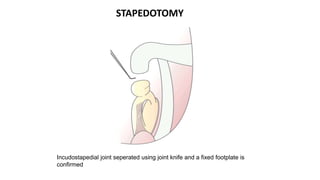
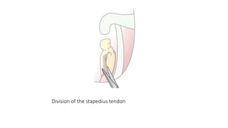
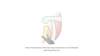
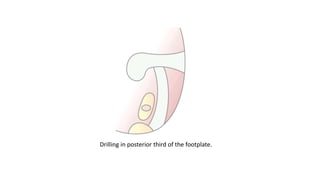
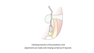
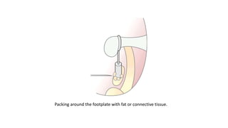
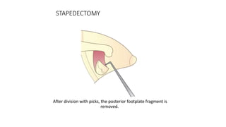
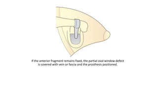
OTOSCLEROSIS
Dr. Anish Narayan Sur
INTRODUCTION
• Otosclerosis is a localized hereditary disorder of bone metabolism of
otic capsule enchondral bone that is characterized by disordered
resorption and deposition of bone
RELEVANT ANATOMY
• LABYRINTH:
1.Membrabous Labyrinth
2.Periotic Labyrinth
3.Bony Labyrinth
-Endosteal layer
-Enchondral layer
-Periosteal layer
ETIOLOGY
1.GENETIC
•OTSC(Otosclerosis)1,OTSC2,OTSC3,OTSC4,OTSC5, and OTSC7 are monogenetic
otosclerosis loci.
•A recent study has shown mutations and altered expression of the SERPINF1
(Serpin Peptidase Inhibitor-F1)gene in patients with familial otosclerosis. This
gene encodes PEDF (pigment epithelium-derived factor) which is a known
regulator of bone density and is the first disease associated gene to be
identified in otosclerosis.
•Other genes showing statistically significant association with otosclerosis are
the ACE(Angiotensin Converting Enzyme) gene,the AGT(Angiotensinogen)
gene,the RELN(Reelin) gene, the TGFB1(Transforming Growth Factor Beta 1)
gene
2.VIRAL INFECTION
• Immunohistochemical evidence of measles virus proteins and
antigenicity in active otosclerotic lesions.
• More recently, two studies have shown an increased expression of
specific measles virus receptor CD46 isoforms in otosclerotic
footplates
• Lower levels of anti-measles virus IgG were found in serum from
otosclerosis patients
• Assessment of anti-measles virus IgG serum level and the presence of
conductive hearing loss results in high diagnostic specificity of 90%
and high sensitivity of 96%
3.AUTOIMMUNE
• It has been suggested that otosclerosis represents a form of
autoimmune disease with humoral autoimmunity to type II collagen
or a closely related antigen that is abundantly present in the regions
of predilection
• Elevated circulating antibodies against type II collagen and type IX
collagen in the blood of patients with otosclerosis have been
reported.
4.CYTOKINES
• Bone morphogenetic protein(BMP), an inflammatory cytokine that is
part of the TGF-β superfamily, may be involved in pathological bone
remodelling in otosclerosis.
• BMPs play a pivotal role in bone formation as well as the healing
cascade of bone.
• Various isoforms, such as BMP2, BMP4, BMP5 and BMP7 have been
detected in fresh frozen footplates with active otosclerotic foci.
5.HORMONAL FACTOR
• Since angiotensin II stimulates the secretion of TNF-α(Tumour
Necrosis Factor Alpha), the renin–angiotensin-aldosterone system
(RAAS) may play a role in the regulation of bone remodelling.
EPIDEMIOLOGY
• Most common age of presentation-15-45 years
• Gender -M:F-1:2
RACE % INCIDENCE OF OTOSCLEROSIS
1.Caucasian 10
2.Asian 5
3.African American 1
PATHOGENESIS
Resorption of enchondral bone around blood vessels
consequent enlargement of perivascular spaces
followed by deposition of immature (woven) bone.
Through continuous remodelling, more mature bone (lamellar bone)
is deposited
ACTIVE PHASE
• Deposition of immature woven bone
• Increased Cellularity
• High Vascularity and Dilatation of vessels
INACTIVE PHASE
• Deposition of mature bone(thick and cellular)
• Vascular spaces are narrowed
TYPES
• Clinical otosclerosis refers to lesions that affect the stapes, stapediovestibular
joint or round window membrane and thus cause conductive hearing loss. In
cases of mixed hearing loss it is assumed that there are lesions affecting the
cochlear endosteum as well.
• Cochlear otosclerosis refers to lesions involving the cochlear endosteum
without affecting the stapes or the stapediovestibular joint, thus causing pure
sensorineural hearing loss, with no conductive element.
• Histologic otosclerosis refers to histopathological lesions that do not affect
the stapes, stapediovestibular joint or cochlear endosteum, and thus remain
asymptomatic during life.
CLINICAL PRESENTATION
• Typically, patients with otosclerosis will complain of a progressive
hearing loss.
• Approximately half of the patients will report a positive family
history of hearing loss.
• Paracusis Willisii-Patient hears better in noisy environment
• Vestibular symptom like vertigo is reported by 10 to 30% of patients.
• A ‘flamingo flush’ or Schwartz sign, a red blush of the tympanic
membrane over the promontory, is said to be due to the vascularity
of an active otosclerotic focus but is rarely seen.
INVESTIGTIONS
1.PURE TONE AUDIOMETRY
• Shows Conductive Hearing Loss
• Carhart's Notch
2.IMPEDANCE AUDIOMETRY
• Type As curve
3.HRCT Temporal Bone
• Not a routine part of the evaluation
of a patient with suspected
otosclerosis; it is helpful in
confirming or excluding the
presence of other pathologies
causing conductive loss
• Shows hypodense or lucent areas
within the otic capsule
• Double ring sign around cochlea
MEDICAL TREATMENT
1.SODIUM FLUORIDE
• Sodium fluoride is a known inhibitor of osteoclast activity and so a stabilizer
of bone turnover. It also leads to increased calcium deposition in
otospongiotic foci and decreased bone remodelling
• Dose-50-75 mg/day
2.BISPHOSPHONATES
• Reduce bone remodelling
• Recent study using new third generation biphosponates showed a reduction
in rate of progressive sensory hearing loss in patients with cochlear
otosclerosis
INDICATIONS
• When there is a reasonable expectations that surgery will result in a perceptible benefit to the
patient
• An air-bone gap of 25 dB or more at frequencies of 250 Hz to l kHz and a negative Rinne at 512 Hz
are considered to be good indicators
• In cases of bilateral involvement, the worse hearing ear is usually operated first.
• The patient's preference can be used to guide side selection in symmetric loss.
SURGICAL TREATMENT
CONTRAINDICATIONS
• Patients with infected middle or external ear, perforation of the tympanic membrane
• Surgery is not to be done in an only hearing ear
• In cases in which the contralateral ear has disease that may threaten hearing in the
future, surgery is relatively contraindicated.
• Advanced age is not a contraindication for surgery,although the likelihood of success
might be slightly reduced in patients older than 70 years.
ANAESTHESIA
•Choice of anaesthesia depends on patient's and surgeon's preferences and the nature of surgery
planned.
•Local anesthesia has the advantage of saving time compared to general anesthesia.
•Intraoperative patient reports of vestibular stimulation may be used as a safety measure to
prevent excessive inner ear irritation;
•However, operations under general anesthesia do not carry increased risk for vestibular
complication. General anesthesia provides assurance against pain and head Movement.
STAPEDOTOMY IS PREFERRED AS:
Less chances of trauma to labyrinth
Incidence of complication is very less
STAPEDECTOMY IS PREFERRED IN:
Floating Footplate
Comminuted fracture of the footplate
Footplate that is inadvertently removed during suprastructure dislocation
Revision Suregery
PROCEDURE
Incudostapedial joint seperated using joint knife and a fixed footplate is
confirmed
STAPEDOTOMY
Division of the stapedius tendon
Division of the posterior crus of the stapes using crurotomy scissors or a laser followed by
down fracture of the crura.
Drilling in posterior third of the footplate.
Following insertion of the prosthesis, final
adjustments are made and crimping carried out if required.
Packing around the footplate with fat or connective tissue.
STAPEDECTOMY
After division with picks, the posterior footplate fragment is
removed.
If the anterior fragment remains fixed, the partial oval window defect
is covered with vein or fascia and the prosthesis positioned.
If the anterior
footplate has become unstable, it is removed and a larger graft used to
cover the oval window.
• A recent variation is the laser stapedotomy minus
prosthesis (STAMP) procedure.
• All these techniques depend on a lack of ankylosis at the
posterior stapediovestibular joint.
• The laser procedure described by Silverstein et al. uses a
handheld laser probe to vaporize the anterior crus of
the stapes and perform a linear stapedotomy across
the anterior third of the footplate. The stapes mobilizes if
otosclerosis is confined to the anterior third of the
footplate
COMPLICATIONS
INTRAOPERATIVE
• Tear in the tympanic membrane
• Subluxation of the incus
• Persistent stapedial artery
• Malleus ankylosis
• Perilymph gusher
• Floating or depressed footplate
POSTOPERATIVE
• Facial palsy
• Chorda tympani nerve dysfunction
• Otitis media
• Vertigo
• Reperative granuloma
• Sensorineural hearing loss
• Conductive hearing loss
THANK YOU

OTOSCLEROSIS.ppt

  • 1.
  • 2.
    INTRODUCTION • Otosclerosis isa localized hereditary disorder of bone metabolism of otic capsule enchondral bone that is characterized by disordered resorption and deposition of bone
  • 3.
    RELEVANT ANATOMY • LABYRINTH: 1.MembrabousLabyrinth 2.Periotic Labyrinth 3.Bony Labyrinth -Endosteal layer -Enchondral layer -Periosteal layer
  • 4.
    ETIOLOGY 1.GENETIC •OTSC(Otosclerosis)1,OTSC2,OTSC3,OTSC4,OTSC5, and OTSC7are monogenetic otosclerosis loci. •A recent study has shown mutations and altered expression of the SERPINF1 (Serpin Peptidase Inhibitor-F1)gene in patients with familial otosclerosis. This gene encodes PEDF (pigment epithelium-derived factor) which is a known regulator of bone density and is the first disease associated gene to be identified in otosclerosis. •Other genes showing statistically significant association with otosclerosis are the ACE(Angiotensin Converting Enzyme) gene,the AGT(Angiotensinogen) gene,the RELN(Reelin) gene, the TGFB1(Transforming Growth Factor Beta 1) gene
  • 5.
    2.VIRAL INFECTION • Immunohistochemicalevidence of measles virus proteins and antigenicity in active otosclerotic lesions. • More recently, two studies have shown an increased expression of specific measles virus receptor CD46 isoforms in otosclerotic footplates • Lower levels of anti-measles virus IgG were found in serum from otosclerosis patients • Assessment of anti-measles virus IgG serum level and the presence of conductive hearing loss results in high diagnostic specificity of 90% and high sensitivity of 96%
  • 6.
    3.AUTOIMMUNE • It hasbeen suggested that otosclerosis represents a form of autoimmune disease with humoral autoimmunity to type II collagen or a closely related antigen that is abundantly present in the regions of predilection • Elevated circulating antibodies against type II collagen and type IX collagen in the blood of patients with otosclerosis have been reported.
  • 7.
    4.CYTOKINES • Bone morphogeneticprotein(BMP), an inflammatory cytokine that is part of the TGF-β superfamily, may be involved in pathological bone remodelling in otosclerosis. • BMPs play a pivotal role in bone formation as well as the healing cascade of bone. • Various isoforms, such as BMP2, BMP4, BMP5 and BMP7 have been detected in fresh frozen footplates with active otosclerotic foci. 5.HORMONAL FACTOR • Since angiotensin II stimulates the secretion of TNF-α(Tumour Necrosis Factor Alpha), the renin–angiotensin-aldosterone system (RAAS) may play a role in the regulation of bone remodelling.
  • 8.
    EPIDEMIOLOGY • Most commonage of presentation-15-45 years • Gender -M:F-1:2 RACE % INCIDENCE OF OTOSCLEROSIS 1.Caucasian 10 2.Asian 5 3.African American 1
  • 9.
    PATHOGENESIS Resorption of enchondralbone around blood vessels consequent enlargement of perivascular spaces followed by deposition of immature (woven) bone. Through continuous remodelling, more mature bone (lamellar bone) is deposited
  • 10.
    ACTIVE PHASE • Depositionof immature woven bone • Increased Cellularity • High Vascularity and Dilatation of vessels INACTIVE PHASE • Deposition of mature bone(thick and cellular) • Vascular spaces are narrowed
  • 11.
    TYPES • Clinical otosclerosisrefers to lesions that affect the stapes, stapediovestibular joint or round window membrane and thus cause conductive hearing loss. In cases of mixed hearing loss it is assumed that there are lesions affecting the cochlear endosteum as well. • Cochlear otosclerosis refers to lesions involving the cochlear endosteum without affecting the stapes or the stapediovestibular joint, thus causing pure sensorineural hearing loss, with no conductive element. • Histologic otosclerosis refers to histopathological lesions that do not affect the stapes, stapediovestibular joint or cochlear endosteum, and thus remain asymptomatic during life.
  • 13.
    CLINICAL PRESENTATION • Typically,patients with otosclerosis will complain of a progressive hearing loss. • Approximately half of the patients will report a positive family history of hearing loss. • Paracusis Willisii-Patient hears better in noisy environment • Vestibular symptom like vertigo is reported by 10 to 30% of patients. • A ‘flamingo flush’ or Schwartz sign, a red blush of the tympanic membrane over the promontory, is said to be due to the vascularity of an active otosclerotic focus but is rarely seen.
  • 14.
    INVESTIGTIONS 1.PURE TONE AUDIOMETRY •Shows Conductive Hearing Loss • Carhart's Notch 2.IMPEDANCE AUDIOMETRY • Type As curve 3.HRCT Temporal Bone • Not a routine part of the evaluation of a patient with suspected otosclerosis; it is helpful in confirming or excluding the presence of other pathologies causing conductive loss • Shows hypodense or lucent areas within the otic capsule • Double ring sign around cochlea
  • 15.
    MEDICAL TREATMENT 1.SODIUM FLUORIDE •Sodium fluoride is a known inhibitor of osteoclast activity and so a stabilizer of bone turnover. It also leads to increased calcium deposition in otospongiotic foci and decreased bone remodelling • Dose-50-75 mg/day 2.BISPHOSPHONATES • Reduce bone remodelling • Recent study using new third generation biphosponates showed a reduction in rate of progressive sensory hearing loss in patients with cochlear otosclerosis
  • 16.
    INDICATIONS • When thereis a reasonable expectations that surgery will result in a perceptible benefit to the patient • An air-bone gap of 25 dB or more at frequencies of 250 Hz to l kHz and a negative Rinne at 512 Hz are considered to be good indicators • In cases of bilateral involvement, the worse hearing ear is usually operated first. • The patient's preference can be used to guide side selection in symmetric loss. SURGICAL TREATMENT
  • 17.
    CONTRAINDICATIONS • Patients withinfected middle or external ear, perforation of the tympanic membrane • Surgery is not to be done in an only hearing ear • In cases in which the contralateral ear has disease that may threaten hearing in the future, surgery is relatively contraindicated. • Advanced age is not a contraindication for surgery,although the likelihood of success might be slightly reduced in patients older than 70 years.
  • 18.
    ANAESTHESIA •Choice of anaesthesiadepends on patient's and surgeon's preferences and the nature of surgery planned. •Local anesthesia has the advantage of saving time compared to general anesthesia. •Intraoperative patient reports of vestibular stimulation may be used as a safety measure to prevent excessive inner ear irritation; •However, operations under general anesthesia do not carry increased risk for vestibular complication. General anesthesia provides assurance against pain and head Movement.
  • 19.
    STAPEDOTOMY IS PREFERREDAS: Less chances of trauma to labyrinth Incidence of complication is very less STAPEDECTOMY IS PREFERRED IN: Floating Footplate Comminuted fracture of the footplate Footplate that is inadvertently removed during suprastructure dislocation Revision Suregery
  • 20.
  • 21.
    Incudostapedial joint seperatedusing joint knife and a fixed footplate is confirmed STAPEDOTOMY
  • 22.
    Division of thestapedius tendon
  • 23.
    Division of theposterior crus of the stapes using crurotomy scissors or a laser followed by down fracture of the crura.
  • 24.
    Drilling in posteriorthird of the footplate.
  • 25.
    Following insertion ofthe prosthesis, final adjustments are made and crimping carried out if required.
  • 26.
    Packing around thefootplate with fat or connective tissue.
  • 27.
    STAPEDECTOMY After division withpicks, the posterior footplate fragment is removed.
  • 28.
    If the anteriorfragment remains fixed, the partial oval window defect is covered with vein or fascia and the prosthesis positioned.
  • 29.
    If the anterior footplatehas become unstable, it is removed and a larger graft used to cover the oval window.
  • 30.
    • A recentvariation is the laser stapedotomy minus prosthesis (STAMP) procedure. • All these techniques depend on a lack of ankylosis at the posterior stapediovestibular joint. • The laser procedure described by Silverstein et al. uses a handheld laser probe to vaporize the anterior crus of the stapes and perform a linear stapedotomy across the anterior third of the footplate. The stapes mobilizes if otosclerosis is confined to the anterior third of the footplate
  • 31.
    COMPLICATIONS INTRAOPERATIVE • Tear inthe tympanic membrane • Subluxation of the incus • Persistent stapedial artery • Malleus ankylosis • Perilymph gusher • Floating or depressed footplate
  • 32.
    POSTOPERATIVE • Facial palsy •Chorda tympani nerve dysfunction • Otitis media • Vertigo • Reperative granuloma • Sensorineural hearing loss • Conductive hearing loss
  • 33.