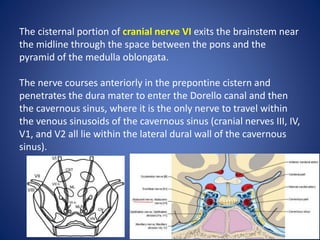
Downloaded 267 times
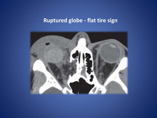
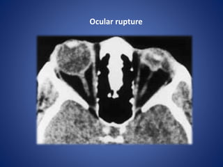
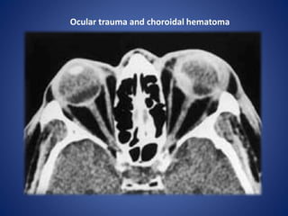
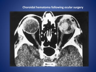
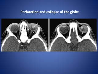
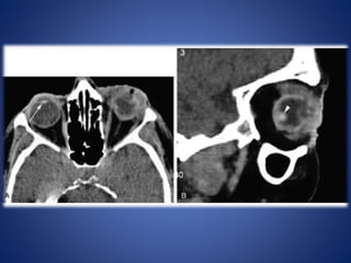
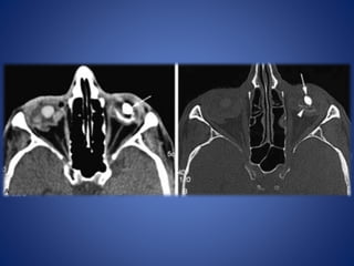
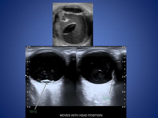
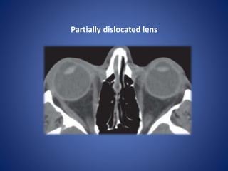
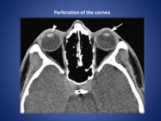
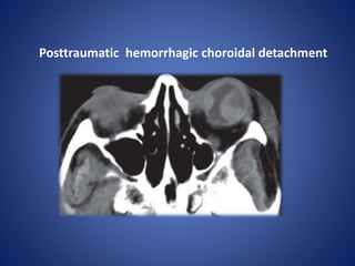
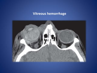
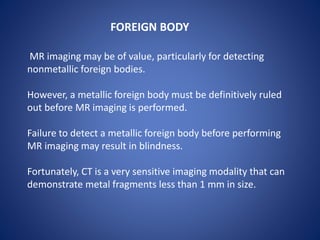
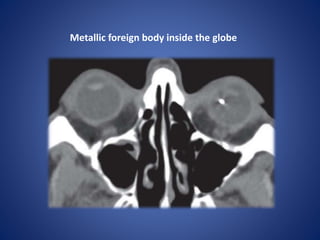
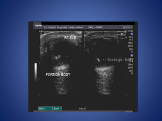
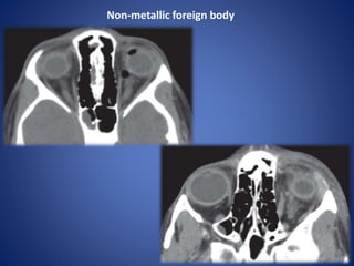
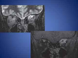
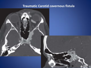
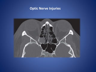
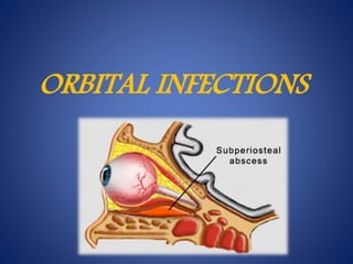
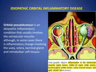
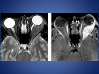
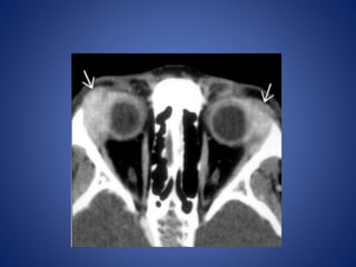

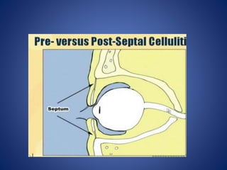
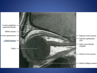
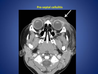

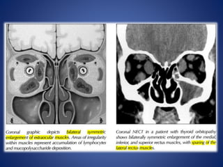

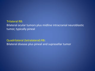
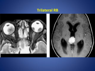
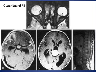
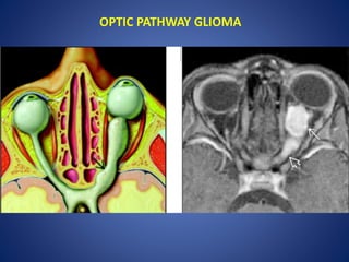
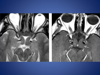
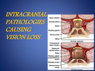
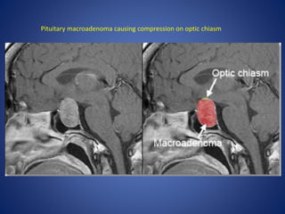
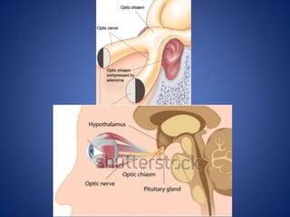
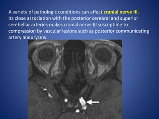
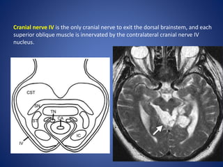
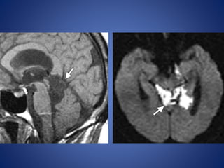


This document summarizes various orbital and intracranial pathologies that can cause vision loss or eye abnormalities. It describes conditions such as ruptured globe, retinal detachment, choroidal detachment, optic neuritis, thyroid orbitopathy, retinoblastoma, optic pathway glioma, and pituitary macroadenomas, among others. Diagnostic imaging findings are provided for many conditions. Orbital cellulitis and inflammatory diseases are distinguished. The document also outlines anatomical details of certain cranial nerves that are susceptible to compression or injury.


