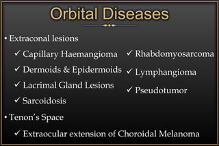
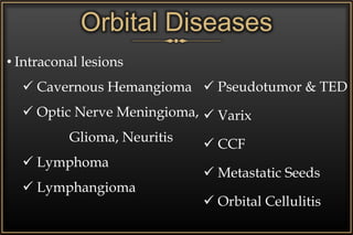
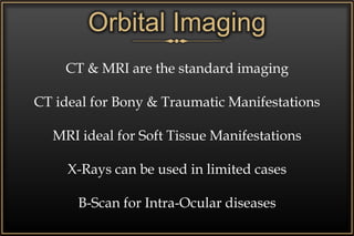
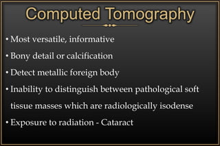
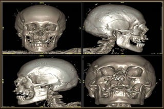
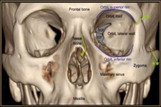
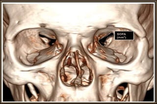
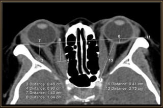
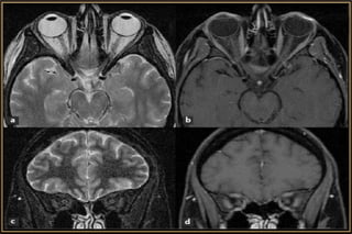
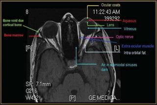
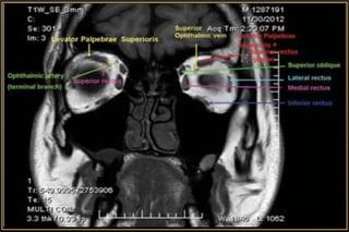
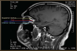
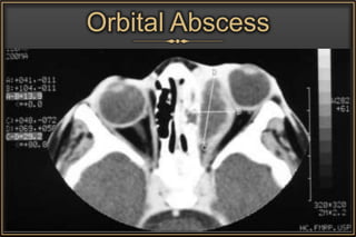
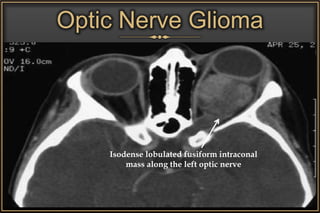
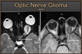
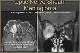
Chairman
Dr. Syed Nabil Bin Maruf
FCPS - II Course Student
NIO&H
Moderator
Dr. Zinat
Nasrin
Assistant Professor
Department of Radiology
NIO&H
Dr. Shafiul
Mostafiz
Assistant Professor
Fellow - Department of Oculoplasty
NIO&H
Imaging inOrbital
Diseases
• Pair of Confined Spaces in the Skull
• Volume : 30 cc
• Direct Link with Cranial Vault
• Contains the Eyeballs along with other important
structures
Orbits
Imaging Modalities
 Radiography
 Ultrasonography
 Computed Tomography
 Magnetic Resonance Imaging
Anatomical Considerations
• Size, Shape & Relations
• 4 Walls, Base, Apex & Passages of Orbit
• Fascial Compartments / Surgical Spaces
• Contents & their Anatomy
• Paranasal Sinuses
Orbital Diseases
• Congenital Malformations
• Inflammatory & Infective
• Vascular Lesions
• Cystic Lesions & Tumours
• Ocular Diseases
• Orbital Trauma
Orbital Diseases
Surgical Spaces
• Extraorbital Space
 Bony Lesions & Sinus Lesions
• Subperiosteal Space
 Subperiosteal haematoma, abscess
 Inflammatory & Neoplastic lesions of PNS
Orbital Diseases
• Extraconal lesions
 Capillary Haemangioma
 Dermoids & Epidermoids
 Lacrimal Gland Lesions
 Sarcoidosis
• Tenon’s Space
 Extraocular extension of Choroidal Melanoma
 Rhabdomyosarcoma
 Lymphangioma
 Pseudotumor
Orbital Diseases
• Intraconal lesions
 Cavernous Hemangioma
 Optic Nerve Meningioma,
Glioma, Neuritis
 Lymphoma
 Lymphangioma
 Pseudotumor & TED
 Varix
 CCF
 Metastatic Seeds
 Orbital Cellulitis
Orbital Imaging
CT & MRI are the standard imaging
CT ideal for Bony & Traumatic Manifestations
MRI ideal for Soft Tissue Manifestations
X-Rays can be used in limited cases
B-Scan for Intra-Ocular diseases
• Most versatile, informative
• Bony detail or calcification
• Detect metallic foreign body
• Inability to distinguish between pathological soft
tissue masses which are radiologically isodense
• Exposure to radiation - Cataract
Computed Tomography
Computed Tomography
Major Considerations
• Slice thickness
• Imaging plane – Axial / Coronal / Sagittal
• Tissue window – Soft or Brain / Bone
• Contrast enhancement – Iohexol (46% Iodine)
• Modification of CT procedure
• Orbit with Brain CT
• Better for soft tissue
• Not ideal to visualise Bony manifestations
• Better resolution of Optic Nerve & Orbital Apex
• No radiation
• Must screen for metallic foreign bodies in orbit
Magnetic Resonance Imaging
Basic Image Sequences
T1- weighted (T1W)
• Fat appear brighter than water/vitreous/CSF
T2- weighted (T2W)
• Water/vitreous/CSF appear brighter than tissues
like blood products
Magnetic Resonance Imaging
Fluid attenuation inversion recovery (FLAIR)
• Signal from fluid suppressed
• Useful in demyelinating conditions as white matter
hyperintensities on T2W images are better
appreciated with adjacent CSF bright signals nulled
Magnetic Resonance Imaging
Post contrast images
• Gadolinium-T1W
Fat-suppressed images
• Bright signal from intraorbital fat can mask the signal
and enhancement of pathology
Magnetic Resonance Imaging
Orbital
Diseases
CT-Scan & MRI
Thyroid Associated
Orbitopathy
Idiopathic Orbital
Inflammatory Disease
Enlargement of Muscle including tendon
Hilal & Trokel
Orbital Abscess
Optic Nerve Glioma
Isodense lobulated fusiform intraconal
mass along the left optic nerve
Optic Nerve Glioma
Hyperintense
Hypointense
Optic Nerve Sheath
Meningioma
Globular Configuration of Optic
Nerve Sheath Meningioma with
“Tram Track Sign”
Fat-suppressed T1 axial (A) coronal (B) MRI, with Gadolinium
Optic Nerve Sheath
Meningioma
Hyperdense mass surrounding the
left optic nerve with slightly
lobulated contour and nerve
compression
Orbital Schwannoma
Orbital Neurofibroma
Lacrimal Gland Tumour
Pleomorphic Adenoma
showing rounded well-
defined lacrimal gland
lesion
Bilateral Benign
Lymphoid Hyperplasia
showing oblong
diffuse enlargement of
the lacrimal gland
Rhabdomyosarcoma
Retinoblastoma
Cavernous Haemangioma
Thrombosis
Capillary Haemangioma
Dermoid
Optic Neuritis
Axial T1W Axial T1W-FLAIR Axial T1W
Angiography & Venograpy
MRA / MRV & CTA / CTV
• Indicated for assessing vascular lesions:
 Arteriovenous Malformation
 Haemangioma
 Lymphangioma
 Venous Malformation
 Varix
 Arterial Aneurysm
Radiography
Plain X-Ray of the Orbit
• Very limited role
• Indications:
 Traumatic Injuries
 Foreign Bodies
Radiography
X-Ray Views
• Waters View
• Caldwell’s View
• Lateral View
• Submentovertex View
• Rhese View
Water’s View
a - Frontal sinus
b - Medial Orbital Wall
c - Innominate Line
d - Inferior Orbital Rim
e - Orbital Floor
f - Maxillary Antrum
g - Superior Orbital Fissure
h - Zygomaticofrontal Suture
i - Zygomatic Arch
Caldwell’s View
a - Frontal sinus
b - Innominate line
c - Inferior orbital rim
d - Posterior orbital floor
e - Superior orbital
fissure
f - Greater wing of
sphenoid
g - Ethmoid air sinus
h - Medial orbital wall
i - Petrous ridge
j - Zygomaticofrontal
suture
k - Foramen rotundum
Lateral View
Orbital Roof
Ethmoid Sinus
Anterior Clinoid
Process
Sella Turcica
Planum
Sphenoidale
Frontal Sinus
Maxillary Sinus
Submentovertex View
a - Zygomatic arch
b - Orbit
c - Lateral orbital wall
d - Posterior wall of maxillary
sinus
e - Pterygoid plate
f - Sphenoid sinus
Rhese View
a - Right Optic Canal
b - Optic Strut
c - Superior Orbital Fissure
d - Ethmoid Sinus
e - Planum Sphenoidale
f - Greater Wing of Sphenoid
Orbital Fat
Herniation
Orbital
Emphysema
Orbital Fracture
Water’s View
Radiography
X-Ray Requisition for
Intraorbital / Intraocular Foreign Body
• X-Ray (A/P) of Orbits
• If Intraocular Foreign Body noted -
 Lateral View of Respective Side with Eyes
looking Up & Down
Orbital Foreign Body
Ultrasonography
• A- & B-Scan can play an important role in differential
diagnoses of orbital masses
• Site, morphology and structure of the lesion (B-scan)
and on the acoustic structure, internal reflectivity,
vascularization and margins of the lesion (A-scan)
• Poor transmission is characteristic in any Solid,
Infiltrative or Inflammatory mass of the orbit
• Real-Time & Dynamic
Acoustically Abnormal Orbit
Mass Lesion
Foreign Body
Inflammatory Lesion
• Cellulitis
• Abscess
• Pseudotumour
Rounded
 Hyper-reflective
• Meningioma, Glioma
• Neurofibroma
Hyporeflective
• Mucocele
• Dermoid Cyst
• Cavernous Haemangioma
Irregular
 Hyper-reflective
• Lymphoma
• Metastatic Cancer
Hyporeflective
• Haemangioma
• Lymphangioma
Ultrasonography
Cornea
Orbital Lymphoma
Lens Retina
Vitreous
Optic Nerve
Hyper-reflective retrobulbar round
solid mass
Cavernous Haemangioma
• 10-MHz A & B-scan
• Well encapsulated
lesion
• Typical hemangioma
Advanced Modalities
MRI-DWI (Diffusion Weighted Imaging)
MR Spectroscopy
Perfusion Studies
Positron Emission Tomography (PET) – CT / MRI
• Can characterize lesions at molecular level
• Distinguish malignant from benign
• Metastatic evaluation
Conclusion
• CT-Scan is the Imaging of Choice
• MRI in case of Orbital Apex, Optic Canal or
Intracranial extension of Orbital Neoplasm
• Coordinate with the Radiologist for best outcome
• Diagnose on the basis of Clinical Correlation
Thank You

Orbital Imaging

  • 1.
    Chairman Dr. Syed NabilBin Maruf FCPS - II Course Student NIO&H Moderator Dr. Zinat Nasrin Assistant Professor Department of Radiology NIO&H Dr. Shafiul Mostafiz Assistant Professor Fellow - Department of Oculoplasty NIO&H Imaging inOrbital Diseases
  • 2.
    • Pair ofConfined Spaces in the Skull • Volume : 30 cc • Direct Link with Cranial Vault • Contains the Eyeballs along with other important structures Orbits
  • 3.
    Imaging Modalities  Radiography Ultrasonography  Computed Tomography  Magnetic Resonance Imaging
  • 4.
    Anatomical Considerations • Size,Shape & Relations • 4 Walls, Base, Apex & Passages of Orbit • Fascial Compartments / Surgical Spaces • Contents & their Anatomy • Paranasal Sinuses
  • 5.
    Orbital Diseases • CongenitalMalformations • Inflammatory & Infective • Vascular Lesions • Cystic Lesions & Tumours • Ocular Diseases • Orbital Trauma
  • 6.
    Orbital Diseases Surgical Spaces •Extraorbital Space  Bony Lesions & Sinus Lesions • Subperiosteal Space  Subperiosteal haematoma, abscess  Inflammatory & Neoplastic lesions of PNS
  • 7.
    Orbital Diseases • Extraconallesions  Capillary Haemangioma  Dermoids & Epidermoids  Lacrimal Gland Lesions  Sarcoidosis • Tenon’s Space  Extraocular extension of Choroidal Melanoma  Rhabdomyosarcoma  Lymphangioma  Pseudotumor
  • 8.
    Orbital Diseases • Intraconallesions  Cavernous Hemangioma  Optic Nerve Meningioma, Glioma, Neuritis  Lymphoma  Lymphangioma  Pseudotumor & TED  Varix  CCF  Metastatic Seeds  Orbital Cellulitis
  • 9.
    Orbital Imaging CT &MRI are the standard imaging CT ideal for Bony & Traumatic Manifestations MRI ideal for Soft Tissue Manifestations X-Rays can be used in limited cases B-Scan for Intra-Ocular diseases
  • 10.
    • Most versatile,informative • Bony detail or calcification • Detect metallic foreign body • Inability to distinguish between pathological soft tissue masses which are radiologically isodense • Exposure to radiation - Cataract Computed Tomography
  • 13.
    Computed Tomography Major Considerations •Slice thickness • Imaging plane – Axial / Coronal / Sagittal • Tissue window – Soft or Brain / Bone • Contrast enhancement – Iohexol (46% Iodine) • Modification of CT procedure • Orbit with Brain CT
  • 19.
    • Better forsoft tissue • Not ideal to visualise Bony manifestations • Better resolution of Optic Nerve & Orbital Apex • No radiation • Must screen for metallic foreign bodies in orbit Magnetic Resonance Imaging
  • 20.
    Magnetic Resonance Imaging BasicImage Sequences T1- weighted (T1W) • Fat appear brighter than water/vitreous/CSF T2- weighted (T2W) • Water/vitreous/CSF appear brighter than tissues like blood products
  • 21.
    Magnetic Resonance Imaging Fluidattenuation inversion recovery (FLAIR) • Signal from fluid suppressed • Useful in demyelinating conditions as white matter hyperintensities on T2W images are better appreciated with adjacent CSF bright signals nulled
  • 22.
    Magnetic Resonance Imaging Postcontrast images • Gadolinium-T1W Fat-suppressed images • Bright signal from intraorbital fat can mask the signal and enhancement of pathology
  • 23.
  • 27.
  • 28.
  • 29.
  • 30.
  • 31.
  • 32.
    Optic Nerve Glioma Isodenselobulated fusiform intraconal mass along the left optic nerve
  • 33.
  • 34.
    Optic Nerve Sheath Meningioma GlobularConfiguration of Optic Nerve Sheath Meningioma with “Tram Track Sign” Fat-suppressed T1 axial (A) coronal (B) MRI, with Gadolinium
  • 35.
    Optic Nerve Sheath Meningioma Hyperdensemass surrounding the left optic nerve with slightly lobulated contour and nerve compression
  • 36.
  • 37.
  • 38.
    Lacrimal Gland Tumour PleomorphicAdenoma showing rounded well- defined lacrimal gland lesion Bilateral Benign Lymphoid Hyperplasia showing oblong diffuse enlargement of the lacrimal gland
  • 39.
  • 40.
  • 41.
  • 42.
  • 43.
  • 44.
    Optic Neuritis Axial T1WAxial T1W-FLAIR Axial T1W
  • 45.
    Angiography & Venograpy MRA/ MRV & CTA / CTV • Indicated for assessing vascular lesions:  Arteriovenous Malformation  Haemangioma  Lymphangioma  Venous Malformation  Varix  Arterial Aneurysm
  • 46.
    Radiography Plain X-Ray ofthe Orbit • Very limited role • Indications:  Traumatic Injuries  Foreign Bodies
  • 47.
    Radiography X-Ray Views • WatersView • Caldwell’s View • Lateral View • Submentovertex View • Rhese View
  • 48.
    Water’s View a -Frontal sinus b - Medial Orbital Wall c - Innominate Line d - Inferior Orbital Rim e - Orbital Floor f - Maxillary Antrum g - Superior Orbital Fissure h - Zygomaticofrontal Suture i - Zygomatic Arch
  • 49.
    Caldwell’s View a -Frontal sinus b - Innominate line c - Inferior orbital rim d - Posterior orbital floor e - Superior orbital fissure f - Greater wing of sphenoid g - Ethmoid air sinus h - Medial orbital wall i - Petrous ridge j - Zygomaticofrontal suture k - Foramen rotundum
  • 50.
    Lateral View Orbital Roof EthmoidSinus Anterior Clinoid Process Sella Turcica Planum Sphenoidale Frontal Sinus Maxillary Sinus
  • 51.
    Submentovertex View a -Zygomatic arch b - Orbit c - Lateral orbital wall d - Posterior wall of maxillary sinus e - Pterygoid plate f - Sphenoid sinus
  • 52.
    Rhese View a -Right Optic Canal b - Optic Strut c - Superior Orbital Fissure d - Ethmoid Sinus e - Planum Sphenoidale f - Greater Wing of Sphenoid
  • 54.
  • 55.
    Radiography X-Ray Requisition for Intraorbital/ Intraocular Foreign Body • X-Ray (A/P) of Orbits • If Intraocular Foreign Body noted -  Lateral View of Respective Side with Eyes looking Up & Down
  • 56.
  • 57.
    Ultrasonography • A- &B-Scan can play an important role in differential diagnoses of orbital masses • Site, morphology and structure of the lesion (B-scan) and on the acoustic structure, internal reflectivity, vascularization and margins of the lesion (A-scan) • Poor transmission is characteristic in any Solid, Infiltrative or Inflammatory mass of the orbit • Real-Time & Dynamic
  • 58.
    Acoustically Abnormal Orbit MassLesion Foreign Body Inflammatory Lesion • Cellulitis • Abscess • Pseudotumour Rounded  Hyper-reflective • Meningioma, Glioma • Neurofibroma Hyporeflective • Mucocele • Dermoid Cyst • Cavernous Haemangioma Irregular  Hyper-reflective • Lymphoma • Metastatic Cancer Hyporeflective • Haemangioma • Lymphangioma Ultrasonography
  • 59.
    Cornea Orbital Lymphoma Lens Retina Vitreous OpticNerve Hyper-reflective retrobulbar round solid mass
  • 60.
    Cavernous Haemangioma • 10-MHzA & B-scan • Well encapsulated lesion • Typical hemangioma
  • 61.
    Advanced Modalities MRI-DWI (DiffusionWeighted Imaging) MR Spectroscopy Perfusion Studies Positron Emission Tomography (PET) – CT / MRI • Can characterize lesions at molecular level • Distinguish malignant from benign • Metastatic evaluation
  • 62.
    Conclusion • CT-Scan isthe Imaging of Choice • MRI in case of Orbital Apex, Optic Canal or Intracranial extension of Orbital Neoplasm • Coordinate with the Radiologist for best outcome • Diagnose on the basis of Clinical Correlation
  • 63.

Editor's Notes

  • #14 Slide Thickness • Spatial resolution of a CT depends on slice thickness. • The thinner the slice, the higher the resolution. • Usually, 2mm cuts are optimal for the eye and orbit. • In special situations (like evaluation of the orbital apex/Optic Nerve), thinner slices of 1mm/1.5mm can be more informative. Modification of CT procedure • Certain cases may require special modifications during the scanning procedure to aid diagnosis. • In a case of orbital venous varix, it is important to request for special scans (with contrast) while the patient performs a Valsalva maneuver. Simultaneous brain CT • Suspected neurocysticercosis with orbital involvement. • Head injury with orbital trauma • Optic nerve meningiomas
  • #30 A&B – CT C&D – Fat supressed T1W