Downloaded 142 times

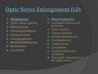
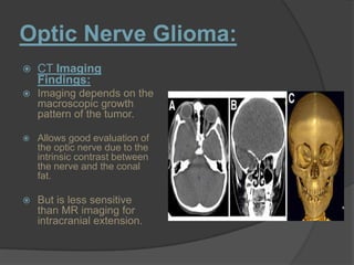
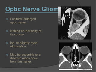
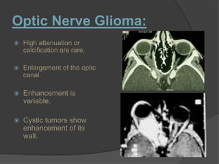
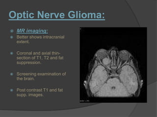
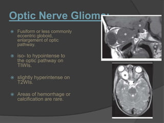
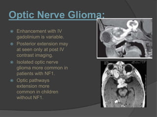
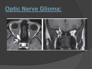
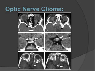
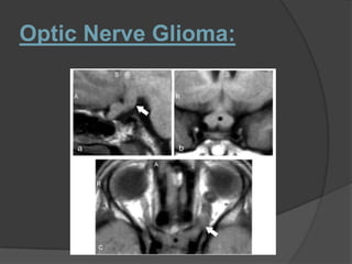
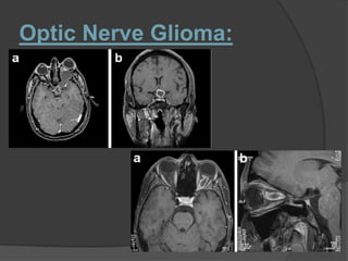
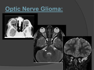
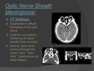
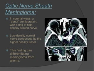
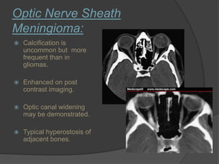
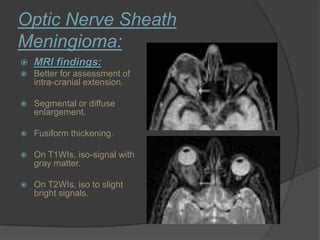
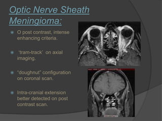
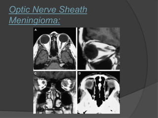
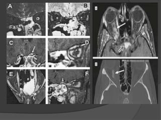
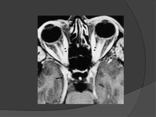


This document discusses imaging findings of optic nerve enlargement. It focuses on optic nerve glioma and optic nerve sheath meningioma. For optic nerve glioma, CT can evaluate the optic nerve but MR imaging is more sensitive for intracranial extension. Findings include fusiform nerve enlargement that may enhance variably. For optic nerve sheath meningioma, CT can show tram-track or donut appearances, while MRI is best to assess intra-cranial spread, showing segmental nerve thickening and enhancement. Differential diagnosis includes tumors, increased intracranial pressure, and other non-neoplastic causes.


