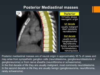
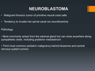
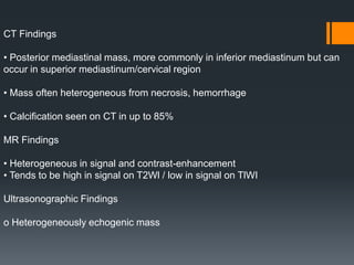
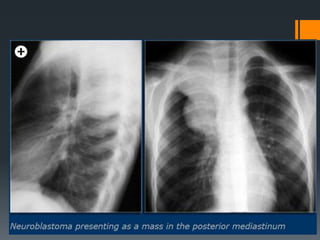
SlideShare a Scribd company logo
PEDIATRIC
MEDIASTINAL MASSES
Dr Mohit Goel
21 Nov. 2012
Paediatric Mediastinal Masses:
Lymphoma
Lymphoma is one of the most common mediastinal neoplasms and may affect
any mediastinal location. Non-Hodgkin’s lymphoma usually manifests as
generalised disease whereas Hodgkin’s disease tends to present as primary
mediastinal lesions.
Hodgkin lymphoma
Hodgkin lymphoma in children is more common in the second decade of life.
It can exhibit as multiple rounded soft tissue masses, a dominant bulky soft
tissue mass, or a discrete or infiltrating thymic mass.
Lesions may comprise homogenous or heterogeneous soft tissue attenuation
depending on the presence of necrosis, haemorrhage, or cystic degeneration.
Foci of calcifications may be seen post-therapy.
The CT-images shows a large soft tissue mass in the anterior mediastinum, which
arises in the thymus. There is associated paratracheal adenopathy (arrow).
Non-Hodgkin lymphoma
Non-Hodgkin disease in children occurs in the first and second decade of life.
It is associated with extranodal disease
and has a greater predilection for
noncontiguous and/or haematogenous
spread to thoracic and distant nodal
and extranodal sites.
Non-Hodgkin disease, in contrast to
Hodgkin disease, often spares the
thymus.
In this case, enlarged lymph nodes are
seen in the right paratracheal , hilar and subcarinal areas without thymus
involvement..
Thymic hyperplasia
In childhood, thymic hyperplasia is most often 'rebound' hyperplasia associated
with chemotherapy, particularly therapy with corticosteroids.
The mechanism of hyperplasia is believed to be initial depletion of lymphocytes
from the cortical portion of the gland due to high serum levels of
glucocorticoids, followed by repopulation of the cortical lymphocytes when the
cortisone levels return to normal.
On CT, hyperplasia appears as diffuse enlargement of the thymus, with
preservation of the normal triangular shape.
CT, MRI of PET cannot differentiate rebound hyperplasia from infiltration of the
thymus by tumor.
The absence of other active disease and a gradual decrease in thymus size on
serial CT's supports the diagnosis of rebound hyperplasia.
The thymus usually returns to its normal size in 3 to 6 months.
Thymic Hyperplasia
THYMOMA
Thymoma is the commonest primary tumour of the anterior
mediastinum. It occurs most frequently in adults older than 40
years and is rare in children and adolescents.
Thymoma appears as a well-defined, rounded or lobulated
anterior-superior mediastinal mass anterior to the aortic root.
The mass contains either homogenous or heterogeneous
contents depending on the presence of haemorrhage, necrosis,
or cyst formation.
Calcific foci are seen on CT in a minority of patients
Thymic Carcinoma
Squamous cell and lymphoepithelioma-like carcinoma are the most common
histological types. These occur most commonly in middle-aged adults. The
appearance is of a large poorly defined infiltrative anterior mediastinal
mass and it is commonly associated with pleural and pericardial effusions, and
regional lymph node and distant metastasis.
Thymic carcinoma.
CT shows large anterior mediastinal mass with
ill-defined medial border. The superior
vena cava is compressed.
Thymolipoma
Thymolipoma is an uncommon benign slow growing neoplasm of the thymus
gland composed of mature adipose cells and thymic tissue. It is typically a
large soft anterior mediastinal mass and is able to conform to adjacent
structures simulating cardiomegaly, lobar collapse, and diaphragmatic
elevation.
Calcifications are absent.
Thymolipoma does not
have a capsule and does
not have any mass effect.
Non-neoplastic thymic cyst may be congenital or acquired secondary to
inflammation. It is seen as a well
Circumscribed antero-superior
Mediastinal mass with low
attenuation contents. Typically,
they are thin walled, homogeneous
masses of near water attenuation
On CT the attenuation value may
be higher than that of simple cysts
when the contents are
proteinaceous
or hemorrhagic rather than serous.
The cystic mass may be uni- or
multiloculated and may show
curvilinear calcification of the
cystic wall or septa .
Non-neoplastic Thymic Cyst
Germ Cell Tumour
Germ-cell tumors are the most common cause of a fat containing lesions in the
anterior mediastinum and the second most common cause of an anterior
mediastinal mass in children.
Approximately 90 % are benign germ-cell tumors.
Most arise in the thymus.
Mediastinal teratoma occurs in children and young adults with no sex
predilection.
On CT, the teratoma appears as a multi-locular cystic tumour with walls of
variable thickness. The combination of fluid, soft tissue, calcium, and fat
attenuation in an anterior mediastinal mass is a highly specific finding that
allows the prospective diagnosis of mature teratoma. Mature teratomas can be
very large and still be benign.
A fat-fluid level produced by high lipid content in the cyst fluid is a rare but
diagnostic sign.
Anterior mediastinal teratoma - A large heterogenous left anterior mediastinal mass
containing soft tissue , fatty and calcific components.
Epicardial fat pad.
(a) PA chest radiograph shows loss of the cardiac silhouette at the border of the right
side of the heart and an epicardial fat pad with relatively low density (arrow)
(b) CT scan shows the fat pad (arrow) as an area of homogeneous fat attenuation
adjacent to the right border of the heart.
Right-sided retrosternal
goiter. (a) PAchest
radiograph demonstrates a
goiter (arrow) extending
into the middle
mediastinum, obliterating
the right paratracheal
stripe, and causing
deviation of the trachea to
the left (black arrowhead).
Above the level of the
clavicles, the margins of
the mass are not sharp
(white arrowhead),
indicating that the mass
has an anterior mediastinal
component.
Posterior mediastinal masses above the level of the clavicles have an interface with
lung and therefore typically have sharp, well-defined margins; in contrast, anterior
masses above the level of the clavicles do not have an interface with lung, so that their
margins are not usually sharp.
(b) CT scan shows the mass (arrow) between the trachea and right lung, a location
that explains the obliteration of the right paratracheal stripe seen in a. There is no
contact between the anterior component of the mass and the lung (arrowhead) at the
level of the clavicular heads, a relationship that continues above the level of the
clavicles. This finding explains why the lateral border of the anterior mediastinal
component above the level of the clavicles is not sharp in a.
Cystic Hygroma/Lymphangioma
Cystic hygroma / lymphangioma is a benign proliferation of interconnecting
lymphatic vessels and sacs that may grow in an infiltrative fashion. It typically
affects infants younger than 6 months of age.
Mediastinal lymphangioma typically occurs in the superior aspect of the
anterior mediastinum and is usually contiguous with a cervical or axillary
component.
The mass usually appears as rounded, lobulated, multi-cystic tumour that can
reach a massive size. It tends to surround and displace mediastinal structures
and may infiltrate across tissue planes. The thin or thick septa may enhance
minimally after contrast administration .
Due to its infiltrating nature, complete surgical resection may be difficult and
close follow-up is needed to check for recurrence.
Cystic hygroma in a 1-year-old baby boy with respiratory distress
The great vessels are encased and displaced although they are all patent. There is
no evidence of superior vena cava obstruction (arrows show the brachiocephalic
veins).
Pericardial Cyst
It is seen as a well marginated , spherical, or teardrop shaped mass that
characteristically abuts the heart, the anterior chest wall, and the diaphragm.
The right anterior cardiophrenic
angle is the most common site.
A pericardial cyst is typically
shown as a unilocular,
non-enhancing mass
with water attenuation contents
and an imperceptible wall .
.
Middle Mediastinal masses
Foregut cysts in the middle mediastinum are classified as bronchogenic or
enteric.
Bronchogenic cysts are lined by respiratory epithelium and most are located
in the subcarinal or right paratracheal area in close proximity to the trachea
or bronchus.
Enteric cysts are lined by gastrointestinal mucosa and are located in a
paraspinal position in the middle to posterior mediastinum near the
esophagus
BRONCHOGENIC CYST
They are developmental lesions that result from abnormal ventral budding of the
tracheobronchial tree between the 26th and 40th days of gestation.
• Location
Mediastinal location is more common than pulmonary
o Mediastinal 65-90%
Majority in the middle mediastinum
Typically para tracheal, carinal, or hilar
Pericarinal most common
o Pulmonary: Majority in the medial third of the lungs, More frequent in the lower lobes
Typically do not communicate with airway and do not contain air, Air presence indicates
infection.
CT Findings
• NECT
o Homogeneous well circumscribed lesion
o Cyst contents variable: Water to proteinaceous
o Hence CT attenuation is variable
• CECT
o Well-defined, typically with nonenhancing or minimally enhancing thin wall
o More prominent wall enhancement and wall thickening may be seen with infection
o No central enhancement
MR Findings
• TlWI : o Well-circumscribed lesion
o Homogeneous signal intensity unless infected
o Variable signal due to varying amounts of proteinaceous material, but usually
water signal
o Imperceptible wall
• T2WI: Signal is almost always equal to or greater than cerebrospinal fluid (CSF)
• STIR: Markedly increased signal, equal to or greater than CSF
• Tl C+ : o May have a thin rim of mild enhancement
o Thicker enhancing wall implies infection
o No central enhancement
(Left) Axial T2WI MR shows homogeneous, well circumscribed ovoid mass (arrow)
with signal greater than CSF (curved arrow).
(Right) AP radiograph shows large, smooth, homogeneous, left retrocardiac
parenchymal mass (arrows).
Enteric foregut cyst
The images show a well defined lesion of water attenuation in the lower mediastinum in
close proximity to the esophagus, which is typical for an enteric foregut cyst.
Posterior Mediastinal masses
Posterior mediastinal masses are of neural origin in approximately 95 % of cases and
may arise from sympathetic ganglion cells (neuroblastoma, ganglioneuroblastoma or
ganglioneuroma) or from nerve sheaths (neurofibroma or schwannoma).
In the first decade of life they are usually malignant, most commonly neuroblastoma.
In the second decade or life they are usually benign (ganglioneuroma, neurofibroma,
rarely schwanoma).
• Malignant thoracic tumor of primitive neural crest cells
• Tendency to invade into spinal canal via neuroforamina
NEUROBLASTOMA
Pathology
• Most commonly arises from the adrenal gland but can arise anywhere along
sympathetic chain, including posterior mediastinum
• Third most common pediatric malignancy behind leukemia and central
nervous system tumors
• Radiography
o Soft tissue mass in posterior mediastinum
o Rib involvement
• Widening of intercostal spaces
• Erosion/destruction of ribs
o Calcifications: Common (up to 30% by radiography)
o Paravertebral soft tissue widening
o Bone metastasis
• Lucent or sclerotic lesions
o Pedicle erosion from intraspinal extension
CT Findings
• Posterior mediastinal mass, more commonly in inferior mediastinum but can
occur in superior mediastinum/cervical region
• Mass often heterogeneous from necrosis, hemorrhage
• Calcification seen on CT in up to 85%
MR Findings
• Heterogeneous in signal and contrast-enhancement
• Tends to be high in signal on T2Wl / low in signal on TlWI
Ultrasonographic Findings
o Heterogeneously echogenic mass
The CT-images show a calcified mass in the posterior mediastinum extending over
several vertebrae, which grows into the vertebral canal.
THANK YOU

More Related Content

What's hot

Ct chest pneumonias and neoplasms
Ct chest pneumonias and neoplasmsCt chest pneumonias and neoplasms
Ct chest pneumonias and neoplasms
Rikin Hasnani
 
Liver segments on ultrasound
Liver segments on ultrasoundLiver segments on ultrasound
Liver segments on ultrasound
Durre Sabih
 
Gloved finger sign and cervicothoracic sign
Gloved finger sign and cervicothoracic signGloved finger sign and cervicothoracic sign
Gloved finger sign and cervicothoracic sign
Minstry of health ,Ibn alnafis hoapital, Damascus
 
The elbow joint in concern of diagnostic imaging .pptx 1
The elbow joint in concern of diagnostic imaging .pptx 1The elbow joint in concern of diagnostic imaging .pptx 1
The elbow joint in concern of diagnostic imaging .pptx 1
DR Laith
 
Pediatric chest part 2
Pediatric chest part 2Pediatric chest part 2
Pediatric chest part 2
Anish Choudhary
 
Radioanatomy of mediastinum and approach to mediastinal masses
Radioanatomy of mediastinum and approach to mediastinal massesRadioanatomy of mediastinum and approach to mediastinal masses
Radioanatomy of mediastinum and approach to mediastinal masses
AkankshaMalviya3
 
Diagnostic Imaging of Mediastinal Masses
Diagnostic Imaging of Mediastinal MassesDiagnostic Imaging of Mediastinal Masses
Diagnostic Imaging of Mediastinal Masses
Mohamed M.A. Zaitoun
 
Presentation1, radiological imaging of barium studies.
Presentation1, radiological imaging of barium studies.Presentation1, radiological imaging of barium studies.
Presentation1, radiological imaging of barium studies.
Abdellah Nazeer
 
Computed Tomography of Liver and Pancreas- Avinesh Shrestha
Computed Tomography of Liver and Pancreas- Avinesh ShresthaComputed Tomography of Liver and Pancreas- Avinesh Shrestha
Computed Tomography of Liver and Pancreas- Avinesh Shrestha
Avinesh Shrestha
 
Cross sectional anatomy of chest by Dr. Milan Silwal, Resident, NAMS, Kathman...
Cross sectional anatomy of chest by Dr. Milan Silwal, Resident, NAMS, Kathman...Cross sectional anatomy of chest by Dr. Milan Silwal, Resident, NAMS, Kathman...
Cross sectional anatomy of chest by Dr. Milan Silwal, Resident, NAMS, Kathman...
Milan Silwal
 
Presentation2.pptx, radiological imaging of gastric lesions.
Presentation2.pptx, radiological imaging of gastric lesions.Presentation2.pptx, radiological imaging of gastric lesions.
Presentation2.pptx, radiological imaging of gastric lesions.Abdellah Nazeer
 
Presentation1.pptx, radiological imaging of small bowel disease.
Presentation1.pptx, radiological imaging of small bowel disease.Presentation1.pptx, radiological imaging of small bowel disease.
Presentation1.pptx, radiological imaging of small bowel disease.Abdellah Nazeer
 
Anal perianal imaging part 1 CT MRI anatomy Dr Ahmed Esawy
Anal perianal imaging part 1 CT MRI anatomy Dr Ahmed EsawyAnal perianal imaging part 1 CT MRI anatomy Dr Ahmed Esawy
Anal perianal imaging part 1 CT MRI anatomy Dr Ahmed Esawy
AHMED ESAWY
 
Presentation1.pptx, radiological imaging of prostatic diseases
Presentation1.pptx, radiological imaging of prostatic diseasesPresentation1.pptx, radiological imaging of prostatic diseases
Presentation1.pptx, radiological imaging of prostatic diseasesAbdellah Nazeer
 
Presentation1.pptx, radiological imaging of congenital abnormalities of the l...
Presentation1.pptx, radiological imaging of congenital abnormalities of the l...Presentation1.pptx, radiological imaging of congenital abnormalities of the l...
Presentation1.pptx, radiological imaging of congenital abnormalities of the l...Abdellah Nazeer
 
Ct anatomy of orbit
Ct anatomy of orbitCt anatomy of orbit
Ct anatomy of orbit
Maajid Mohi ud din
 
Anatomy of duodenum
Anatomy of duodenumAnatomy of duodenum
Anatomy of duodenum
anwaradil4
 
Presentation1.pptx, radiological imaging of cholangiocarcinoma.
Presentation1.pptx, radiological imaging of cholangiocarcinoma.Presentation1.pptx, radiological imaging of cholangiocarcinoma.
Presentation1.pptx, radiological imaging of cholangiocarcinoma.Abdellah Nazeer
 
Presentation1, radiological imaging of gastro intestinal stromal tumour(gist).
Presentation1, radiological imaging of gastro intestinal stromal tumour(gist).Presentation1, radiological imaging of gastro intestinal stromal tumour(gist).
Presentation1, radiological imaging of gastro intestinal stromal tumour(gist).
Abdellah Nazeer
 
Imaging of aortic pathologies
Imaging of aortic pathologies Imaging of aortic pathologies
Imaging of aortic pathologies
Pankaj Kaira
 

What's hot (20)

Ct chest pneumonias and neoplasms
Ct chest pneumonias and neoplasmsCt chest pneumonias and neoplasms
Ct chest pneumonias and neoplasms
 
Liver segments on ultrasound
Liver segments on ultrasoundLiver segments on ultrasound
Liver segments on ultrasound
 
Gloved finger sign and cervicothoracic sign
Gloved finger sign and cervicothoracic signGloved finger sign and cervicothoracic sign
Gloved finger sign and cervicothoracic sign
 
The elbow joint in concern of diagnostic imaging .pptx 1
The elbow joint in concern of diagnostic imaging .pptx 1The elbow joint in concern of diagnostic imaging .pptx 1
The elbow joint in concern of diagnostic imaging .pptx 1
 
Pediatric chest part 2
Pediatric chest part 2Pediatric chest part 2
Pediatric chest part 2
 
Radioanatomy of mediastinum and approach to mediastinal masses
Radioanatomy of mediastinum and approach to mediastinal massesRadioanatomy of mediastinum and approach to mediastinal masses
Radioanatomy of mediastinum and approach to mediastinal masses
 
Diagnostic Imaging of Mediastinal Masses
Diagnostic Imaging of Mediastinal MassesDiagnostic Imaging of Mediastinal Masses
Diagnostic Imaging of Mediastinal Masses
 
Presentation1, radiological imaging of barium studies.
Presentation1, radiological imaging of barium studies.Presentation1, radiological imaging of barium studies.
Presentation1, radiological imaging of barium studies.
 
Computed Tomography of Liver and Pancreas- Avinesh Shrestha
Computed Tomography of Liver and Pancreas- Avinesh ShresthaComputed Tomography of Liver and Pancreas- Avinesh Shrestha
Computed Tomography of Liver and Pancreas- Avinesh Shrestha
 
Cross sectional anatomy of chest by Dr. Milan Silwal, Resident, NAMS, Kathman...
Cross sectional anatomy of chest by Dr. Milan Silwal, Resident, NAMS, Kathman...Cross sectional anatomy of chest by Dr. Milan Silwal, Resident, NAMS, Kathman...
Cross sectional anatomy of chest by Dr. Milan Silwal, Resident, NAMS, Kathman...
 
Presentation2.pptx, radiological imaging of gastric lesions.
Presentation2.pptx, radiological imaging of gastric lesions.Presentation2.pptx, radiological imaging of gastric lesions.
Presentation2.pptx, radiological imaging of gastric lesions.
 
Presentation1.pptx, radiological imaging of small bowel disease.
Presentation1.pptx, radiological imaging of small bowel disease.Presentation1.pptx, radiological imaging of small bowel disease.
Presentation1.pptx, radiological imaging of small bowel disease.
 
Anal perianal imaging part 1 CT MRI anatomy Dr Ahmed Esawy
Anal perianal imaging part 1 CT MRI anatomy Dr Ahmed EsawyAnal perianal imaging part 1 CT MRI anatomy Dr Ahmed Esawy
Anal perianal imaging part 1 CT MRI anatomy Dr Ahmed Esawy
 
Presentation1.pptx, radiological imaging of prostatic diseases
Presentation1.pptx, radiological imaging of prostatic diseasesPresentation1.pptx, radiological imaging of prostatic diseases
Presentation1.pptx, radiological imaging of prostatic diseases
 
Presentation1.pptx, radiological imaging of congenital abnormalities of the l...
Presentation1.pptx, radiological imaging of congenital abnormalities of the l...Presentation1.pptx, radiological imaging of congenital abnormalities of the l...
Presentation1.pptx, radiological imaging of congenital abnormalities of the l...
 
Ct anatomy of orbit
Ct anatomy of orbitCt anatomy of orbit
Ct anatomy of orbit
 
Anatomy of duodenum
Anatomy of duodenumAnatomy of duodenum
Anatomy of duodenum
 
Presentation1.pptx, radiological imaging of cholangiocarcinoma.
Presentation1.pptx, radiological imaging of cholangiocarcinoma.Presentation1.pptx, radiological imaging of cholangiocarcinoma.
Presentation1.pptx, radiological imaging of cholangiocarcinoma.
 
Presentation1, radiological imaging of gastro intestinal stromal tumour(gist).
Presentation1, radiological imaging of gastro intestinal stromal tumour(gist).Presentation1, radiological imaging of gastro intestinal stromal tumour(gist).
Presentation1, radiological imaging of gastro intestinal stromal tumour(gist).
 
Imaging of aortic pathologies
Imaging of aortic pathologies Imaging of aortic pathologies
Imaging of aortic pathologies
 

Viewers also liked

Double Aortic Arch with TOF
Double Aortic Arch with TOFDouble Aortic Arch with TOF
Double Aortic Arch with TOF
Jb Dhull
 
Pediatric chest pain
Pediatric chest painPediatric chest pain
Pediatric chest painShama
 
Pediatric stroke radiology
Pediatric stroke radiologyPediatric stroke radiology
Pediatric stroke radiology
Dr. Mohit Goel
 
Approach to Chest Pain in Children
Approach to Chest Pain in ChildrenApproach to Chest Pain in Children
Approach to Chest Pain in Children
Dr Padmesh Vadakepat
 
Preoperative assesment in cochlear implantation
Preoperative assesment in cochlear implantationPreoperative assesment in cochlear implantation
Preoperative assesment in cochlear implantation
Sunil Kumar
 
Dr. RAHUL VC TIWARI, SIBAR INSTITUTE OF DENTAL SCIENCES, GUNTUR, ANDHRA PRADE...
Dr. RAHUL VC TIWARI, SIBAR INSTITUTE OF DENTAL SCIENCES, GUNTUR, ANDHRA PRADE...Dr. RAHUL VC TIWARI, SIBAR INSTITUTE OF DENTAL SCIENCES, GUNTUR, ANDHRA PRADE...
Dr. RAHUL VC TIWARI, SIBAR INSTITUTE OF DENTAL SCIENCES, GUNTUR, ANDHRA PRADE...
CLOVE Dental OMNI Hospitals Andhra Hospital
 
Ser 2016 30 mts k sodhi
Ser 2016 30 mts k sodhiSer 2016 30 mts k sodhi
Ser 2016 30 mts k sodhi
Teleradiology Solutions
 
Clinical case base of tongue cancer
Clinical case base of tongue cancerClinical case base of tongue cancer
Clinical case base of tongue cancer
crsalim
 
Imaging of demyelinating diseases final
Imaging of demyelinating diseases finalImaging of demyelinating diseases final
Imaging of demyelinating diseases final
Sunil Kumar
 
Imaging and pathology of larynx (2)
Imaging and pathology of larynx (2)Imaging and pathology of larynx (2)
Imaging and pathology of larynx (2)
Sunil Kumar
 
Mediastinal mass
Mediastinal massMediastinal mass
Mediastinal mass
Vikram Patil
 
Case presentation
Case presentationCase presentation
Case presentationairwave12
 
Pediatric chest infection imaging considerations
Pediatric chest infection imaging considerationsPediatric chest infection imaging considerations
Pediatric chest infection imaging considerations
Ahmed Bahnassy
 
Diagnostic Imaging of Congenital Pulmonary Abnormalities
Diagnostic Imaging of Congenital Pulmonary AbnormalitiesDiagnostic Imaging of Congenital Pulmonary Abnormalities
Diagnostic Imaging of Congenital Pulmonary Abnormalities
Mohamed M.A. Zaitoun
 
Oral cavity, pharynx radio-anatomy
Oral cavity, pharynx radio-anatomyOral cavity, pharynx radio-anatomy
Oral cavity, pharynx radio-anatomy
Dr. Mohit Goel
 
Nephrotic Syndrome
Nephrotic SyndromeNephrotic Syndrome
Nephrotic Syndrome
HIRANGER
 

Viewers also liked (20)

Double Aortic Arch with TOF
Double Aortic Arch with TOFDouble Aortic Arch with TOF
Double Aortic Arch with TOF
 
Pediatric chest
Pediatric chestPediatric chest
Pediatric chest
 
Pediatric chest pain
Pediatric chest painPediatric chest pain
Pediatric chest pain
 
Pediatric stroke radiology
Pediatric stroke radiologyPediatric stroke radiology
Pediatric stroke radiology
 
Approach to Chest Pain in Children
Approach to Chest Pain in ChildrenApproach to Chest Pain in Children
Approach to Chest Pain in Children
 
Preoperative assesment in cochlear implantation
Preoperative assesment in cochlear implantationPreoperative assesment in cochlear implantation
Preoperative assesment in cochlear implantation
 
Dr. RAHUL VC TIWARI, SIBAR INSTITUTE OF DENTAL SCIENCES, GUNTUR, ANDHRA PRADE...
Dr. RAHUL VC TIWARI, SIBAR INSTITUTE OF DENTAL SCIENCES, GUNTUR, ANDHRA PRADE...Dr. RAHUL VC TIWARI, SIBAR INSTITUTE OF DENTAL SCIENCES, GUNTUR, ANDHRA PRADE...
Dr. RAHUL VC TIWARI, SIBAR INSTITUTE OF DENTAL SCIENCES, GUNTUR, ANDHRA PRADE...
 
Poster chest masses
Poster   chest massesPoster   chest masses
Poster chest masses
 
A Case of Mediastinal Mass
A Case of Mediastinal MassA Case of Mediastinal Mass
A Case of Mediastinal Mass
 
Ser 2016 30 mts k sodhi
Ser 2016 30 mts k sodhiSer 2016 30 mts k sodhi
Ser 2016 30 mts k sodhi
 
Clinical case base of tongue cancer
Clinical case base of tongue cancerClinical case base of tongue cancer
Clinical case base of tongue cancer
 
Imaging of demyelinating diseases final
Imaging of demyelinating diseases finalImaging of demyelinating diseases final
Imaging of demyelinating diseases final
 
Imaging and pathology of larynx (2)
Imaging and pathology of larynx (2)Imaging and pathology of larynx (2)
Imaging and pathology of larynx (2)
 
Mediastinal mass
Mediastinal massMediastinal mass
Mediastinal mass
 
Case presentation
Case presentationCase presentation
Case presentation
 
A Case of Pancoast's tumour
A Case of Pancoast's tumourA Case of Pancoast's tumour
A Case of Pancoast's tumour
 
Pediatric chest infection imaging considerations
Pediatric chest infection imaging considerationsPediatric chest infection imaging considerations
Pediatric chest infection imaging considerations
 
Diagnostic Imaging of Congenital Pulmonary Abnormalities
Diagnostic Imaging of Congenital Pulmonary AbnormalitiesDiagnostic Imaging of Congenital Pulmonary Abnormalities
Diagnostic Imaging of Congenital Pulmonary Abnormalities
 
Oral cavity, pharynx radio-anatomy
Oral cavity, pharynx radio-anatomyOral cavity, pharynx radio-anatomy
Oral cavity, pharynx radio-anatomy
 
Nephrotic Syndrome
Nephrotic SyndromeNephrotic Syndrome
Nephrotic Syndrome
 

Similar to Pediatric chest (part 2)

Cystic masses of neck
Cystic masses of neckCystic masses of neck
Cystic masses of neck
PRAMODG11
 
Cystic neck masses
Cystic neck massesCystic neck masses
Cystic neck masses
Navni Garg
 
Congenital neck mass radiology pk final
Congenital neck mass radiology pk finalCongenital neck mass radiology pk final
Congenital neck mass radiology pk final
Dr pradeep Kumar
 
Amol cardiac tumours
Amol cardiac tumoursAmol cardiac tumours
Amol cardiac tumours
Amol Gulhane
 
RADIOIMAGING IN MEDIASTINAL MASSES YN.pptx
RADIOIMAGING IN MEDIASTINAL MASSES YN.pptxRADIOIMAGING IN MEDIASTINAL MASSES YN.pptx
RADIOIMAGING IN MEDIASTINAL MASSES YN.pptx
dypradio
 
Pediatric abdominal tumors
Pediatric abdominal tumorsPediatric abdominal tumors
Pediatric abdominal tumors
passant dorgham
 
Presentation1.pptx, radiological imaging of extra nodal lymphoma.
Presentation1.pptx, radiological imaging of extra nodal lymphoma.Presentation1.pptx, radiological imaging of extra nodal lymphoma.
Presentation1.pptx, radiological imaging of extra nodal lymphoma.Abdellah Nazeer
 
Benign liver lesions
Benign liver lesionsBenign liver lesions
Benign liver lesionsairwave12
 
Abdominal masses in children
Abdominal masses in childrenAbdominal masses in children
Abdominal masses in children
aliebrahimi60
 
Imaging of Pleural tumors Dr Alekya
Imaging of Pleural tumors Dr Alekya Imaging of Pleural tumors Dr Alekya
Imaging of Pleural tumors Dr Alekya
Alex Allu
 
Presentation1.pptx, chest film reading. lecture 2
Presentation1.pptx, chest film reading. lecture 2Presentation1.pptx, chest film reading. lecture 2
Presentation1.pptx, chest film reading. lecture 2Abdellah Nazeer
 
Mediastinal and lung masses xrays images radiology.pdf
Mediastinal and lung masses xrays images radiology.pdfMediastinal and lung masses xrays images radiology.pdf
Mediastinal and lung masses xrays images radiology.pdf
Nagasai Pelala
 
spotters
spotters spotters
spotters
JineshJain285582
 
spotters
spotters spotters
spotters
JineshJain285582
 
spotters
spotters spotters
spotters
JineshJain285582
 
Inraventricular mases
Inraventricular masesInraventricular mases
Inraventricular mases
Ali Jiwani
 
Chest diseases
Chest diseasesChest diseases
Chest diseases
FaizahMohdZakiPPUKM
 
Atypical lung neoplasms1
Atypical lung neoplasms1Atypical lung neoplasms1
Atypical lung neoplasms1
Jayanth Hiremagalur
 
Surgery 5th year, 6th lecture (Dr. Ahmed Al-Azzawi)
Surgery 5th year, 6th lecture (Dr. Ahmed Al-Azzawi)Surgery 5th year, 6th lecture (Dr. Ahmed Al-Azzawi)
Surgery 5th year, 6th lecture (Dr. Ahmed Al-Azzawi)
College of Medicine, Sulaymaniyah
 
Scortal Ultrasound
Scortal UltrasoundScortal Ultrasound
Scortal Ultrasound
Double M
 

Similar to Pediatric chest (part 2) (20)

Cystic masses of neck
Cystic masses of neckCystic masses of neck
Cystic masses of neck
 
Cystic neck masses
Cystic neck massesCystic neck masses
Cystic neck masses
 
Congenital neck mass radiology pk final
Congenital neck mass radiology pk finalCongenital neck mass radiology pk final
Congenital neck mass radiology pk final
 
Amol cardiac tumours
Amol cardiac tumoursAmol cardiac tumours
Amol cardiac tumours
 
RADIOIMAGING IN MEDIASTINAL MASSES YN.pptx
RADIOIMAGING IN MEDIASTINAL MASSES YN.pptxRADIOIMAGING IN MEDIASTINAL MASSES YN.pptx
RADIOIMAGING IN MEDIASTINAL MASSES YN.pptx
 
Pediatric abdominal tumors
Pediatric abdominal tumorsPediatric abdominal tumors
Pediatric abdominal tumors
 
Presentation1.pptx, radiological imaging of extra nodal lymphoma.
Presentation1.pptx, radiological imaging of extra nodal lymphoma.Presentation1.pptx, radiological imaging of extra nodal lymphoma.
Presentation1.pptx, radiological imaging of extra nodal lymphoma.
 
Benign liver lesions
Benign liver lesionsBenign liver lesions
Benign liver lesions
 
Abdominal masses in children
Abdominal masses in childrenAbdominal masses in children
Abdominal masses in children
 
Imaging of Pleural tumors Dr Alekya
Imaging of Pleural tumors Dr Alekya Imaging of Pleural tumors Dr Alekya
Imaging of Pleural tumors Dr Alekya
 
Presentation1.pptx, chest film reading. lecture 2
Presentation1.pptx, chest film reading. lecture 2Presentation1.pptx, chest film reading. lecture 2
Presentation1.pptx, chest film reading. lecture 2
 
Mediastinal and lung masses xrays images radiology.pdf
Mediastinal and lung masses xrays images radiology.pdfMediastinal and lung masses xrays images radiology.pdf
Mediastinal and lung masses xrays images radiology.pdf
 
spotters
spotters spotters
spotters
 
spotters
spotters spotters
spotters
 
spotters
spotters spotters
spotters
 
Inraventricular mases
Inraventricular masesInraventricular mases
Inraventricular mases
 
Chest diseases
Chest diseasesChest diseases
Chest diseases
 
Atypical lung neoplasms1
Atypical lung neoplasms1Atypical lung neoplasms1
Atypical lung neoplasms1
 
Surgery 5th year, 6th lecture (Dr. Ahmed Al-Azzawi)
Surgery 5th year, 6th lecture (Dr. Ahmed Al-Azzawi)Surgery 5th year, 6th lecture (Dr. Ahmed Al-Azzawi)
Surgery 5th year, 6th lecture (Dr. Ahmed Al-Azzawi)
 
Scortal Ultrasound
Scortal UltrasoundScortal Ultrasound
Scortal Ultrasound
 

More from Dr. Mohit Goel

Utrasound Gall-bladder & biliary tract
Utrasound Gall-bladder & biliary tractUtrasound Gall-bladder & biliary tract
Utrasound Gall-bladder & biliary tract
Dr. Mohit Goel
 
Ultrasoud hernia
Ultrasoud herniaUltrasoud hernia
Ultrasoud hernia
Dr. Mohit Goel
 
TVS image gallery
TVS image galleryTVS image gallery
TVS image gallery
Dr. Mohit Goel
 
Transitional vertebrae radiology
Transitional vertebrae radiologyTransitional vertebrae radiology
Transitional vertebrae radiology
Dr. Mohit Goel
 
Shoulder ultrasound
Shoulder ultrasoundShoulder ultrasound
Shoulder ultrasound
Dr. Mohit Goel
 
Shoulder labral tears MRI
Shoulder labral tears MRIShoulder labral tears MRI
Shoulder labral tears MRI
Dr. Mohit Goel
 
Sectional radio-anatomy of abdomen
Sectional radio-anatomy of abdomenSectional radio-anatomy of abdomen
Sectional radio-anatomy of abdomen
Dr. Mohit Goel
 
Renal doppler usg
Renal doppler usgRenal doppler usg
Renal doppler usg
Dr. Mohit Goel
 
Retroperitoneal masses radiology
Retroperitoneal masses radiologyRetroperitoneal masses radiology
Retroperitoneal masses radiology
Dr. Mohit Goel
 
Prostate carcinoma raiology
Prostate carcinoma raiologyProstate carcinoma raiology
Prostate carcinoma raiology
Dr. Mohit Goel
 
Precocious puberty - Imaging
Precocious puberty - ImagingPrecocious puberty - Imaging
Precocious puberty - Imaging
Dr. Mohit Goel
 
Pre-FESS PNS CT
Pre-FESS PNS CTPre-FESS PNS CT
Pre-FESS PNS CT
Dr. Mohit Goel
 
PNS (Para-nasal-sinuses) anatomy and variants
PNS (Para-nasal-sinuses) anatomy and variantsPNS (Para-nasal-sinuses) anatomy and variants
PNS (Para-nasal-sinuses) anatomy and variants
Dr. Mohit Goel
 
Pineal region masses - radiology
Pineal region masses - radiologyPineal region masses - radiology
Pineal region masses - radiology
Dr. Mohit Goel
 
Peripheral arterial doppler
Peripheral  arterial dopplerPeripheral  arterial doppler
Peripheral arterial doppler
Dr. Mohit Goel
 
Patello femoral joint - MRI
Patello femoral joint - MRIPatello femoral joint - MRI
Patello femoral joint - MRI
Dr. Mohit Goel
 
Orbital pathologies radiology
Orbital pathologies radiologyOrbital pathologies radiology
Orbital pathologies radiology
Dr. Mohit Goel
 
Osteopetrosis
OsteopetrosisOsteopetrosis
Osteopetrosis
Dr. Mohit Goel
 
Orbital pathologies.pptx (part 1)
Orbital pathologies.pptx (part 1)Orbital pathologies.pptx (part 1)
Orbital pathologies.pptx (part 1)
Dr. Mohit Goel
 
Orbit imaging anatomy
Orbit imaging anatomyOrbit imaging anatomy
Orbit imaging anatomy
Dr. Mohit Goel
 

More from Dr. Mohit Goel (20)

Utrasound Gall-bladder & biliary tract
Utrasound Gall-bladder & biliary tractUtrasound Gall-bladder & biliary tract
Utrasound Gall-bladder & biliary tract
 
Ultrasoud hernia
Ultrasoud herniaUltrasoud hernia
Ultrasoud hernia
 
TVS image gallery
TVS image galleryTVS image gallery
TVS image gallery
 
Transitional vertebrae radiology
Transitional vertebrae radiologyTransitional vertebrae radiology
Transitional vertebrae radiology
 
Shoulder ultrasound
Shoulder ultrasoundShoulder ultrasound
Shoulder ultrasound
 
Shoulder labral tears MRI
Shoulder labral tears MRIShoulder labral tears MRI
Shoulder labral tears MRI
 
Sectional radio-anatomy of abdomen
Sectional radio-anatomy of abdomenSectional radio-anatomy of abdomen
Sectional radio-anatomy of abdomen
 
Renal doppler usg
Renal doppler usgRenal doppler usg
Renal doppler usg
 
Retroperitoneal masses radiology
Retroperitoneal masses radiologyRetroperitoneal masses radiology
Retroperitoneal masses radiology
 
Prostate carcinoma raiology
Prostate carcinoma raiologyProstate carcinoma raiology
Prostate carcinoma raiology
 
Precocious puberty - Imaging
Precocious puberty - ImagingPrecocious puberty - Imaging
Precocious puberty - Imaging
 
Pre-FESS PNS CT
Pre-FESS PNS CTPre-FESS PNS CT
Pre-FESS PNS CT
 
PNS (Para-nasal-sinuses) anatomy and variants
PNS (Para-nasal-sinuses) anatomy and variantsPNS (Para-nasal-sinuses) anatomy and variants
PNS (Para-nasal-sinuses) anatomy and variants
 
Pineal region masses - radiology
Pineal region masses - radiologyPineal region masses - radiology
Pineal region masses - radiology
 
Peripheral arterial doppler
Peripheral  arterial dopplerPeripheral  arterial doppler
Peripheral arterial doppler
 
Patello femoral joint - MRI
Patello femoral joint - MRIPatello femoral joint - MRI
Patello femoral joint - MRI
 
Orbital pathologies radiology
Orbital pathologies radiologyOrbital pathologies radiology
Orbital pathologies radiology
 
Osteopetrosis
OsteopetrosisOsteopetrosis
Osteopetrosis
 
Orbital pathologies.pptx (part 1)
Orbital pathologies.pptx (part 1)Orbital pathologies.pptx (part 1)
Orbital pathologies.pptx (part 1)
 
Orbit imaging anatomy
Orbit imaging anatomyOrbit imaging anatomy
Orbit imaging anatomy
 

Recently uploaded

Biological Screening of Herbal Drugs in detailed.
Biological Screening of Herbal Drugs in detailed.Biological Screening of Herbal Drugs in detailed.
Biological Screening of Herbal Drugs in detailed.
Ashokrao Mane college of Pharmacy Peth-Vadgaon
 
678020731-Sumas-y-Restas-Para-Colorear.pdf
678020731-Sumas-y-Restas-Para-Colorear.pdf678020731-Sumas-y-Restas-Para-Colorear.pdf
678020731-Sumas-y-Restas-Para-Colorear.pdf
CarlosHernanMontoyab2
 
The Roman Empire A Historical Colossus.pdf
The Roman Empire A Historical Colossus.pdfThe Roman Empire A Historical Colossus.pdf
The Roman Empire A Historical Colossus.pdf
kaushalkr1407
 
Home assignment II on Spectroscopy 2024 Answers.pdf
Home assignment II on Spectroscopy 2024 Answers.pdfHome assignment II on Spectroscopy 2024 Answers.pdf
Home assignment II on Spectroscopy 2024 Answers.pdf
Tamralipta Mahavidyalaya
 
CACJapan - GROUP Presentation 1- Wk 4.pdf
CACJapan - GROUP Presentation 1- Wk 4.pdfCACJapan - GROUP Presentation 1- Wk 4.pdf
CACJapan - GROUP Presentation 1- Wk 4.pdf
camakaiclarkmusic
 
Chapter 3 - Islamic Banking Products and Services.pptx
Chapter 3 - Islamic Banking Products and Services.pptxChapter 3 - Islamic Banking Products and Services.pptx
Chapter 3 - Islamic Banking Products and Services.pptx
Mohd Adib Abd Muin, Senior Lecturer at Universiti Utara Malaysia
 
Welcome to TechSoup New Member Orientation and Q&A (May 2024).pdf
Welcome to TechSoup   New Member Orientation and Q&A (May 2024).pdfWelcome to TechSoup   New Member Orientation and Q&A (May 2024).pdf
Welcome to TechSoup New Member Orientation and Q&A (May 2024).pdf
TechSoup
 
Additional Benefits for Employee Website.pdf
Additional Benefits for Employee Website.pdfAdditional Benefits for Employee Website.pdf
Additional Benefits for Employee Website.pdf
joachimlavalley1
 
CLASS 11 CBSE B.St Project AIDS TO TRADE - INSURANCE
CLASS 11 CBSE B.St Project AIDS TO TRADE - INSURANCECLASS 11 CBSE B.St Project AIDS TO TRADE - INSURANCE
CLASS 11 CBSE B.St Project AIDS TO TRADE - INSURANCE
BhavyaRajput3
 
Instructions for Submissions thorugh G- Classroom.pptx
Instructions for Submissions thorugh G- Classroom.pptxInstructions for Submissions thorugh G- Classroom.pptx
Instructions for Submissions thorugh G- Classroom.pptx
Jheel Barad
 
special B.ed 2nd year old paper_20240531.pdf
special B.ed 2nd year old paper_20240531.pdfspecial B.ed 2nd year old paper_20240531.pdf
special B.ed 2nd year old paper_20240531.pdf
Special education needs
 
Thesis Statement for students diagnonsed withADHD.ppt
Thesis Statement for students diagnonsed withADHD.pptThesis Statement for students diagnonsed withADHD.ppt
Thesis Statement for students diagnonsed withADHD.ppt
EverAndrsGuerraGuerr
 
Supporting (UKRI) OA monographs at Salford.pptx
Supporting (UKRI) OA monographs at Salford.pptxSupporting (UKRI) OA monographs at Salford.pptx
Supporting (UKRI) OA monographs at Salford.pptx
Jisc
 
The Accursed House by Émile Gaboriau.pptx
The Accursed House by Émile Gaboriau.pptxThe Accursed House by Émile Gaboriau.pptx
The Accursed House by Émile Gaboriau.pptx
DhatriParmar
 
Model Attribute Check Company Auto Property
Model Attribute  Check Company Auto PropertyModel Attribute  Check Company Auto Property
Model Attribute Check Company Auto Property
Celine George
 
Palestine last event orientationfvgnh .pptx
Palestine last event orientationfvgnh .pptxPalestine last event orientationfvgnh .pptx
Palestine last event orientationfvgnh .pptx
RaedMohamed3
 
2024.06.01 Introducing a competency framework for languag learning materials ...
2024.06.01 Introducing a competency framework for languag learning materials ...2024.06.01 Introducing a competency framework for languag learning materials ...
2024.06.01 Introducing a competency framework for languag learning materials ...
Sandy Millin
 
Guidance_and_Counselling.pdf B.Ed. 4th Semester
Guidance_and_Counselling.pdf B.Ed. 4th SemesterGuidance_and_Counselling.pdf B.Ed. 4th Semester
Guidance_and_Counselling.pdf B.Ed. 4th Semester
Atul Kumar Singh
 
"Protectable subject matters, Protection in biotechnology, Protection of othe...
"Protectable subject matters, Protection in biotechnology, Protection of othe..."Protectable subject matters, Protection in biotechnology, Protection of othe...
"Protectable subject matters, Protection in biotechnology, Protection of othe...
SACHIN R KONDAGURI
 
aaaaaaaaaaaaaaaaaaaaaaaaaaaaaaaaaaaaaaaaa
aaaaaaaaaaaaaaaaaaaaaaaaaaaaaaaaaaaaaaaaaaaaaaaaaaaaaaaaaaaaaaaaaaaaaaaaaaaaaaaaaa
aaaaaaaaaaaaaaaaaaaaaaaaaaaaaaaaaaaaaaaaa
siemaillard
 

Recently uploaded (20)

Biological Screening of Herbal Drugs in detailed.
Biological Screening of Herbal Drugs in detailed.Biological Screening of Herbal Drugs in detailed.
Biological Screening of Herbal Drugs in detailed.
 
678020731-Sumas-y-Restas-Para-Colorear.pdf
678020731-Sumas-y-Restas-Para-Colorear.pdf678020731-Sumas-y-Restas-Para-Colorear.pdf
678020731-Sumas-y-Restas-Para-Colorear.pdf
 
The Roman Empire A Historical Colossus.pdf
The Roman Empire A Historical Colossus.pdfThe Roman Empire A Historical Colossus.pdf
The Roman Empire A Historical Colossus.pdf
 
Home assignment II on Spectroscopy 2024 Answers.pdf
Home assignment II on Spectroscopy 2024 Answers.pdfHome assignment II on Spectroscopy 2024 Answers.pdf
Home assignment II on Spectroscopy 2024 Answers.pdf
 
CACJapan - GROUP Presentation 1- Wk 4.pdf
CACJapan - GROUP Presentation 1- Wk 4.pdfCACJapan - GROUP Presentation 1- Wk 4.pdf
CACJapan - GROUP Presentation 1- Wk 4.pdf
 
Chapter 3 - Islamic Banking Products and Services.pptx
Chapter 3 - Islamic Banking Products and Services.pptxChapter 3 - Islamic Banking Products and Services.pptx
Chapter 3 - Islamic Banking Products and Services.pptx
 
Welcome to TechSoup New Member Orientation and Q&A (May 2024).pdf
Welcome to TechSoup   New Member Orientation and Q&A (May 2024).pdfWelcome to TechSoup   New Member Orientation and Q&A (May 2024).pdf
Welcome to TechSoup New Member Orientation and Q&A (May 2024).pdf
 
Additional Benefits for Employee Website.pdf
Additional Benefits for Employee Website.pdfAdditional Benefits for Employee Website.pdf
Additional Benefits for Employee Website.pdf
 
CLASS 11 CBSE B.St Project AIDS TO TRADE - INSURANCE
CLASS 11 CBSE B.St Project AIDS TO TRADE - INSURANCECLASS 11 CBSE B.St Project AIDS TO TRADE - INSURANCE
CLASS 11 CBSE B.St Project AIDS TO TRADE - INSURANCE
 
Instructions for Submissions thorugh G- Classroom.pptx
Instructions for Submissions thorugh G- Classroom.pptxInstructions for Submissions thorugh G- Classroom.pptx
Instructions for Submissions thorugh G- Classroom.pptx
 
special B.ed 2nd year old paper_20240531.pdf
special B.ed 2nd year old paper_20240531.pdfspecial B.ed 2nd year old paper_20240531.pdf
special B.ed 2nd year old paper_20240531.pdf
 
Thesis Statement for students diagnonsed withADHD.ppt
Thesis Statement for students diagnonsed withADHD.pptThesis Statement for students diagnonsed withADHD.ppt
Thesis Statement for students diagnonsed withADHD.ppt
 
Supporting (UKRI) OA monographs at Salford.pptx
Supporting (UKRI) OA monographs at Salford.pptxSupporting (UKRI) OA monographs at Salford.pptx
Supporting (UKRI) OA monographs at Salford.pptx
 
The Accursed House by Émile Gaboriau.pptx
The Accursed House by Émile Gaboriau.pptxThe Accursed House by Émile Gaboriau.pptx
The Accursed House by Émile Gaboriau.pptx
 
Model Attribute Check Company Auto Property
Model Attribute  Check Company Auto PropertyModel Attribute  Check Company Auto Property
Model Attribute Check Company Auto Property
 
Palestine last event orientationfvgnh .pptx
Palestine last event orientationfvgnh .pptxPalestine last event orientationfvgnh .pptx
Palestine last event orientationfvgnh .pptx
 
2024.06.01 Introducing a competency framework for languag learning materials ...
2024.06.01 Introducing a competency framework for languag learning materials ...2024.06.01 Introducing a competency framework for languag learning materials ...
2024.06.01 Introducing a competency framework for languag learning materials ...
 
Guidance_and_Counselling.pdf B.Ed. 4th Semester
Guidance_and_Counselling.pdf B.Ed. 4th SemesterGuidance_and_Counselling.pdf B.Ed. 4th Semester
Guidance_and_Counselling.pdf B.Ed. 4th Semester
 
"Protectable subject matters, Protection in biotechnology, Protection of othe...
"Protectable subject matters, Protection in biotechnology, Protection of othe..."Protectable subject matters, Protection in biotechnology, Protection of othe...
"Protectable subject matters, Protection in biotechnology, Protection of othe...
 
aaaaaaaaaaaaaaaaaaaaaaaaaaaaaaaaaaaaaaaaa
aaaaaaaaaaaaaaaaaaaaaaaaaaaaaaaaaaaaaaaaaaaaaaaaaaaaaaaaaaaaaaaaaaaaaaaaaaaaaaaaaa
aaaaaaaaaaaaaaaaaaaaaaaaaaaaaaaaaaaaaaaaa
 

Pediatric chest (part 2)

  • 3. Lymphoma Lymphoma is one of the most common mediastinal neoplasms and may affect any mediastinal location. Non-Hodgkin’s lymphoma usually manifests as generalised disease whereas Hodgkin’s disease tends to present as primary mediastinal lesions. Hodgkin lymphoma Hodgkin lymphoma in children is more common in the second decade of life. It can exhibit as multiple rounded soft tissue masses, a dominant bulky soft tissue mass, or a discrete or infiltrating thymic mass. Lesions may comprise homogenous or heterogeneous soft tissue attenuation depending on the presence of necrosis, haemorrhage, or cystic degeneration. Foci of calcifications may be seen post-therapy.
  • 4. The CT-images shows a large soft tissue mass in the anterior mediastinum, which arises in the thymus. There is associated paratracheal adenopathy (arrow).
  • 5. Non-Hodgkin lymphoma Non-Hodgkin disease in children occurs in the first and second decade of life. It is associated with extranodal disease and has a greater predilection for noncontiguous and/or haematogenous spread to thoracic and distant nodal and extranodal sites. Non-Hodgkin disease, in contrast to Hodgkin disease, often spares the thymus. In this case, enlarged lymph nodes are seen in the right paratracheal , hilar and subcarinal areas without thymus involvement..
  • 6. Thymic hyperplasia In childhood, thymic hyperplasia is most often 'rebound' hyperplasia associated with chemotherapy, particularly therapy with corticosteroids. The mechanism of hyperplasia is believed to be initial depletion of lymphocytes from the cortical portion of the gland due to high serum levels of glucocorticoids, followed by repopulation of the cortical lymphocytes when the cortisone levels return to normal. On CT, hyperplasia appears as diffuse enlargement of the thymus, with preservation of the normal triangular shape. CT, MRI of PET cannot differentiate rebound hyperplasia from infiltration of the thymus by tumor. The absence of other active disease and a gradual decrease in thymus size on serial CT's supports the diagnosis of rebound hyperplasia. The thymus usually returns to its normal size in 3 to 6 months.
  • 8. THYMOMA Thymoma is the commonest primary tumour of the anterior mediastinum. It occurs most frequently in adults older than 40 years and is rare in children and adolescents. Thymoma appears as a well-defined, rounded or lobulated anterior-superior mediastinal mass anterior to the aortic root. The mass contains either homogenous or heterogeneous contents depending on the presence of haemorrhage, necrosis, or cyst formation. Calcific foci are seen on CT in a minority of patients
  • 9.
  • 10. Thymic Carcinoma Squamous cell and lymphoepithelioma-like carcinoma are the most common histological types. These occur most commonly in middle-aged adults. The appearance is of a large poorly defined infiltrative anterior mediastinal mass and it is commonly associated with pleural and pericardial effusions, and regional lymph node and distant metastasis. Thymic carcinoma. CT shows large anterior mediastinal mass with ill-defined medial border. The superior vena cava is compressed.
  • 11. Thymolipoma Thymolipoma is an uncommon benign slow growing neoplasm of the thymus gland composed of mature adipose cells and thymic tissue. It is typically a large soft anterior mediastinal mass and is able to conform to adjacent structures simulating cardiomegaly, lobar collapse, and diaphragmatic elevation. Calcifications are absent. Thymolipoma does not have a capsule and does not have any mass effect.
  • 12. Non-neoplastic thymic cyst may be congenital or acquired secondary to inflammation. It is seen as a well Circumscribed antero-superior Mediastinal mass with low attenuation contents. Typically, they are thin walled, homogeneous masses of near water attenuation On CT the attenuation value may be higher than that of simple cysts when the contents are proteinaceous or hemorrhagic rather than serous. The cystic mass may be uni- or multiloculated and may show curvilinear calcification of the cystic wall or septa . Non-neoplastic Thymic Cyst
  • 13. Germ Cell Tumour Germ-cell tumors are the most common cause of a fat containing lesions in the anterior mediastinum and the second most common cause of an anterior mediastinal mass in children. Approximately 90 % are benign germ-cell tumors. Most arise in the thymus. Mediastinal teratoma occurs in children and young adults with no sex predilection. On CT, the teratoma appears as a multi-locular cystic tumour with walls of variable thickness. The combination of fluid, soft tissue, calcium, and fat attenuation in an anterior mediastinal mass is a highly specific finding that allows the prospective diagnosis of mature teratoma. Mature teratomas can be very large and still be benign. A fat-fluid level produced by high lipid content in the cyst fluid is a rare but diagnostic sign.
  • 14. Anterior mediastinal teratoma - A large heterogenous left anterior mediastinal mass containing soft tissue , fatty and calcific components.
  • 15.
  • 16. Epicardial fat pad. (a) PA chest radiograph shows loss of the cardiac silhouette at the border of the right side of the heart and an epicardial fat pad with relatively low density (arrow)
  • 17. (b) CT scan shows the fat pad (arrow) as an area of homogeneous fat attenuation adjacent to the right border of the heart.
  • 18. Right-sided retrosternal goiter. (a) PAchest radiograph demonstrates a goiter (arrow) extending into the middle mediastinum, obliterating the right paratracheal stripe, and causing deviation of the trachea to the left (black arrowhead). Above the level of the clavicles, the margins of the mass are not sharp (white arrowhead), indicating that the mass has an anterior mediastinal component. Posterior mediastinal masses above the level of the clavicles have an interface with lung and therefore typically have sharp, well-defined margins; in contrast, anterior masses above the level of the clavicles do not have an interface with lung, so that their margins are not usually sharp.
  • 19. (b) CT scan shows the mass (arrow) between the trachea and right lung, a location that explains the obliteration of the right paratracheal stripe seen in a. There is no contact between the anterior component of the mass and the lung (arrowhead) at the level of the clavicular heads, a relationship that continues above the level of the clavicles. This finding explains why the lateral border of the anterior mediastinal component above the level of the clavicles is not sharp in a.
  • 20. Cystic Hygroma/Lymphangioma Cystic hygroma / lymphangioma is a benign proliferation of interconnecting lymphatic vessels and sacs that may grow in an infiltrative fashion. It typically affects infants younger than 6 months of age. Mediastinal lymphangioma typically occurs in the superior aspect of the anterior mediastinum and is usually contiguous with a cervical or axillary component. The mass usually appears as rounded, lobulated, multi-cystic tumour that can reach a massive size. It tends to surround and displace mediastinal structures and may infiltrate across tissue planes. The thin or thick septa may enhance minimally after contrast administration . Due to its infiltrating nature, complete surgical resection may be difficult and close follow-up is needed to check for recurrence.
  • 21. Cystic hygroma in a 1-year-old baby boy with respiratory distress The great vessels are encased and displaced although they are all patent. There is no evidence of superior vena cava obstruction (arrows show the brachiocephalic veins).
  • 22. Pericardial Cyst It is seen as a well marginated , spherical, or teardrop shaped mass that characteristically abuts the heart, the anterior chest wall, and the diaphragm. The right anterior cardiophrenic angle is the most common site. A pericardial cyst is typically shown as a unilocular, non-enhancing mass with water attenuation contents and an imperceptible wall .
  • 24. Foregut cysts in the middle mediastinum are classified as bronchogenic or enteric. Bronchogenic cysts are lined by respiratory epithelium and most are located in the subcarinal or right paratracheal area in close proximity to the trachea or bronchus. Enteric cysts are lined by gastrointestinal mucosa and are located in a paraspinal position in the middle to posterior mediastinum near the esophagus
  • 25. BRONCHOGENIC CYST They are developmental lesions that result from abnormal ventral budding of the tracheobronchial tree between the 26th and 40th days of gestation. • Location Mediastinal location is more common than pulmonary o Mediastinal 65-90% Majority in the middle mediastinum Typically para tracheal, carinal, or hilar Pericarinal most common o Pulmonary: Majority in the medial third of the lungs, More frequent in the lower lobes Typically do not communicate with airway and do not contain air, Air presence indicates infection. CT Findings • NECT o Homogeneous well circumscribed lesion o Cyst contents variable: Water to proteinaceous o Hence CT attenuation is variable
  • 26. • CECT o Well-defined, typically with nonenhancing or minimally enhancing thin wall o More prominent wall enhancement and wall thickening may be seen with infection o No central enhancement MR Findings • TlWI : o Well-circumscribed lesion o Homogeneous signal intensity unless infected o Variable signal due to varying amounts of proteinaceous material, but usually water signal o Imperceptible wall • T2WI: Signal is almost always equal to or greater than cerebrospinal fluid (CSF) • STIR: Markedly increased signal, equal to or greater than CSF • Tl C+ : o May have a thin rim of mild enhancement o Thicker enhancing wall implies infection o No central enhancement
  • 27. (Left) Axial T2WI MR shows homogeneous, well circumscribed ovoid mass (arrow) with signal greater than CSF (curved arrow). (Right) AP radiograph shows large, smooth, homogeneous, left retrocardiac parenchymal mass (arrows).
  • 28. Enteric foregut cyst The images show a well defined lesion of water attenuation in the lower mediastinum in close proximity to the esophagus, which is typical for an enteric foregut cyst.
  • 29. Posterior Mediastinal masses Posterior mediastinal masses are of neural origin in approximately 95 % of cases and may arise from sympathetic ganglion cells (neuroblastoma, ganglioneuroblastoma or ganglioneuroma) or from nerve sheaths (neurofibroma or schwannoma). In the first decade of life they are usually malignant, most commonly neuroblastoma. In the second decade or life they are usually benign (ganglioneuroma, neurofibroma, rarely schwanoma).
  • 30. • Malignant thoracic tumor of primitive neural crest cells • Tendency to invade into spinal canal via neuroforamina NEUROBLASTOMA Pathology • Most commonly arises from the adrenal gland but can arise anywhere along sympathetic chain, including posterior mediastinum • Third most common pediatric malignancy behind leukemia and central nervous system tumors
  • 31. • Radiography o Soft tissue mass in posterior mediastinum o Rib involvement • Widening of intercostal spaces • Erosion/destruction of ribs o Calcifications: Common (up to 30% by radiography) o Paravertebral soft tissue widening o Bone metastasis • Lucent or sclerotic lesions o Pedicle erosion from intraspinal extension
  • 32. CT Findings • Posterior mediastinal mass, more commonly in inferior mediastinum but can occur in superior mediastinum/cervical region • Mass often heterogeneous from necrosis, hemorrhage • Calcification seen on CT in up to 85% MR Findings • Heterogeneous in signal and contrast-enhancement • Tends to be high in signal on T2Wl / low in signal on TlWI Ultrasonographic Findings o Heterogeneously echogenic mass
  • 33.
  • 34.
  • 35. The CT-images show a calcified mass in the posterior mediastinum extending over several vertebrae, which grows into the vertebral canal.