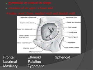
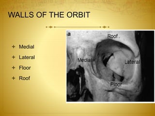
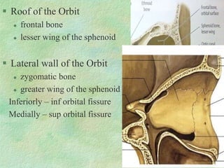
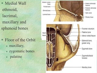
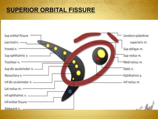
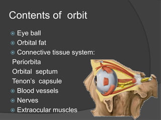
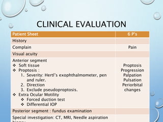
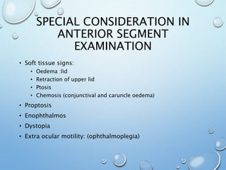
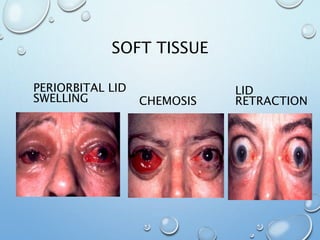
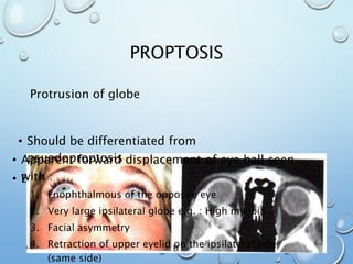
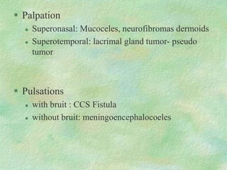
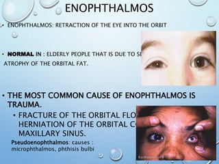
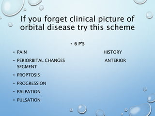
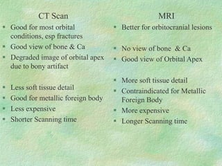
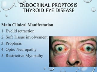
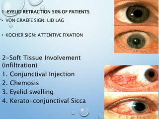
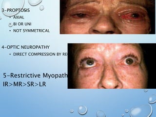
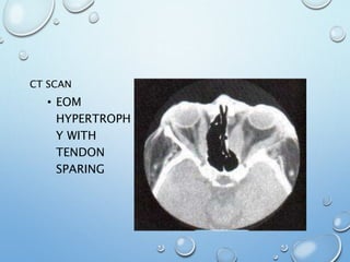
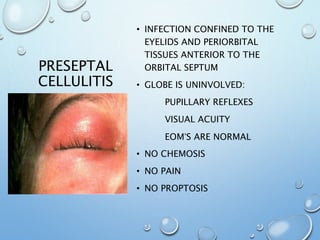
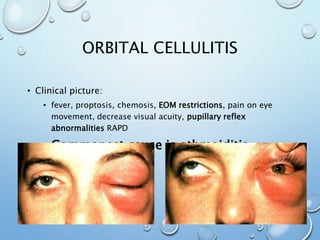
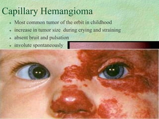
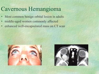
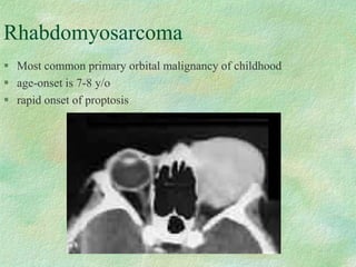
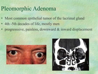
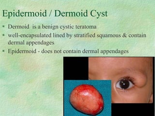
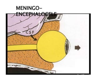
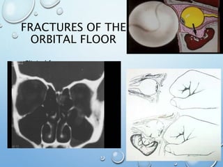
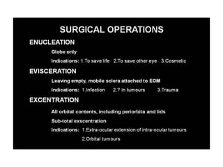
The document provides an overview of orbital anatomy and diseases. It describes the bones that make up the walls of the orbit, as well as the contents of the orbit such as the eyeball, muscles, nerves and blood vessels. Common orbital diseases are discussed including thyroid eye disease, infections, tumors and cysts, and trauma. Clinical evaluation involves assessing features like proptosis, extraocular motility, and imaging studies. Specific conditions like dermoid cysts, hemangiomas and fractures are also outlined.
3. Frontal Ethmoid Sphenoid
Lacrimal Palatine
Maxillary Zygomatic
pyramidal or conical in shape
consists of an apex, a base and
4 sides: roof, floor, medial wall and lateral wall
4. WALLS OF THE ORBIT
Medial
Lateral
Floor
Roof
5. Roof of the Orbit
frontal bone
lesser wing of the sphenoid
Lateral wall of the Orbit
zygomatic bone
greater wing of the sphenoid
Inferiorly – inf orbital fissure
Medially – sup orbital fissure
8. Contents of orbit
Eye ball
Orbital fat
Connective tissue system:
Periorbita
Orbital septum
Tenon’s capsule
Blood vessels
Nerves
Extraocular muscles
9. Orbital septum:
Interconnecting / circumferential radial
webs of fascial system
support and transmit forces in trauma
Compressive optic neuropathy following
trauma
10. CLINICAL EVALUATION
Patient Sheet 6 P’s
History
Complain Pain
Visual acuity
Anterior segment
Soft tissue
Proptosis :
1. Severity: Hertl’s exophthalmometer, pen
and ruler.
2. Direction
3. Exclude pseudoproptosis.
Extra Ocular Motility
Forced duction test
Differential IOP
Proptosis
Progression
Palpation
Pulsation
Periorbital
changes
Posterior segment : fundus examination
Special investigation: CT, MRI, Needle aspiration
11. SPECIAL CONSIDERATION IN
ANTERIOR SEGMENT
EXAMINATION
• Soft tissue signs:
• Oedema :lid
• Retraction of upper lid
• Ptosis
• Chemosis (conjunctival and caruncle oedema)
• Proptosis
• Enophthalmos
• Dystopia
• Extra ocular motility: (ophthalmoplegia)
13. PROPTOSIS
• Examining A case of proptosis entails:
• Degree or severity
• Direction
• Exclude psuedoproptosis
Protrusion of globe
• Apparent forward displacement of eye ball seen
with :
1. Enophthalmous of the opposite eye
2. Very large ipsilateral globe e.g. : High myopia
3. Facial asymmetry
4. Retraction of upper eyelid on the ipsilateral side
(same side)
• Should be differentiated from
psuedoproptosis
14. MEASUREMENT OF PROPTOSIS
• The normal distance between the apex of the
cornea and the lateral orbital rim is usually less
than 20 mm .
• A reading of 21 mm or more is regarded abnormal.
• A difference of 2 mm between 2 eyes is suspicious.
• The amount of proptosis is measured with a hertel
exophthalmometer or a plastic ruler placed at the
lateral canthus and resting on the bone.
15.
16.
17. Axial e.g. Cavernous haemangioma
Non Axial Displacement - outside the muscle cone
-Superior Displacement: maxillary tumor invading the
floor of the orbit
-Inferomedial displacement:dermoid cyst and lacrimal
gland tumor
-Days to weeks: inflammatory diseases. Infectious
diseases, metastatic tumors
-Months to years: dermoids
DIRECTION OF
PROPTOSIS
PROGRESSION
19. ENOPHTHALMOS
• ENOPHTHALMOS: RETRACTION OF THE EYE INTO THE ORBIT
• NORMAL IN : ELDERLY PEOPLE THAT IS DUE TO SENILE
ATROPHY OF THE ORBITAL FAT.
• THE MOST COMMON CAUSE OF ENOPHTHALMOS IS
TRAUMA.
• FRACTURE OF THE ORBITAL FLOOR RESULTS IN
HERNIATION OF THE ORBITAL CONTENT INTO THE
MAXILLARY SINUS.
Pseudoenophthalmos: causes :
microphthalmos, phthisis bulbi
20. If you forget clinical picture of
orbital disease try this scheme
• 6 P’S
• PAIN HISTORY
• PERIORBITAL CHANGES ANTERIOR
SEGMENT
• PROPTOSIS
• PROGRESSION
• PALPATION
• PULSATION
22. CT Scan
Good for most orbital
conditions, esp fractures
Good view of bone & Ca
Degraded image of orbital apex
due to bony artifact
Less soft tissue detail
Good for metallic foreign body
Less expensive
Shorter Scanning time
MRI
Better for orbitocranial lesions
No view of bone & Ca
Good view of Orbital Apex
More soft tissue detail
Contraindicated for Metallic
Foreign Body
More expensive
Longer Scanning time
23. ORBITAL DISEASES
1. INFLAMMATORY:
1. THYROID EYE DISEASE (ALSO CAN BE CLASSIFIED AS
ENDOCRINAL)
2. IDIOPATHIC ORBITAL INFLAMMATORY DISEASE
2. TUMORS AND CYSTS
3. INFECTION
4. TRAUMA
28. PRESEPTAL
CELLULITIS
• INFECTION CONFINED TO THE
EYELIDS AND PERIORBITAL
TISSUES ANTERIOR TO THE
ORBITAL SEPTUM
• GLOBE IS UNINVOLVED:
PUPILLARY REFLEXES
VISUAL ACUITY
EOM’S ARE NORMAL
• NO CHEMOSIS
• NO PAIN
• NO PROPTOSIS
29. ORBITAL CELLULITIS
• Clinical picture:
• fever, proptosis, chemosis, EOM restrictions, pain on eye
movement, decrease visual acuity, pupillary reflex
abnormalities RAPD
• Commonest cause is ethmoiditis
30. Capillary Hemangioma
Most common tumor of the orbit in childhood
increase in tumor size during crying and straining
absent bruit and pulsation
involute spontaneously
31. Cavernous Hemangioma
Most common benign orbital lesion in adults
middle-aged women commonly affected
enhanced well-encapsulated mass on CT scan
32. Rhabdomyosarcoma
Most common primary orbital malignancy of childhood
age-onset is 7-8 y/o
rapid onset of proptosis
33. Pleomorphic Adenoma
Most common epithelial tumor of the lacrimal gland
4th -5th decades of life, mostly men
progresssive, painless, downward & inward displacement
34. Epidermoid / Dermoid Cyst
Dermoid is a benign cystic teratoma
well-encapsulated lined by stratified squamous & contain
dermal appendages
Epidermoid - does not contain dermal appendages