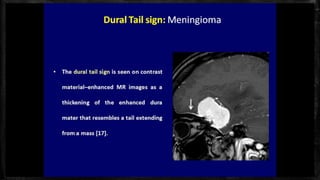
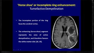
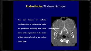
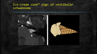
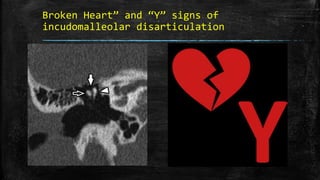
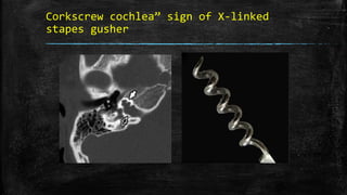
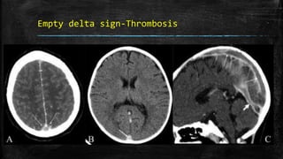
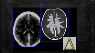
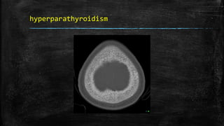
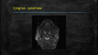
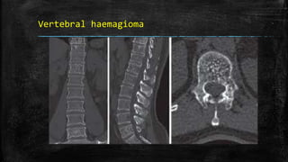
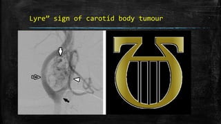
Neuroradiology spotters
DR.Kannan gunasekaran
Paediatric Radiologist
Kmch
Ice-cream cone” sign of vestibular
schwannoma
Broken Heart” and “Y” signs of
incudomalleolar disarticulation
Corkscrew cochlea” sign of X-linked
stapes gusher
Empty delta sign-Thrombosis
▪ Hyperdense subarachnoid hemorrhage, subdural empyema, or
hematoma surrounding the sinus may result in the ‘pseudodelta’ sign
on unenhanced CT scan.
Cord sign
Dural tail sign- meningioma
Medusa head sign- DVA
Lemon sign- spina bifida
Pancake brain-alobar holoprosencephaly
Reversal sign-cerebral edema
Hyperdense MCA sign
Salt and pepper sign –jugular
paraganglioma
hyperparathyroidism
Sjögren syndrome
Vertebral haemagioma
Lyre” sign of carotid body tumour
Optic nerve tram-track sign- meningioma
vs glioma
▪ not specific for meningiomas
▪ Orbital pseudotumor,
▪ Perioptic neuritis,
▪ Sarcoidosis,
▪ Leukemia, lymphomas,
▪ Metastases,
▪ Perioptic hemorrhages,
▪ Erdheim-Chester disease
Martini glass” sign- PHPV
Bare orbit sign- NF1
Moya moya appearance
Hot nose sign- obstruction to ICA
eye-of-the-tiger sign-Hallervorden-Spatz
syndrome
Molar tooth sign-Joubert syndrome
Tigroid pattern-MLD
Tiger stripe” sign of non-suppurative
tonsillopharyngitis
Tau sign-persistent trigeminal artery
Kissing carotids” sign of medialised
course of the internal carotid arteries
Radial band sign-TS
Harlequin appearance- Apert syndrome
Hot cross bun sign-MSP
▪ Thank you

neuro radiology spotters

Editor's Notes

  • #64 Ice-cream cone” sign of vestibular schwannoma. (a) Axial T1 SPGR post-contrast image shows an avidly enhancing mass (white solid arrow) extending from the CPA to the fundus of the IAC. (b) When large enough, vestibular schwannomas viewed in the axial plane resemble an “ice-cream cone” with the bulbous extension of tumour in the CPA resembling the ice-cream sitting upon a cone of tumour in the IAC.
  • #65 “Broken Heart” and “Y” signs of incudomalleolar disarticulation. (a) Coronal non-contrast enhanced CT shows widening of the incudomalleolar joint (white solid arrow) with lateral displacement of the short process of the incus (white open arrow) relative to the head of the malleus (white arrowhead). (b) This configuration of the ossicles observed on coronal CT has been likened to the appearance of a “broken heart” or “Y”.
  • #66 Corkscrew cochlea” sign of X-linked stapes gusher. (a) Axial non-contrast CT of the temporal bones demonstrates an abnormal morphology of the cochlea (white solid arrow) with absent interscalar septum and modiolus, as well as a bulbous configuration of the lateral internal auditory canal (white open arrow). (b) This abnormal cochlear configuration resembles a corkscrew and is typical of X-linked stapes gusher.
  • #67 Empty delta sign. Postcontrast axial CT scan of the brain (A) shows the nonenhancing lumen of the superior sagittal sinus surrounded by enhancing dura (arrows). Nonenhanced axial CT scan of the brain (B) shows a hyperdense superior sagittal sinus (arrow) suggestive of thrombus within. This is called the ‘triangle sign.’ Sagittal reconstruction of a contrast-enhanced CT scan (C) shows filling defects in the superior sagittal sinus (long arrows) and in the straight sinus (thick arrow), suggestive of thrombus
  • #69 g
  • #71 Dural tail sign. Axial (A) and sagittal (B) contrast-enhanced MRI images of the brain show an avidly enhancing lesion in the left frontal region. The lesion is dural based and there is thickening and enhancement of the dura, which tapers away from the mass (arrows). This thickening and enhancement of the dura adjacent to the mass is the ‘dural tail sign’ that is seen in meningiomas
  • #72 Medusa head sign. Postcontrast axial T1W MRI image of the brain (A) shows a developmental venous anomaly (arrow), with multiple, small, radiating veins forming a ‘Medusa head’ in the left cerebellar hemisphere. Postcontrast axial T1W MRI image of the supratentorial brain (B) shows a large developmental venous anomaly with multiple radiating veins (arrows) draining into it. Postcontrast axial T1W image of the brain (C) shows a large developmental venous anomaly and a round hyperintense lesion with a dark rim (arrow), suggestive of a cavernoma, anterior to it
  • #73 Lemon sign. Axial USG image of the fetal head at the level of the biparietal diameter (A) shows inward concavity of both frontal bones (arrows) instead of the normally seen convexity. This gives the head the appearance of a lemon. Sagittal USG image of the fetus (B) shows a meningomyelocele (thick arrow) in the lumbar region
  • #74 Pancake brain. Axial T1W MRI images (A, B) of the brain in alobar holoprosencephaly show fusion of the cerebral hemispheres, with a single ventricular cavity (star) in the center; this gives the brain the appearance of a pancake. Note the fusion of the thalami (arrows)
  • #75 Reversal sign. Axial noncontrast CT scan of the brain in a child with severe head injury shows low attenuation of the cerebral hemispheres with complete loss of gray matter–white matter differentiation, small temporal horns, and effacement of the basal cisterns. The findings are suggestive of cerebral edema. There is relative preservation of the cerebellum (arrows) with hyperattenuation (‘white cerebellum’)
  • #76 Hyperdense MCA sign. Noncontrast axial CT scans (A, B) of the brain in a patient with sudden-onset right hemiparesis show hyperattenuation of the left middle cerebral artery (arrows). This is suggestive of occlusion of the artery. This is one of the signs of acute stroke but does not necessarily represent infarction
  • #77 Salt and pepper sign. FLAIR axial MRI image of the left temporal bone shows an isointense mass (arrows) in the region of the jugular foramen. The mass is heterogeneous, with dark and slightly bright areas within it. Axial CT scan (B) in the same patient shows the mass causing destruction of the surrounding bone (arrows). An external carotid angiogram (C) shows tumor blush (black arrow) due to hypervascularity. These findings are suggestive of a glomus jugulare tumor of the jugular foramen
  • #78 Salt and pepper” sign of paraganglioma. (a) Axial non-contrast enhanced T1-weighted image demonstrates a circumscribed mass (white arrowhead) splaying the internal and external carotid arteries and demonstrating a speckled appearance with foci of intrinsic bright T1 signal (white open arrows). (b) Axial T2-weighted imaging shows the mass (white arrowhead) to have areas of peripheral dark T2 signal (white solid arrows). (c) This pattern has been likened to the appearance of “salt and pepper” and is pathognomonic for paraganglioma.
  • #82 Lyre” sign of carotid body tumour. (a) Lateral digital subtraction angiography image demonstrates a hypervascular mass (white solid arrow) located at the carotid bifurcation (black solid arrow). (b) Characteristic splaying of the internal carotid artery (white open arrow) and external carotid artery (white arrowhead) by a paraganglioma of the carotid body resembles the lyre, a harp-like instrument of classic Greek antiquity.
  • #83 Optic nerve tram-track sign. Postcontrast T1W axial MRI image of the orbits (A) shows an enhancing mass (thick small arrows) around the left optic nerve. The optic nerve (thin long arrow) itself is not enlarged and is nonenhancing. This is suggestive of a mass, such as a meningioma, arising from the optic nerve sheath. Axial T2W MRI image of the orbits in a different patient (B) shows fusiform enlargement of the left optic nerve itself (arrows), in a case of optic nerve glioma
  • #84 Tram-track” sign of optic nerve sheath meningioma. (a) Axial post-contrast T1-weighted fat-saturated image shows parallel linear enhancement along the orbital segment of the optic nerve sheath (white solid arrows), which resembles the parallel orientation of light rail or train tracks (Powell/Hyde cable car line, San Francisco, California). This pattern of enhancement is sensitive, but not specific for optic nerve sheath meningioma. (b) Non-contrast enhanced CT reveals “tram-track” calcification of the optic nerve sheath (white open arrows), which, in comparison, is highly specific for meningioma.
  • #89 “Martini glass” sign of PHPV. (a) Axial fat-saturated T2-weighted image demonstrates a small right globe with abnormal triangularshaped retrolenticular soft tissue (white open arrow) in a child with PHPV. (b) This configuration of the retrolenticular soft tissue resembles the shape of a martini glass.
  • #90 Bare orbit sign. Frontal CT scan scannogram of the head (A) shows a bare and large orbit on the left, with absence of the greater wing of the left sphenoid bone, in this patient with neurofibromatosis type 1. Note the normal lesser (thick small arrow) and greater (thin long arrow) wings of the sphenoid on the right side. Frontal surface-shaded display, 3D view of the skull (B) shows a large left orbit with dysplasia of the sphenoid wing. Axial CT scan image (C) shows anterior herniation of the left temporal lobe (thin long arrow) because of the dysplastic sphenoid bone. The left globe is small and calcified (phthisis bulbi) (thick short arrow) due to plexiform neurofibromatosis of the left orbit
  • #92 Moya moya appearance. Lateral anterior oblique view (A) of an internal carotid artery (thick short arrow) angiogram shows multiple, small, tortuous collateral vessels in the distribution of the middle cerebral artery (arrows), suggestive of the moya moya (puff of smoke) appearance. Axial view of the MRI angiogram (B) shows complete occlusion of the middle cerebral arteries bilaterally. Arrows indicate the internal carotid arteries
  • #94 Hot nose sign. Frontal view of a radionuclide scan shows increased tracer activity in the nasopharyngeal region (arrows) suggestive of increased blood flow. Obstruction of the internal carotid artery from any cause leads to increased flow in the external carotid artery and increased tracer activity in the nasopharyngeal region (hot nose)
  • #95 The eye-of-the-tiger sign. Axial T2W MRI image of the brain shows hypointensity of the globus palladi (arrows). There is relative hyperintensity of the central part, giving the globus palladi the appearance of the eyes of a tiger. This appearance is seen in Hallervorden-Spatz syndrome
  • #96 Molar tooth sign. Axial T1W MRI image of the brain (A) in a child with Joubert syndrome shows elongated superior cerebellar peduncles (arrows) giving the midbrain and superior peduncles the appearance of a molar tooth. Axial T2W image of the brain (B) in another child shows the molar tooth appearance of the midbrain and superior cerebellar peduncles. There is also hypoplasia of the cerebellar vermis (arrows)
  • #97 Tigroid pattern. Axial T2W image of the brain in a child with metachromatic leukodystrophy shows symmetric, increased signal intensity of the white matter, with sparing of the subcortical U fibers. Linear low signal intensity areas radiating away from the ventricular margin (arrows) represent areas of white matter around the vessels that have been spared from the process of demyelination. These low signal linear areas within the hyperintense white matter resemble the skin of a leopard and hence the term ‘tigroid’ pattern. FLAIR axial MRI image (B) shows symmetric hyperintensity of the white matter sparing the subcortical U fibers
  • #99 Tiger stripe” sign of non-suppurative tonsillopharyngitis. (a) Coronal contrast-enhanced CT image demonstrates enlargement of the palatine tonsils, which are hyperaemic and show a striated pattern of enhancement (white open arrows). (b) This pattern of enhancement resembles the striped coat of a tiger and when present usually excludes the diagnosis of tonsillar abscess.
  • #100 Tau sign. Sagittal T1-W image of the brain shows (A) flow voids of the internal carotid artery (ICA) in the precavernous segment (thick short arrow), in the cavernous segment (medium-sized arrow), and a persistent trigeminal artery (thin long arrow). Together, these flow voids form the Greek letter ‘τ’ (tau). Sagittal view of the MRI angiogram shows the persistent trigeminal artery (arrow) arising from the ICA and joining the basilar artery in its mid segment
  • #101 Kissing carotids” sign of medialised course of the internal carotid arteries. (a) Axial contrast-enhanced CT image shows a markedly medialised course of the internal carotid arteries, which nearly contact at midline (white open arrow). (b) The close proximity of the carotids has been described as resembling the kissing embrace of lovers (Unconditional Surrender sculpture, San Diego, California). Although typically an incidental finding, a medialised course of the internal carotid arteries may place them at increased risk of iatrogenic injury during surgery or instrumentation.
  • #102 Radial band sign. Proton density-weighted axial image of the brain (A) in a patient with tuberous sclerosis shows hyperintense bands in the white matter radiating from the ventricular margin to the subcortical region (arrows). FLAIR axial MRI image (B) shows irregular hyperintense areas in the subcortical white matter suggestive of subcortical tubers. A large hyperintense nodule is seen in the region of the foramen of Monroe, representing a subependymal giant cell astrocytoma (thick arrow)
  • #103 Harlequin appearance. Frontal view of the skull (A) in a child with Apert syndrome shows elevated superolateral angles of both orbits giving the appearance of a ‘harlequin mask.’ Frontal (B) and lateral (C) surface-shaded display 3D CT views of the skull show the harlequin appearance of the orbits. The sagittal suture (arrow in B) and the lambdoid suture (black arrows in C) are wide open. The coronal suture (thin arrows in C) is fused, which is suggestive of coronal craniosynostosis
  • #105 Hot cross bun sign. Axial T2W MRI image of the pons (A) in a patient with multiple system atrophy shows a hyperintense linear area forming a ‘cross’ (arrow) in the pons. Sagittal T1W image (B) shows atrophy of the cerebellum and cerebrum (prominence of sulci)