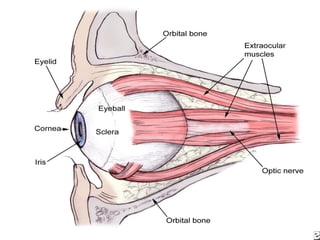
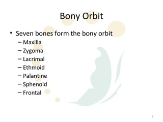
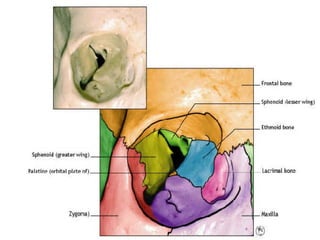
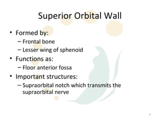
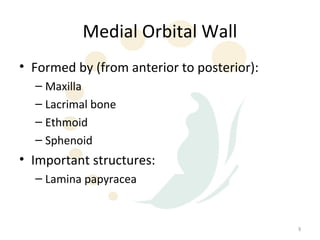
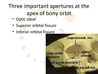
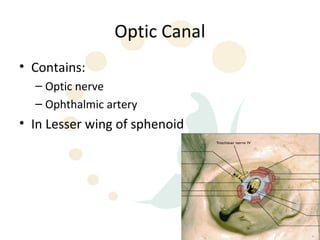
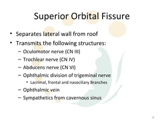
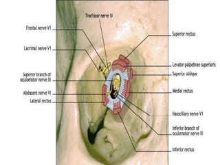
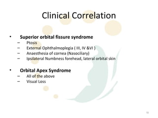
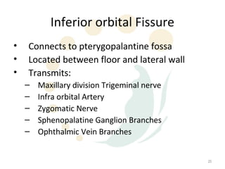
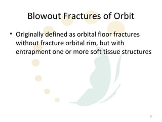
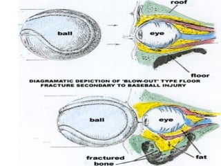
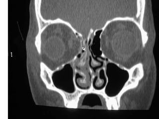
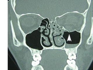
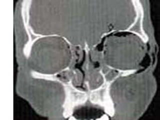
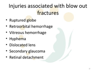
SlideShare a Scribd company logo
Orbital Fractures


                    2
Topics for Discussion
•   Orbital anatomy
•   Types of fractures
•   Signs and symptoms
•   Management


                                   3
Orbital Anatomy
• The bony orbit refers to the shell of bone
  which surrounds and protects the eye.
• The bony orbit is a pyramidal cavity with an
  elliptical base presenting anteriorly and the
  apex posteriorly


                                                  4
5
Bony Orbit
• Seven bones form the bony orbit
  – Maxilla
  – Zygoma
  – Lacrimal
  – Ethmoid
  – Palantine
  – Sphenoid
  – Frontal


                                    6
7
Superior Orbital Wall
• Formed by:
  – Frontal bone
  – Lesser wing of sphenoid
• Functions as:
  – Floor anterior fossa
• Important structures:
  – Supraorbital notch which transmits the
    supraorbital nerve


                                             8
Medial Orbital Wall
• Formed by (from anterior to posterior):
  – Maxilla
  – Lacrimal bone
  – Ethmoid
  – Sphenoid
• Important structures:
  – Lamina papyracea


                                            9
Lamina Papyracea
• Thin segment of the medial orbital wall
• Separates the orbit from the ethmoid air cells


                                                   10
Lateral Orbital Wall
• Formed by:
  – Zygomatic bone
  – Greater wing of sphenoid


                                 11
Orbital Floor
• Formed by:
  – Maxilla
  – Palatine
• Important structures:
  – Infraorbital groove
     • Transverses floor from lateral to medial
     • Location of infraorbital nerve which supplies sensation
       to check and ipsilateral upper alveolus and teeth


                                                                 12
Orbital Floor
• Forms roof of maxillary sinus
• Location of more blow out fractures due to
  inherent weakness of bone overlying maxillary
  sinus


                                              13
Three important apertures at the
       apex of bony orbit
 • Optic canal
 • Superior orbital fissure
 • Inferior orbital fissure


                                   14
Optic Canal
• Contains:
  – Optic nerve
  – Ophthalmic artery
• In Lesser wing of sphenoid


                               15
Superior Orbital Fissure
• Separates lateral wall from roof
• Transmits the following structures:
   –   Oculomotor nerve (CN III)
   –   Trochlear nerve (CN IV)
   –   Abducens nerve (CN VI)
   –   Ophthalmic division of trigeminal nerve
        • Lacrimal, frontal and nasociliary Branches
   – Ophthalmic vein
   – Sympathetics from cavernous sinus


                                                       16
17
18
Clinical Correlation

•       Superior orbital fissure syndrome
    –     Ptosis
    –     External Ophthalmoplegia ( III, IV &VI )
    –     Anaesthesia of cornea (Nasociliary)
    –     Ipsilateral Numbness forehead, lateral orbital skin

•       Orbital Apex Syndrome
    –     All of the above
    –     Visual Loss


                                                                19
Inferior orbital Fissure
•   Connects to pterygopalantine fossa
•   Located between floor and lateral wall
•   Transmits:
    –   Maxillary division Trigeminal nerve
    –   Infra orbital Artery
    –   Zygomatic Nerve
    –   Sphenopalatine Ganglion Branches
    –   Ophthalmic Vein Branches


                                              20
Blowout Fractures of Orbit
• Originally defined as orbital floor fractures
  without fracture orbital rim, but with
  entrapment one or more soft tissue structures


                                              21
Blowout Fractures
• Blowout fractures now refer to fractures of the:
   –   Orbital floor
   –   Medial wall
   –   Lateral wall
   –   Superior wall
• “pure” blowout fractures – trapdoor rotation to
  bone fragments involving central area of bone
• “impure” fracture – fracture line extends to orbital
  rim


                                                         22
Physiology of Blowout Fracture
• The bony defect is filled with soft tissue and
  fat from the orbit
• Alters support mechanisms for EOM
• EOM can become entrapped
• Direct muscle damage can result


                                                   23
Common causes of orbital
              fractures
•   Falling
•   Aggression
•   Sporting events
•   MVAs


                                   24
25
26
Initial Evaluation
• History
  – Time and mechanism of injury
  – Change in appearance of eye
  – State of vision immediately after injury
     • Immediate loss of vision – severe damage to retina
     • Loss of light perception - vascular occlusion or optic nerve
       compression
     • Initial good vision – compression optic neuropathy


                                                                      27
Initial Evaluation
• Physical Exam
  – Cranial nerve examination
       • EOM
       • Numbness check
  –   Palpation orbital rim
  –   Papillary function
  –   Visual acuity
  –   Fundus examine
  –   Ophthalmologic evaluation


                                     28
Visual Acuity
• Light perception
• Finger counting
• Visual acuity


                               29
Consultation


Do not hesitate to obtain an ophthalmologic
 consultation


                                              30
Common physical signs
•   Periorbital eccyhmosis
•   Impaired extraocular muscles
•   Hypoesthesia in V2 distribution
•   Intraorbital emphysema


                                      31
Common Symptoms
• Diplopia
• Pain with eye movement


                           32
Radiographic Evaluation
• CT scan of the orbits
• Plain films not useful due to a high rate of
  false negatives and non-diagnostic studies


                                                 33
34
35
36
Injuries associated with blow out
                 fractures
•   Ruptured globe
•   Retroorbital hemorrhage
•   Vitreous hemorrhage
•   Hyphema
•   Dislocated lens
•   Secondary glaucoma
•   Retinal detachment

                                        37
Treatment Options
• Nonsurgical
• Surgical


                              38
Initial Management
•   ABC
•   C-Spine
•   Analgesia
•   Nurse Head up
•   Ice affected area
•   Broad spectrum antibiotics
•   Steroids
•   No nose blowing

                                  39
Indications for Surgery
• Retrobulbar haematoma
• Diplopia
• Enophthalmos >2 mm
• Substantial soft tissue herniation into
  maxillary sinus
• Displaced fracture esp if palpable step at rim


                                                   40
Contraindications to surgery
•   Hyphema
•   Retinal detachment
•   Globe perforation
•   Only seeing eye
•   Medically unstable patient


                                     41
Surgical Approaches
• Transconjunctival approach
• Transcutaneous
• Subciliary


                               42
Factors to consider for surgery
•   Site
•   Location
•   Severity
•   What needs to be corrected


                                       43
Orbital Implants
• Use of implants based on degree of
  comminution and size of fracture
• Various implant material used
  – Autogenous bone and cartilage
  – Alloplastic material
     • Teflon
     • Marlex
     • PDS


                                       44
Complications of Surgery
•   Ectropion
•   Lid retraction
•   Persistent diplopia
•   Malposition of eye
•   Hypoaesthesia of V2
•   Extrusion of orbital floor implant
•   BLINDNESS

                                         45

More Related Content

What's hot

Zygomatic complex fractures
Zygomatic complex fracturesZygomatic complex fractures
Zygomatic complex fractures
Mahak Ralli
 
Orbital trauma
Orbital traumaOrbital trauma
Orbital trauma
DrFirdousMulla
 
Orbital Fractures
Orbital FracturesOrbital Fractures
Orbital Fractureshashem10177
 
Surgical Anatomy of Orbit
Surgical Anatomy of OrbitSurgical Anatomy of Orbit
Surgical Anatomy of Orbit
Dibya Falgoon Sarkar
 
Orbital Fractures - The Role of an Ophthalmologist
Orbital Fractures - The Role of an OphthalmologistOrbital Fractures - The Role of an Ophthalmologist
Orbital Fractures - The Role of an Ophthalmologist
Ankit Punjabi
 
Management of Mandibular Fractures
Management of Mandibular FracturesManagement of Mandibular Fractures
Management of Mandibular Fractures
Dr. Tshewang Gyeltshen
 
Management of condylar fractures
Management of condylar fracturesManagement of condylar fractures
Management of condylar fracturesdralimohammedhasan
 
Frontal sinus fracture
Frontal sinus fractureFrontal sinus fracture
Frontal sinus fracture
Jamil Kifayatullah
 
Arthrocentesis of the temporomandibular joint
Arthrocentesis of the temporomandibular jointArthrocentesis of the temporomandibular joint
Arthrocentesis of the temporomandibular joint
Ahmed Adawy
 
Osteomyelitis in maxillofacial region
Osteomyelitis  in maxillofacial regionOsteomyelitis  in maxillofacial region
Osteomyelitis in maxillofacial region
Cathrine Diana
 
NOE fractures
NOE fractures NOE fractures
NOE fractures
anchalag8
 
Orbital floor blow out fractures
Orbital floor blow out fracturesOrbital floor blow out fractures
Orbital floor blow out fractures
Ahmed Adawy
 
Zygomatic maxillary complex fracture
Zygomatic maxillary complex fractureZygomatic maxillary complex fracture
Zygomatic maxillary complex fracture
josna thankachan
 
Mandibular Fracture.ppt
Mandibular Fracture.pptMandibular Fracture.ppt
Mandibular Fracture.ppt
DentalYoutube
 
Orbital fractures ih
Orbital fractures ihOrbital fractures ih
Orbital fractures ih
itrat hussain
 
Management of oroantral fistula
Management of oroantral fistulaManagement of oroantral fistula
Management of oroantral fistula
Saleh Bakry
 
Mid facial fractures and their management
Mid facial fractures and their managementMid facial fractures and their management
Mid facial fractures and their management
Ruhi Kashmiri
 
NOE FRACTURE PPT
NOE FRACTURE PPTNOE FRACTURE PPT
NOE FRACTURE PPT
VIGNESH PRABHU.T
 
Classification of Mandible, Midface, ZMC and NOE Fractures
Classification of Mandible, Midface, ZMC and NOE FracturesClassification of Mandible, Midface, ZMC and NOE Fractures
Classification of Mandible, Midface, ZMC and NOE Fractures
Arjun Shenoy
 
Fractures of middle third of face
Fractures of middle third of faceFractures of middle third of face
Fractures of middle third of face
Mohammed Shalik
 

What's hot (20)

Zygomatic complex fractures
Zygomatic complex fracturesZygomatic complex fractures
Zygomatic complex fractures
 
Orbital trauma
Orbital traumaOrbital trauma
Orbital trauma
 
Orbital Fractures
Orbital FracturesOrbital Fractures
Orbital Fractures
 
Surgical Anatomy of Orbit
Surgical Anatomy of OrbitSurgical Anatomy of Orbit
Surgical Anatomy of Orbit
 
Orbital Fractures - The Role of an Ophthalmologist
Orbital Fractures - The Role of an OphthalmologistOrbital Fractures - The Role of an Ophthalmologist
Orbital Fractures - The Role of an Ophthalmologist
 
Management of Mandibular Fractures
Management of Mandibular FracturesManagement of Mandibular Fractures
Management of Mandibular Fractures
 
Management of condylar fractures
Management of condylar fracturesManagement of condylar fractures
Management of condylar fractures
 
Frontal sinus fracture
Frontal sinus fractureFrontal sinus fracture
Frontal sinus fracture
 
Arthrocentesis of the temporomandibular joint
Arthrocentesis of the temporomandibular jointArthrocentesis of the temporomandibular joint
Arthrocentesis of the temporomandibular joint
 
Osteomyelitis in maxillofacial region
Osteomyelitis  in maxillofacial regionOsteomyelitis  in maxillofacial region
Osteomyelitis in maxillofacial region
 
NOE fractures
NOE fractures NOE fractures
NOE fractures
 
Orbital floor blow out fractures
Orbital floor blow out fracturesOrbital floor blow out fractures
Orbital floor blow out fractures
 
Zygomatic maxillary complex fracture
Zygomatic maxillary complex fractureZygomatic maxillary complex fracture
Zygomatic maxillary complex fracture
 
Mandibular Fracture.ppt
Mandibular Fracture.pptMandibular Fracture.ppt
Mandibular Fracture.ppt
 
Orbital fractures ih
Orbital fractures ihOrbital fractures ih
Orbital fractures ih
 
Management of oroantral fistula
Management of oroantral fistulaManagement of oroantral fistula
Management of oroantral fistula
 
Mid facial fractures and their management
Mid facial fractures and their managementMid facial fractures and their management
Mid facial fractures and their management
 
NOE FRACTURE PPT
NOE FRACTURE PPTNOE FRACTURE PPT
NOE FRACTURE PPT
 
Classification of Mandible, Midface, ZMC and NOE Fractures
Classification of Mandible, Midface, ZMC and NOE FracturesClassification of Mandible, Midface, ZMC and NOE Fractures
Classification of Mandible, Midface, ZMC and NOE Fractures
 
Fractures of middle third of face
Fractures of middle third of faceFractures of middle third of face
Fractures of middle third of face
 

Similar to Orbital fractures

Orbital trauma 1
Orbital trauma 1Orbital trauma 1
Orbital trauma 1
Gahens13
 
161992-brain-template-16x9.pptx
161992-brain-template-16x9.pptx161992-brain-template-16x9.pptx
161992-brain-template-16x9.pptx
AbhilashVemula4
 
BLOW_OUT_FRACTURE_(1) presentation dhir hospital bhiwani.pptx
BLOW_OUT_FRACTURE_(1) presentation dhir hospital bhiwani.pptxBLOW_OUT_FRACTURE_(1) presentation dhir hospital bhiwani.pptx
BLOW_OUT_FRACTURE_(1) presentation dhir hospital bhiwani.pptx
DHIR EYE HOSPITAL
 
Anaesthesia for ophthalmic surgery
Anaesthesia for ophthalmic surgery Anaesthesia for ophthalmic surgery
Anaesthesia for ophthalmic surgery Thomas Mphanza
 
Orbital structure
Orbital structure Orbital structure
Orbital structure
Frenky Ramiro
 
Anatomy and Visual field defects of optic nerve and chiasma
Anatomy and Visual field defects of optic nerve and chiasmaAnatomy and Visual field defects of optic nerve and chiasma
Anatomy and Visual field defects of optic nerve and chiasma
Sadhwini Harish
 
ANATOMY OF VISUAL PATHWAY - DR.RUTHRA.pptx
ANATOMY OF VISUAL PATHWAY - DR.RUTHRA.pptxANATOMY OF VISUAL PATHWAY - DR.RUTHRA.pptx
ANATOMY OF VISUAL PATHWAY - DR.RUTHRA.pptx
Ruthra Suresh
 
eyeid_disorder_lecture (1).pptx
eyeid_disorder_lecture (1).pptxeyeid_disorder_lecture (1).pptx
eyeid_disorder_lecture (1).pptx
Harshika Malik
 
Orbital anatomy
Orbital anatomy Orbital anatomy
Orbital anatomy
Samten Dorji
 
Anatomy of orbit and its clinical applications.
Anatomy of orbit and its clinical applications.Anatomy of orbit and its clinical applications.
Anatomy of orbit and its clinical applications.
RahulGupta39947
 
5 NOE FRACTURE seminar 5.pptx
5 NOE FRACTURE seminar 5.pptx5 NOE FRACTURE seminar 5.pptx
5 NOE FRACTURE seminar 5.pptx
sneha
 
maxillofacial trauma
maxillofacial traumamaxillofacial trauma
maxillofacial trauma
Dr Harjitpal Singh
 
Orbital apex syndrome
Orbital apex syndromeOrbital apex syndrome
Orbital apex syndrome
Reshma Peter
 
Neuro-Ophthalmology_Dr. Bastola.pptx
Neuro-Ophthalmology_Dr. Bastola.pptxNeuro-Ophthalmology_Dr. Bastola.pptx
Neuro-Ophthalmology_Dr. Bastola.pptx
Dr. Pradeep Bastola
 
visual pathways sivateja
visual pathways sivatejavisual pathways sivateja
visual pathways sivatejaSivateja Challa
 
Visual pathway & optic nerve tumours
Visual pathway & optic nerve tumoursVisual pathway & optic nerve tumours
Visual pathway & optic nerve tumours
Shakti Samir
 
ANATOMY OF EYEBALL MBBS OPTHALMOLOGY CLASS
ANATOMY OF EYEBALL MBBS OPTHALMOLOGY CLASSANATOMY OF EYEBALL MBBS OPTHALMOLOGY CLASS
ANATOMY OF EYEBALL MBBS OPTHALMOLOGY CLASS
Maxpayne485184
 
The neurocranium
The neurocraniumThe neurocranium
The neurocranium
Abdul Hasib Samadi
 
Anatomy OF VISUAL PATHWAY
Anatomy OF VISUAL PATHWAYAnatomy OF VISUAL PATHWAY
Anatomy OF VISUAL PATHWAY
SSSIHMS-PG
 
8 ARTHROSCOPY IN TMJ CONDITIONS seminar 8.pptx
8 ARTHROSCOPY IN TMJ CONDITIONS seminar 8.pptx8 ARTHROSCOPY IN TMJ CONDITIONS seminar 8.pptx
8 ARTHROSCOPY IN TMJ CONDITIONS seminar 8.pptx
sneha
 

Similar to Orbital fractures (20)

Orbital trauma 1
Orbital trauma 1Orbital trauma 1
Orbital trauma 1
 
161992-brain-template-16x9.pptx
161992-brain-template-16x9.pptx161992-brain-template-16x9.pptx
161992-brain-template-16x9.pptx
 
BLOW_OUT_FRACTURE_(1) presentation dhir hospital bhiwani.pptx
BLOW_OUT_FRACTURE_(1) presentation dhir hospital bhiwani.pptxBLOW_OUT_FRACTURE_(1) presentation dhir hospital bhiwani.pptx
BLOW_OUT_FRACTURE_(1) presentation dhir hospital bhiwani.pptx
 
Anaesthesia for ophthalmic surgery
Anaesthesia for ophthalmic surgery Anaesthesia for ophthalmic surgery
Anaesthesia for ophthalmic surgery
 
Orbital structure
Orbital structure Orbital structure
Orbital structure
 
Anatomy and Visual field defects of optic nerve and chiasma
Anatomy and Visual field defects of optic nerve and chiasmaAnatomy and Visual field defects of optic nerve and chiasma
Anatomy and Visual field defects of optic nerve and chiasma
 
ANATOMY OF VISUAL PATHWAY - DR.RUTHRA.pptx
ANATOMY OF VISUAL PATHWAY - DR.RUTHRA.pptxANATOMY OF VISUAL PATHWAY - DR.RUTHRA.pptx
ANATOMY OF VISUAL PATHWAY - DR.RUTHRA.pptx
 
eyeid_disorder_lecture (1).pptx
eyeid_disorder_lecture (1).pptxeyeid_disorder_lecture (1).pptx
eyeid_disorder_lecture (1).pptx
 
Orbital anatomy
Orbital anatomy Orbital anatomy
Orbital anatomy
 
Anatomy of orbit and its clinical applications.
Anatomy of orbit and its clinical applications.Anatomy of orbit and its clinical applications.
Anatomy of orbit and its clinical applications.
 
5 NOE FRACTURE seminar 5.pptx
5 NOE FRACTURE seminar 5.pptx5 NOE FRACTURE seminar 5.pptx
5 NOE FRACTURE seminar 5.pptx
 
maxillofacial trauma
maxillofacial traumamaxillofacial trauma
maxillofacial trauma
 
Orbital apex syndrome
Orbital apex syndromeOrbital apex syndrome
Orbital apex syndrome
 
Neuro-Ophthalmology_Dr. Bastola.pptx
Neuro-Ophthalmology_Dr. Bastola.pptxNeuro-Ophthalmology_Dr. Bastola.pptx
Neuro-Ophthalmology_Dr. Bastola.pptx
 
visual pathways sivateja
visual pathways sivatejavisual pathways sivateja
visual pathways sivateja
 
Visual pathway & optic nerve tumours
Visual pathway & optic nerve tumoursVisual pathway & optic nerve tumours
Visual pathway & optic nerve tumours
 
ANATOMY OF EYEBALL MBBS OPTHALMOLOGY CLASS
ANATOMY OF EYEBALL MBBS OPTHALMOLOGY CLASSANATOMY OF EYEBALL MBBS OPTHALMOLOGY CLASS
ANATOMY OF EYEBALL MBBS OPTHALMOLOGY CLASS
 
The neurocranium
The neurocraniumThe neurocranium
The neurocranium
 
Anatomy OF VISUAL PATHWAY
Anatomy OF VISUAL PATHWAYAnatomy OF VISUAL PATHWAY
Anatomy OF VISUAL PATHWAY
 
8 ARTHROSCOPY IN TMJ CONDITIONS seminar 8.pptx
8 ARTHROSCOPY IN TMJ CONDITIONS seminar 8.pptx8 ARTHROSCOPY IN TMJ CONDITIONS seminar 8.pptx
8 ARTHROSCOPY IN TMJ CONDITIONS seminar 8.pptx
 

More from drpouriamoradi (20)

Zplasty
ZplastyZplasty
Zplasty
 
Skin grafts
Skin graftsSkin grafts
Skin grafts
 
Scc
SccScc
Scc
 
Scaphoid fractures
Scaphoid fracturesScaphoid fractures
Scaphoid fractures
 
Radial nerve-palsy-tendon-transfers
Radial nerve-palsy-tendon-transfersRadial nerve-palsy-tendon-transfers
Radial nerve-palsy-tendon-transfers
 
Radial nerve-anatomy
Radial nerve-anatomyRadial nerve-anatomy
Radial nerve-anatomy
 
Principles of-tendon-transfers
Principles of-tendon-transfersPrinciples of-tendon-transfers
Principles of-tendon-transfers
 
Pipjw
PipjwPipjw
Pipjw
 
Perineal reconstruction
Perineal reconstructionPerineal reconstruction
Perineal reconstruction
 
Parotid gland
Parotid glandParotid gland
Parotid gland
 
Nsw speech-path-talk-flapvs-grafts
Nsw speech-path-talk-flapvs-graftsNsw speech-path-talk-flapvs-grafts
Nsw speech-path-talk-flapvs-grafts
 
Nsw plastic-nurses
Nsw plastic-nursesNsw plastic-nurses
Nsw plastic-nurses
 
Mucous cysts-dipjw
Mucous cysts-dipjwMucous cysts-dipjw
Mucous cysts-dipjw
 
Lower limb-guidelines
Lower limb-guidelinesLower limb-guidelines
Lower limb-guidelines
 
Lower limb-flaps
Lower limb-flapsLower limb-flaps
Lower limb-flaps
 
Intro to-plastics
Intro to-plasticsIntro to-plastics
Intro to-plastics
 
Hand tumours
Hand tumoursHand tumours
Hand tumours
 
Hand infections
Hand infectionsHand infections
Hand infections
 
Hand anatomy
Hand anatomyHand anatomy
Hand anatomy
 
Gps suture-workshop
Gps suture-workshopGps suture-workshop
Gps suture-workshop
 

Orbital fractures

  • 1.
  • 3. Topics for Discussion • Orbital anatomy • Types of fractures • Signs and symptoms • Management 3
  • 4. Orbital Anatomy • The bony orbit refers to the shell of bone which surrounds and protects the eye. • The bony orbit is a pyramidal cavity with an elliptical base presenting anteriorly and the apex posteriorly 4
  • 5. 5
  • 6. Bony Orbit • Seven bones form the bony orbit – Maxilla – Zygoma – Lacrimal – Ethmoid – Palantine – Sphenoid – Frontal 6
  • 7. 7
  • 8. Superior Orbital Wall • Formed by: – Frontal bone – Lesser wing of sphenoid • Functions as: – Floor anterior fossa • Important structures: – Supraorbital notch which transmits the supraorbital nerve 8
  • 9. Medial Orbital Wall • Formed by (from anterior to posterior): – Maxilla – Lacrimal bone – Ethmoid – Sphenoid • Important structures: – Lamina papyracea 9
  • 10. Lamina Papyracea • Thin segment of the medial orbital wall • Separates the orbit from the ethmoid air cells 10
  • 11. Lateral Orbital Wall • Formed by: – Zygomatic bone – Greater wing of sphenoid 11
  • 12. Orbital Floor • Formed by: – Maxilla – Palatine • Important structures: – Infraorbital groove • Transverses floor from lateral to medial • Location of infraorbital nerve which supplies sensation to check and ipsilateral upper alveolus and teeth 12
  • 13. Orbital Floor • Forms roof of maxillary sinus • Location of more blow out fractures due to inherent weakness of bone overlying maxillary sinus 13
  • 14. Three important apertures at the apex of bony orbit • Optic canal • Superior orbital fissure • Inferior orbital fissure 14
  • 15. Optic Canal • Contains: – Optic nerve – Ophthalmic artery • In Lesser wing of sphenoid 15
  • 16. Superior Orbital Fissure • Separates lateral wall from roof • Transmits the following structures: – Oculomotor nerve (CN III) – Trochlear nerve (CN IV) – Abducens nerve (CN VI) – Ophthalmic division of trigeminal nerve • Lacrimal, frontal and nasociliary Branches – Ophthalmic vein – Sympathetics from cavernous sinus 16
  • 17. 17
  • 18. 18
  • 19. Clinical Correlation • Superior orbital fissure syndrome – Ptosis – External Ophthalmoplegia ( III, IV &VI ) – Anaesthesia of cornea (Nasociliary) – Ipsilateral Numbness forehead, lateral orbital skin • Orbital Apex Syndrome – All of the above – Visual Loss 19
  • 20. Inferior orbital Fissure • Connects to pterygopalantine fossa • Located between floor and lateral wall • Transmits: – Maxillary division Trigeminal nerve – Infra orbital Artery – Zygomatic Nerve – Sphenopalatine Ganglion Branches – Ophthalmic Vein Branches 20
  • 21. Blowout Fractures of Orbit • Originally defined as orbital floor fractures without fracture orbital rim, but with entrapment one or more soft tissue structures 21
  • 22. Blowout Fractures • Blowout fractures now refer to fractures of the: – Orbital floor – Medial wall – Lateral wall – Superior wall • “pure” blowout fractures – trapdoor rotation to bone fragments involving central area of bone • “impure” fracture – fracture line extends to orbital rim 22
  • 23. Physiology of Blowout Fracture • The bony defect is filled with soft tissue and fat from the orbit • Alters support mechanisms for EOM • EOM can become entrapped • Direct muscle damage can result 23
  • 24. Common causes of orbital fractures • Falling • Aggression • Sporting events • MVAs 24
  • 25. 25
  • 26. 26
  • 27. Initial Evaluation • History – Time and mechanism of injury – Change in appearance of eye – State of vision immediately after injury • Immediate loss of vision – severe damage to retina • Loss of light perception - vascular occlusion or optic nerve compression • Initial good vision – compression optic neuropathy 27
  • 28. Initial Evaluation • Physical Exam – Cranial nerve examination • EOM • Numbness check – Palpation orbital rim – Papillary function – Visual acuity – Fundus examine – Ophthalmologic evaluation 28
  • 29. Visual Acuity • Light perception • Finger counting • Visual acuity 29
  • 30. Consultation Do not hesitate to obtain an ophthalmologic consultation 30
  • 31. Common physical signs • Periorbital eccyhmosis • Impaired extraocular muscles • Hypoesthesia in V2 distribution • Intraorbital emphysema 31
  • 32. Common Symptoms • Diplopia • Pain with eye movement 32
  • 33. Radiographic Evaluation • CT scan of the orbits • Plain films not useful due to a high rate of false negatives and non-diagnostic studies 33
  • 34. 34
  • 35. 35
  • 36. 36
  • 37. Injuries associated with blow out fractures • Ruptured globe • Retroorbital hemorrhage • Vitreous hemorrhage • Hyphema • Dislocated lens • Secondary glaucoma • Retinal detachment 37
  • 39. Initial Management • ABC • C-Spine • Analgesia • Nurse Head up • Ice affected area • Broad spectrum antibiotics • Steroids • No nose blowing 39
  • 40. Indications for Surgery • Retrobulbar haematoma • Diplopia • Enophthalmos >2 mm • Substantial soft tissue herniation into maxillary sinus • Displaced fracture esp if palpable step at rim 40
  • 41. Contraindications to surgery • Hyphema • Retinal detachment • Globe perforation • Only seeing eye • Medically unstable patient 41
  • 42. Surgical Approaches • Transconjunctival approach • Transcutaneous • Subciliary 42
  • 43. Factors to consider for surgery • Site • Location • Severity • What needs to be corrected 43
  • 44. Orbital Implants • Use of implants based on degree of comminution and size of fracture • Various implant material used – Autogenous bone and cartilage – Alloplastic material • Teflon • Marlex • PDS 44
  • 45. Complications of Surgery • Ectropion • Lid retraction • Persistent diplopia • Malposition of eye • Hypoaesthesia of V2 • Extrusion of orbital floor implant • BLINDNESS 45

Editor's Notes

  1. Ectropion – turing outward eyelid