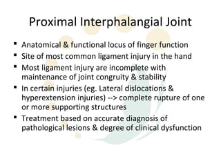
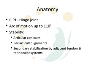
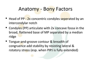
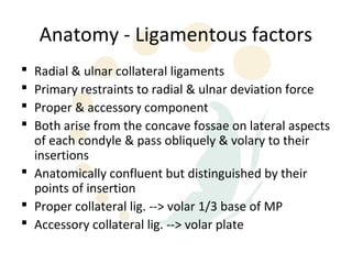
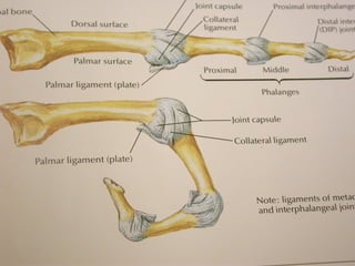
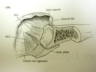
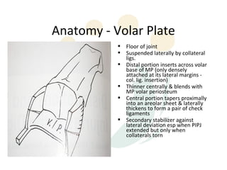
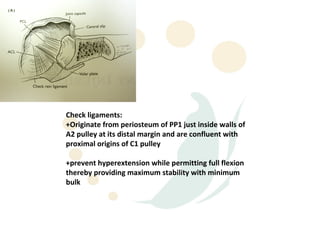
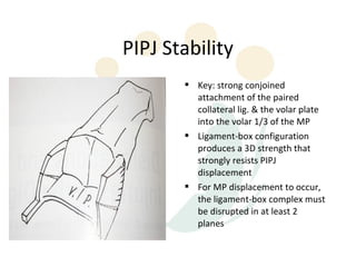
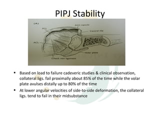
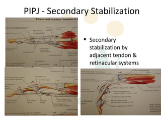
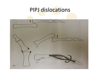
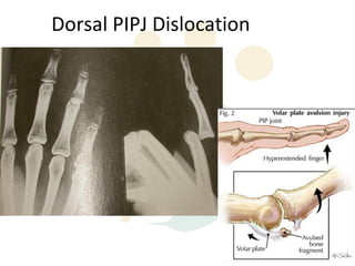
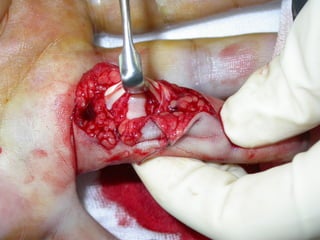
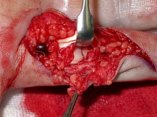
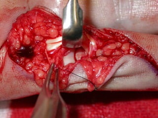
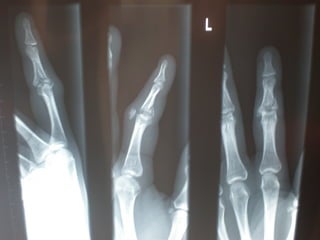
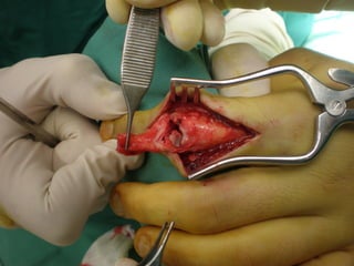
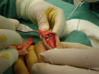
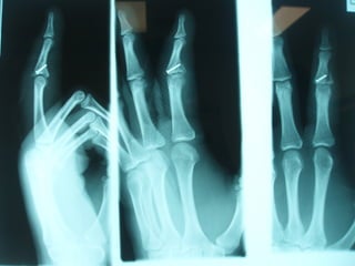
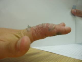
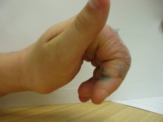
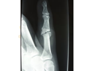
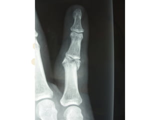
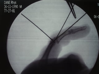
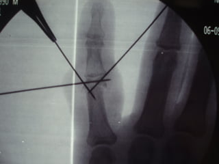
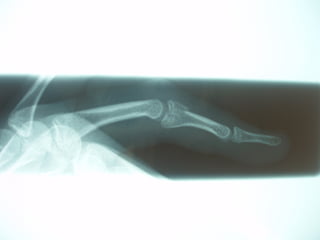
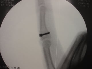
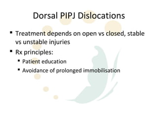
PIPJ Anatomy
Proximal Interphalangial Joint
ď‚§ Anatomical & functional locus of finger function
ď‚§ Site of most common ligament injury in the hand
ď‚§ Most ligament injury are incomplete with
  maintenance of joint congruity & stability
ď‚§ In certain injuries (eg. Lateral dislocations &
  hyperextension injuries) --> complete rupture of one
  or more supporting structures
ď‚§ Treatment based on accurate diagnosis of
  pathological lesions & degree of clinical dysfunction
Anatomy
ď‚§ PIPJ - Hinge joint
ď‚§ Arc of motion up to 1100
ď‚§ Stability:
  ď‚§ Articular contours
  ď‚§ Periarticular ligaments
  ď‚§ Secondary stabilization by adjacent tendon &
    retinacular systems
Anatomy - Bony Factors
ď‚§ Head of PP - 2x concentric condyles seperated by an
  intercondylar notch
ď‚§ Condyles (PP) articulate with 2x concave fossa in the
  broad, flattened base of MP separated by a median
  ridge
ď‚§ Tongue-and-groove contour & breadth of
  congruence add stability by resisting lateral &
  rotatory stress (esp. when PIPJ is fully extended)
Anatomy - Ligamentous factors
ď‚§ Radial & ulnar collateral ligaments
ď‚§ Primary restraints to radial & ulnar deviation force
ď‚§ Proper & accessory component
ď‚§ Both arise from the concave fossae on lateral aspects
  of each condyle & pass obliquely & volary to their
  insertions
ď‚§ Anatomically confluent but distinguished by their
  points of insertion
ď‚§ Proper collateral lig. --> volar 1/3 base of MP
ď‚§ Accessory collateral lig. --> volar plate
Anatomy - Volar Plate
           ď‚§ Floor of joint
           ď‚§ Suspended laterally by collateral
             ligs.
           ď‚§ Distal portion inserts across volar
             base of MP (only densely
             attached at its lateral margins -
             col. lig. insertion)
           ď‚§ Thinner centrally & blends with
             MP volar periosteum
           ď‚§ Central portion tapers proximally
             into an areolar sheet & laterally
             thickens to form a pair of check
             ligaments
           ď‚§ Secondary stabilizer against
             lateral deviation esp when PIPJ
             extended but only when
             collaterals torn
Check ligaments:
+Originate from periosteum of PP1 just inside walls of
A2 pulley at its distal margin and are confluent with
proximal origins of C1 pulley

+prevent hyperextension while permitting full flexion
thereby providing maximum stability with minimum
bulk
PIPJ Stability
       ď‚§ Key: strong conjoined
         attachment of the paired
         collateral lig. & the volar plate
         into the volar 1/3 of the MP
       ď‚§ Ligament-box configuration
         produces a 3D strength that
         strongly resists PIPJ
         displacement
       ď‚§ For MP displacement to occur,
         the ligament-box complex must
         be disrupted in at least 2
         planes
PIPJ Stability


ď‚§ Based on load to failure cadeveric studies & clinical observation,
  collateral ligs. fail proximally about 85% of the time while the volar
  plate avulses distally up to 80% of the time
ď‚§ At lower angular velocities of side-to-side deformation, the collateral
  ligs. tend to fail in their midsubstance
PIPJ - Secondary Stabilization

               ď‚§ Secondary
                 stabilization by
                 adjacent tendon &
                 retinacular systems
PIPJ dislocations
Dorsal PIPJ Dislocation
Dorsal PIPJ Dislocations
ď‚§ Mechanism: PIPJ hyperextension combined with
  some degree of longitudinal compression
ď‚§ Frequently occurs in ball-handling sports
ď‚§ Usually produces soft tissue or bone injury to the
  distal insertions of the 3D ligament-box complex.
ď‚§ The greater the longitudinal force, the more
  likelihood for fracture dislocation
ď‚§ Rarely, VP ruptures volarly & become interposed
  within the PIPJ causing irreducible dislocation
ď‚§ Volar fracture may even become trapped within the
  flexor sheath and inhibit motion.
Dorsal PIPJ Dislocations
            ď‚§ Type I (hyperextension): VP
              avulsed; incomplete
              longitudinal split in col. ligs.;
              articular surfaces remain
              congruous.
            ď‚§ Type II (dorsal dislocation):
              complete rupture VP; complete
              split in col. ligs.; MP resting on
              dorsum of PP.
            ď‚§ Type III (fracture-dislocation):
              disruption at the volar base of
              MP where VP is inserted; stable
              vs unstable injuries
Dorsal PIPJ Dislocations
            ď‚§ Stable Type III:
               ď‚§ fracture < 40% of volar
                 base MP; significant
                 portion of col. ligs. still
                 attached; possible
                 congruous reduction
            ď‚§ Unstable Type III:
               ď‚§ fracture > 40% of volar
                 base MP; little or no col.
                 ligs. attached; congruous
                 reduction unlikely;
                 depressed volar articular
                 defect
Dorsal PIPJ Dislocations
ď‚§ Treatment depends on open vs closed, stable
  vs unstable injuries
ď‚§ Rx principles:
  ď‚§ Patient education
  ď‚§ Avoidance of prolonged immobilisation
Dorsal PIPJ Dislocations
ď‚§ Operative Mx:
  ď‚§   Debridement & joint washout for open injuries
  ď‚§   Dorsal block splinting
  ď‚§   ? Role of primary VP repair
  ď‚§   Other specific techniques for unstable PIPJ injuries:
       ď‚§   Dynamic skeletal traction
       ď‚§   Extension block pinning
       ď‚§   Trans-articular pinning
       ď‚§   ORIF
       ď‚§   Volar plate arthroplasty
       ď‚§   FDS tenodesis (for chronic hyperextension deformity of PIPJ)
Dorsal PIPJ Dislocations
ď‚§ Complications of operative Mx:
  ď‚§   Redisplacement
  ď‚§   Angulation
  ď‚§   Flexion contracture
  ď‚§   DIPJ stiffness

Pipjw

  • 2.
  • 3.
    Proximal Interphalangial Joint ď‚§Anatomical & functional locus of finger function ď‚§ Site of most common ligament injury in the hand ď‚§ Most ligament injury are incomplete with maintenance of joint congruity & stability ď‚§ In certain injuries (eg. Lateral dislocations & hyperextension injuries) --> complete rupture of one or more supporting structures ď‚§ Treatment based on accurate diagnosis of pathological lesions & degree of clinical dysfunction
  • 4.
    Anatomy ď‚§ PIPJ -Hinge joint ď‚§ Arc of motion up to 1100 ď‚§ Stability: ď‚§ Articular contours ď‚§ Periarticular ligaments ď‚§ Secondary stabilization by adjacent tendon & retinacular systems
  • 5.
    Anatomy - BonyFactors ď‚§ Head of PP - 2x concentric condyles seperated by an intercondylar notch ď‚§ Condyles (PP) articulate with 2x concave fossa in the broad, flattened base of MP separated by a median ridge ď‚§ Tongue-and-groove contour & breadth of congruence add stability by resisting lateral & rotatory stress (esp. when PIPJ is fully extended)
  • 7.
    Anatomy - Ligamentousfactors ď‚§ Radial & ulnar collateral ligaments ď‚§ Primary restraints to radial & ulnar deviation force ď‚§ Proper & accessory component ď‚§ Both arise from the concave fossae on lateral aspects of each condyle & pass obliquely & volary to their insertions ď‚§ Anatomically confluent but distinguished by their points of insertion ď‚§ Proper collateral lig. --> volar 1/3 base of MP ď‚§ Accessory collateral lig. --> volar plate
  • 10.
    Anatomy - VolarPlate ď‚§ Floor of joint ď‚§ Suspended laterally by collateral ligs. ď‚§ Distal portion inserts across volar base of MP (only densely attached at its lateral margins - col. lig. insertion) ď‚§ Thinner centrally & blends with MP volar periosteum ď‚§ Central portion tapers proximally into an areolar sheet & laterally thickens to form a pair of check ligaments ď‚§ Secondary stabilizer against lateral deviation esp when PIPJ extended but only when collaterals torn
  • 11.
    Check ligaments: +Originate fromperiosteum of PP1 just inside walls of A2 pulley at its distal margin and are confluent with proximal origins of C1 pulley +prevent hyperextension while permitting full flexion thereby providing maximum stability with minimum bulk
  • 13.
    PIPJ Stability ď‚§ Key: strong conjoined attachment of the paired collateral lig. & the volar plate into the volar 1/3 of the MP ď‚§ Ligament-box configuration produces a 3D strength that strongly resists PIPJ displacement ď‚§ For MP displacement to occur, the ligament-box complex must be disrupted in at least 2 planes
  • 14.
    PIPJ Stability ď‚§ Basedon load to failure cadeveric studies & clinical observation, collateral ligs. fail proximally about 85% of the time while the volar plate avulses distally up to 80% of the time ď‚§ At lower angular velocities of side-to-side deformation, the collateral ligs. tend to fail in their midsubstance
  • 15.
    PIPJ - SecondaryStabilization ď‚§ Secondary stabilization by adjacent tendon & retinacular systems
  • 16.
  • 17.
  • 18.
    Dorsal PIPJ Dislocations ď‚§Mechanism: PIPJ hyperextension combined with some degree of longitudinal compression ď‚§ Frequently occurs in ball-handling sports ď‚§ Usually produces soft tissue or bone injury to the distal insertions of the 3D ligament-box complex. ď‚§ The greater the longitudinal force, the more likelihood for fracture dislocation ď‚§ Rarely, VP ruptures volarly & become interposed within the PIPJ causing irreducible dislocation ď‚§ Volar fracture may even become trapped within the flexor sheath and inhibit motion.
  • 19.
    Dorsal PIPJ Dislocations ď‚§ Type I (hyperextension): VP avulsed; incomplete longitudinal split in col. ligs.; articular surfaces remain congruous. ď‚§ Type II (dorsal dislocation): complete rupture VP; complete split in col. ligs.; MP resting on dorsum of PP. ď‚§ Type III (fracture-dislocation): disruption at the volar base of MP where VP is inserted; stable vs unstable injuries
  • 20.
    Dorsal PIPJ Dislocations ď‚§ Stable Type III: ď‚§ fracture < 40% of volar base MP; significant portion of col. ligs. still attached; possible congruous reduction ď‚§ Unstable Type III: ď‚§ fracture > 40% of volar base MP; little or no col. ligs. attached; congruous reduction unlikely; depressed volar articular defect
  • 37.
    Dorsal PIPJ Dislocations ď‚§Treatment depends on open vs closed, stable vs unstable injuries ď‚§ Rx principles: ď‚§ Patient education ď‚§ Avoidance of prolonged immobilisation
  • 38.
    Dorsal PIPJ Dislocations ď‚§Operative Mx: ď‚§ Debridement & joint washout for open injuries ď‚§ Dorsal block splinting ď‚§ ? Role of primary VP repair ď‚§ Other specific techniques for unstable PIPJ injuries: ď‚§ Dynamic skeletal traction ď‚§ Extension block pinning ď‚§ Trans-articular pinning ď‚§ ORIF ď‚§ Volar plate arthroplasty ď‚§ FDS tenodesis (for chronic hyperextension deformity of PIPJ)
  • 39.
    Dorsal PIPJ Dislocations ď‚§Complications of operative Mx: ď‚§ Redisplacement ď‚§ Angulation ď‚§ Flexion contracture ď‚§ DIPJ stiffness