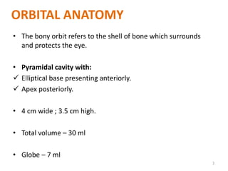
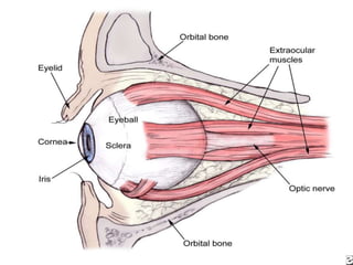
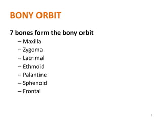
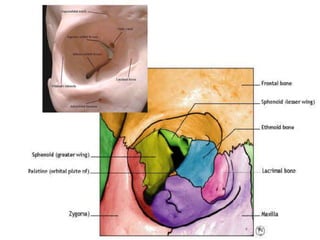
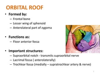
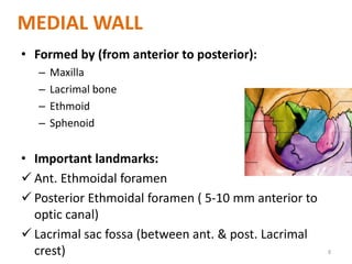
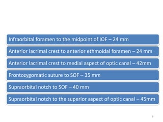
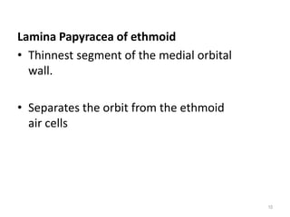
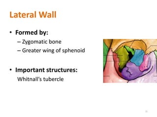
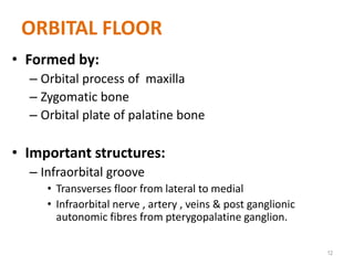
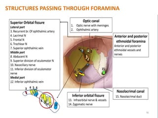
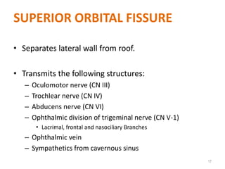
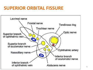
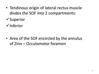
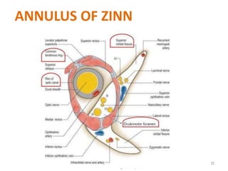
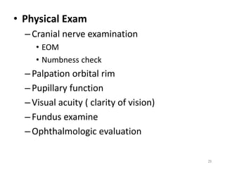
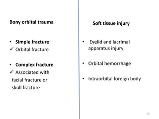
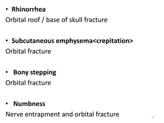
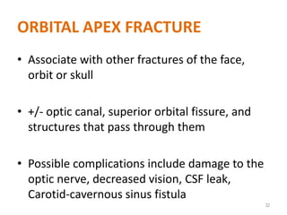
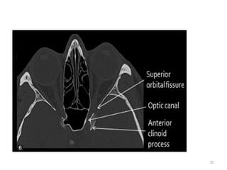
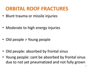
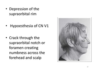
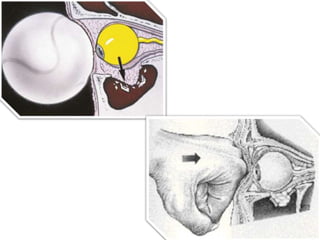
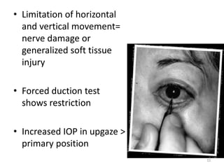
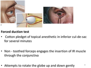
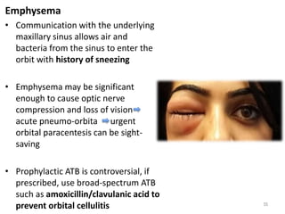
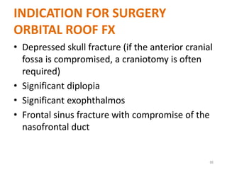
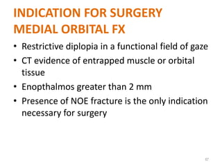
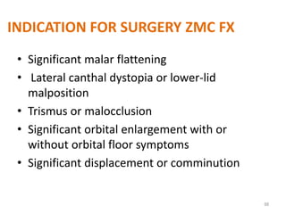
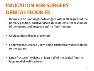
Dr. SNEHA GUPTA, JR II
ORAL AND MAXILLOFACIAL
SURGERY, K.G.M.U.
1
ORBITAL TRAUMA
TOPICS FOR DISCUSSION
• Orbital anatomy
• Types of fractures
• Signs and symptoms
• Management
2
ORBITAL ANATOMY
• The bony orbit refers to the shell of bone which surrounds
and protects the eye.
• Pyramidal cavity with:
 Elliptical base presenting anteriorly.
 Apex posteriorly.
• 4 cm wide ; 3.5 cm high.
• Total volume – 30 ml
• Globe – 7 ml
3
4
BONY ORBIT
7 bones form the bony orbit
– Maxilla
– Zygoma
– Lacrimal
– Ethmoid
– Palantine
– Sphenoid
– Frontal
5
6
ORBITAL ROOF
• Formed by:
– Frontal bone
– Lesser wing of sphenoid
– Anterolateral part of zygoma
• Functions as:
– Floor anterior fossa
• Important structures:
– Supraorbital notch - transmits supraorbital nerve
– Lacrimal fossa ( anterolaterally)
– Trochlear fossa (medially – supratrochlear artery & nerve)
7
MEDIAL WALL
• Formed by (from anterior to posterior):
– Maxilla
– Lacrimal bone
– Ethmoid
– Sphenoid
• Important landmarks:
 Ant. Ethmoidal foramen
 Posterior Ethmoidal foramen ( 5-10 mm anterior to
optic canal)
 Lacrimal sac fossa (between ant. & post. Lacrimal
crest) 8
Infraorbital foramen to the midpoint of IOF – 24 mm
Anterior lacrimal crest to anterior ethmoidal foramen – 24 mm
Anterior lacrimal crest to medial aspect of optic canal – 42mm
Frontozygomatic suture to SOF – 35 mm
Supraorbital notch to SOF – 40 mm
Supraorbital notch to the superior aspect of optic canal – 45mm
9
Lamina Papyracea of ethmoid
• Thinnest segment of the medial orbital
wall.
• Separates the orbit from the ethmoid
air cells
10
Lateral Wall
• Formed by:
– Zygomatic bone
– Greater wing of sphenoid
• Important structures:
Whitnall’s tubercle
11
ORBITAL FLOOR
• Formed by:
– Orbital process of maxilla
– Zygomatic bone
– Orbital plate of palatine bone
• Important structures:
– Infraorbital groove
• Transverses floor from lateral to medial
• Infraorbital nerve , artery , veins & post ganglionic
autonomic fibres from pterygopalatine ganglion.
12
ORBITAL FLOOR
• 0.5 mm thick
• Forms roof of maxillary sinus
• Location of more blow out fractures due to
inherent weakness of bone overlying maxillary
sinus
13
ORBITAL APEX
• Optic canal
• Superior orbital fissure
• Inferior orbital fissure
14
OPTIC CANAL
• Contains:
– Optic nerve
– Ophthalmic artery
• In Lesser wing of sphenoid
15
16
STRUCTURES PASSING THROUGH FORAMINA
SUPERIOR ORBITAL FISSURE
• Separates lateral wall from roof.
• Transmits the following structures:
– Oculomotor nerve (CN III)
– Trochlear nerve (CN IV)
– Abducens nerve (CN VI)
– Ophthalmic division of trigeminal nerve (CN V-1)
• Lacrimal, frontal and nasociliary Branches
– Ophthalmic vein
– Sympathetics from cavernous sinus
17
18
SUPERIOR ORBITAL FISSURE
• Tendinous origin of lateral rectus muscle
divdes the SOF into 2 compartments:
Superior
Inferior
• Area of the SOF encircled by the annulus
of Zinn – Occulomotor foramen
19
20
ANNULUS OF ZINN
CLINICAL CORRELATION
• Superior Orbital Fissure Syndrome
– Ptosis
– External Ophthalmoplegia ( III, IV &VI )
– Anaesthesia of cornea (Nasociliary)
– Ipsilateral Numbness forehead, lateral orbital
skin
• Orbital Apex Syndrome
– All of the above
– Visual Loss
21
INFERIOR ORBITAL FISSURE
• Connects to pterygopalantine fossa
• Located between floor and lateral wall
• Transmits:
– Maxillary division Trigeminal nerve
– Infra orbital Artery
– Zygomatic Nerve
– Sphenopalatine Ganglion Branches
– Ophthalmic Vein Branches 22
ARTERIAL SUPPLY
• Opthalmic a. ( branch of ICA)
 Muscle cone globe, all superior orbital
structures
• ECA – lower orbit through ECA
through maxillary a., infra orbital a.,
zygomaticofacial a., zygomatico temporal a.
23
VENOUS DRAINAGE
• Superior Opthalmic Veins SOF
Cavernous Sinus
• Inferior Opthalmic Vein Pterygoid
plexus Superior Opthalmic Veins
Cavernous Sinus
24
COMMON CAUSES OF ORBITAL
FRACTURES
• Falling
• Aggression
• Sporting events
• RTA
25
INITIAL EVALUATION
• History
–Time and mechanism of injury
–Change in appearance of eye
–State of vision immediately after injury
• Immediate loss of vision – severe damage to
retina
• Loss of light perception - vascular occlusion or
optic nerve compression
• Initial good vision – compression optic
neuropathy 26
EXTERNAL EYE EXAMINATION
Exophthalmos
• Without pulsation
Orbital hemorrhage
Emphysema
• With pulsation
High flow carotid cavernous fistula
Orbital roof fracture with secondary
herniation of anterior cranial fossa content
27
Telecanthus
• Medial canthal tendon
injury
28
• Physical Exam
–Cranial nerve examination
• EOM
• Numbness check
–Palpation orbital rim
–Pupillary function
–Visual acuity ( clarity of vision)
–Fundus examine
–Ophthalmologic evaluation
29
Bony orbital trauma
• Simple fracture
 Orbital fracture
• Complex fracture
 Associated with
facial fracture or
skull fracture
• Eyelid and lacrimal
apparatus injury
• Orbital hemorrhage
• Intraorbital foreign body
30
Soft tissue injury
• Rhinorrhea
Orbital roof / base of skull fracture
• Subcutaneous emphysema<crepitation>
Orbital fracture
• Bony stepping
Orbital fracture
• Numbness
Nerve entrapment and orbital fracture 31
ORBITAL APEX FRACTURE
• Associate with other fractures of the face,
orbit or skull
• +/- optic canal, superior orbital fissure, and
structures that pass through them
• Possible complications include damage to the
optic nerve, decreased vision, CSF leak,
Carotid-cavernous sinus fistula
32
33
ORBITAL ROOF FRACTURES
• Blunt trauma or missile injuries
• Moderate to high energy injuries
• Old people > Young people
• Old people: absorbed by frontal sinus
• Young people: cant be absorbed by frontal sinus
due to not yet pneumatized and not fully grown
34
Epistaxis, CSF rhinorrhea and
anosmia
• Roof fractures extend to the very
thin bones of the ethmoid and
cribriform plates
• If dura is torn in these areas, CSF can
drain from the anterior cranial fossa
through the frontonasal recesses as
clear fluid rhinorrhea
• Fracture at cribriform plate can also
damage the olfactory nerves
anosmia which might never be fully
recovered 35
Restricted up-gaze and
ptosis
• Secondary to the
inward displacement
of the levator/supeior
rectus muscle complex
by the bony fracture
plate and associated
subperiosteal
hematoma
36
• Depression of the
supraorbital rim
• Hypoesthesia of CN V1
• Crack through the
supraorbital notch or
foramen creating
numbness across the
forehead and scalp
37
Hypo-ophthalmos and pulsatile
exophthalmos
• Orbital floors tend to blow out and roofs tend to blow
in because the orbit is more compressible than the
brain bony fracture plate dislocates into the orbit,
displacing the orbital contents anteriorly
(exophthalmos) and inferiorly (hypoophthalmos)
• Open connection to the pulsatile ICP causes the globe
to pulse, best seen during supine position
38
MEDIAL ORBITAL FRACTURES
Naso-Orbital-Ethmoidal (NOE)
• Fractures usually result from the face striking
solid surfaces
• Involve frontal process of the maxilla, the
lacrimal bone, and the ethmoid bones along
the medial wall of the orbit
• Depressed bridge of the nose and traumatic
telecanthus 39
• NOE fractures are the result of high-energy
trauma that impacts on the central midface
• “Crumple zone” absorbing energy as it
collapses internally but mostly not involves
ocular
40
SIGNS & SYMPTOMS
• Horizontal diplopia
Unlike floor fractures with vertical diplopia
• Orbital emphysema
 Fracture into the adjacent sinus allows sinus air and bacteria into the orbit
 Precautions regarding nose blowing and prophylactic ATB
• Orbital hemorrhage
More dramatic than fracture floor due to lack of the natural drainage afforded
by a floor fracture
• Enophthalmos
Sufficiently large medial wall fracture allows prolapse of enough orbital tissue
to create significant loss of globe projection
41
Complications
• Facial flattening
• Cerebral and ocular damage
• Severe epistaxis due to avulsion of
ant.ethmoidal artery
• Orbital hematoma
• Cerebrospinal fluid rhinorrhea
• Damage to the lacrimal drainage system
• Lateral displacement of the medial canthus
42
Treatment
• Repair of the nasal
fracture and plate
stabilization
• Transnasal wiring of
the medial canthus
is seldomly used
• Miniplate fixation
allows precise bony
reduction 43
ORBITAL FLOOR FRACTURE
• History of object
struck at orbital
entrance
• Forceful enough
to cause
ecchymosis (low-
moderate energy)
44
BLOWOUT FRACTURES OF ORBIT
• Originally defined as orbital floor fractures
without fracture orbital rim, but with
entrapment one or more soft tissue
structures.
45
BLOWOUT FRACTURES
• Blowout fractures now refer to fractures of the:
– Orbital floor
– Medial wall
– Lateral wall
– Superior wall
• “pure” blowout fractures – trapdoor rotation to bone
fragments involving central area of bone
• “impure” fracture – fracture line extends to orbital
rim
46
PHYSIOLOGY OF BLOWOUT FRACTURE
• The bony defect is filled with soft tissue and
fat from the orbit.
• Alters support mechanisms for EOM.
• EOM can become entrapped.
• Direct muscle damage can result.
47
48
49
INJURIES ASSOCIATED WITH BLOW
OUT FRACTURES
• Ruptured globe
• Retroorbital hemorrhage
• Vitreous hemorrhage
• Hyphema
• Dislocated lens
• Secondary glaucoma
• Retinal detachment
50
Eyelid sign
• Ecchymosis
and edema
but other
signs of
injury can be
absent
(white-eyed
blowout)
51
Vertical Diplopia with limitation of
upgaze, downgaze, or both
• Limited vertical movement of the
globe, vertical diplopia ad pain in the
inferior orbit on attempted vertical
movement=entrapment of the IR
muscle, the larger and more
comminuted the fracture, the less
likely entrapment and diplopia are to
occur
• Orbital edema and hemorrhage or
damage to EOM or innervation can
result in limit movement but improve
in 1-2 weeks
52
• Limitation of horizontal
and vertical movement=
nerve damage or
generalized soft tissue
injury
• Forced duction test
shows restriction
• Increased IOP in upgaze >
primary position
53
Forced duction test
• Cotton pledget of topical anesthetic in inferior cul-de-sac
for several minutes
• Non - toothed forceps engages the insertion of IR muscle
through the conjunctiva
• Attempts to rotate the globe up and down gently 54
Emphysema
• Communication with the underlying
maxillary sinus allows air and
bacteria from the sinus to enter the
orbit with history of sneezing
• Emphysema may be significant
enough to cause optic nerve
compression and loss of vision
acute pneumo-orbita urgent
orbital paracentesis can be sight-
saving
• Prophylactic ATB is controversial, if
prescribed, use broad-spectrum ATB
such as amoxicillin/clavulanic acid to
prevent orbital cellulitis 55
Enophthalmos and ptosis of the globe
• Large fractures
• Tissue prolapse into maxillary sinus
• Medial wall + orbital floor fracture = significant enopthalmos
• More apparent when edema subsides
• 2mm of enophthalmos is generally regarded as normal variation and not
cosmetically noticeable in most people
• Late correction lead to fibrosis and Volkmann’s contractures within the orbital
tissues
Pupillary abnormalities
• Damage to the pupillary nerve fibers traveling with the inferior oblique muscle
Hypoesthesia of infraorbital cranial nerve V2
• Infraorbital nerve distribution which nerves travel along orbital floor before
exits from the infraorbital foramen
56
Compartment syndrome
• Orbital hemorrhage in patient with loss of
vision+proptosis+increased IOP
Oculocardiac reflex and the white-eyed-blow-out
• Attempting to move an eye that is entrapped by a blowout fracture
may cause increased vagal tone oculocardiac reflex severe
bradycardia or heart bock, syncope - urgent surgical intervention is
warranted
• More common in young patients(<18 y) with greenstick fractures
and trapdoor fractures of the floor
• White-eyed blowout, bony plate don’t fracture completely but
opens long enough to allow orbital tissues to herniate.
• The fracture plate then closes, entrapping the tissues - long term
tissue ischemia may occur - fail in surgery
57
Management
• CT scan coronal , sagittal view
• Diagnosis mainly by clinical examination
• Mostly not require surgical intervention
• Observed for 5-10 days for swelling to
subsides
• Oral steroids (for 7 days) decrease edema
• Pediatric patients with IR muscle trapping
beneath trapdoor fracture - eye movement
aggravate occulocardiac reflex(pain , nausea
and bradycardia)
58
59
60
Herniation of orbital fat into the maxillary sinus
61
Image shows depressed fragment of the left orbital floor (arrow)
and an air-fluid level in the left maxillary sinus
62
TREATMENT OPTIONS
• Nonsurgical
• Surgical
63
64
TAKE HOME MESSAGE
ELLIS’ 6 PRINCIPLES FOR
RECONSTRUCTION
In large defects, a thin, rigid material should be used to maintain the orbital
shape during surgery.
Although all of the defect should be covered by the implant, the minimum size
necessary should be used to avoid injury to the vital structures within the orbit.
Normal anatomy should be achieved by properly shaping the material before
insertion.
Placement should be tension free to avoid trapping periorbital tissues that may
restrict the eye movement.
The material should be stabilized.
Orbital reconstruction should be verified for accuracy.
65
INDICATION FOR SURGERY
ORBITAL ROOF FX
• Depressed skull fracture (if the anterior cranial
fossa is compromised, a craniotomy is often
required)
• Significant diplopia
• Significant exophthalmos
• Frontal sinus fracture with compromise of the
nasofrontal duct
66
INDICATION FOR SURGERY
MEDIAL ORBITAL FX
• Restrictive diplopia in a functional field of gaze
• CT evidence of entrapped muscle or orbital
tissue
• Enopthalmos greater than 2 mm
• Presence of NOE fracture is the only indication
necessary for surgery
67
INDICATION FOR SURGERY ZMC FX
• Significant malar flattening
• Lateral canthal dystopia or lower-lid
malposition
• Trismus or malocclusion
• Significant orbital enlargement with or
without orbital floor symptoms
• Significant displacement or comminution
68
INDICATION FOR SURGERY
ORBITAL FLOOR FX
• Diplopia with limit upgaze/downgaze within 30 degrees of the
primary position, positive forced duction test after resolution
of the edema and imaging confirm floor fracture
• Oculocardiac reflex is presented
• Enophthalmos exceed 2 mm and is cosmetically unacceptable
to the patient
• Large fractures involving at least half of the orbital floor +/-
large medial wall fractures
69
CONTRAINDICATIONS TO
SURGERY
• Hyphema (collection of blood b/n iris &
cornea)
• Retinal detachment
• Globe perforation
• Only seeing eye
• Medically unstable patient
• Recent opthalmologic surgery
70
SURGICAL APPROACHES
• Transconjunctival approach
• Infra Orbital
• Subciliary
• Trans septal
71
Orbital Implants
• Use of implants based on degree of
comminution and size of fracture
• Various implant material used
– Autogenous bone and cartilage
– Alloplastic material
• Teflon
• Marlex
• PEEK
• Titanium
72
• A thin porous polyethylene sheet can be used
in smaller defects.
• Larger defect can be treated with a sheet of titanium-
reinforced porous polyethylene secured with 1 or 2
screws just behind the anterior rim of the orbit.
• No scientific evidence indicates that one material is
better than others.
• In growing orbit (before age 8), the surgeon should be
cautious about any materialmothher than autogenous
bone or resorbable material.
73
COMPLICATIONS OF SURGERY
• Ectropion
• Lid retraction
• Persistent diplopia
• Malposition of eye
• Hypoaesthesia of V2
• Extrusion of orbital floor implant
• BLINDNESS
74
75

Orbital trauma 1

  • 1.
    Dr. SNEHA GUPTA,JR II ORAL AND MAXILLOFACIAL SURGERY, K.G.M.U. 1 ORBITAL TRAUMA
  • 2.
    TOPICS FOR DISCUSSION •Orbital anatomy • Types of fractures • Signs and symptoms • Management 2
  • 3.
    ORBITAL ANATOMY • Thebony orbit refers to the shell of bone which surrounds and protects the eye. • Pyramidal cavity with:  Elliptical base presenting anteriorly.  Apex posteriorly. • 4 cm wide ; 3.5 cm high. • Total volume – 30 ml • Globe – 7 ml 3
  • 4.
  • 5.
    BONY ORBIT 7 bonesform the bony orbit – Maxilla – Zygoma – Lacrimal – Ethmoid – Palantine – Sphenoid – Frontal 5
  • 6.
  • 7.
    ORBITAL ROOF • Formedby: – Frontal bone – Lesser wing of sphenoid – Anterolateral part of zygoma • Functions as: – Floor anterior fossa • Important structures: – Supraorbital notch - transmits supraorbital nerve – Lacrimal fossa ( anterolaterally) – Trochlear fossa (medially – supratrochlear artery & nerve) 7
  • 8.
    MEDIAL WALL • Formedby (from anterior to posterior): – Maxilla – Lacrimal bone – Ethmoid – Sphenoid • Important landmarks:  Ant. Ethmoidal foramen  Posterior Ethmoidal foramen ( 5-10 mm anterior to optic canal)  Lacrimal sac fossa (between ant. & post. Lacrimal crest) 8
  • 9.
    Infraorbital foramen tothe midpoint of IOF – 24 mm Anterior lacrimal crest to anterior ethmoidal foramen – 24 mm Anterior lacrimal crest to medial aspect of optic canal – 42mm Frontozygomatic suture to SOF – 35 mm Supraorbital notch to SOF – 40 mm Supraorbital notch to the superior aspect of optic canal – 45mm 9
  • 10.
    Lamina Papyracea ofethmoid • Thinnest segment of the medial orbital wall. • Separates the orbit from the ethmoid air cells 10
  • 11.
    Lateral Wall • Formedby: – Zygomatic bone – Greater wing of sphenoid • Important structures: Whitnall’s tubercle 11
  • 12.
    ORBITAL FLOOR • Formedby: – Orbital process of maxilla – Zygomatic bone – Orbital plate of palatine bone • Important structures: – Infraorbital groove • Transverses floor from lateral to medial • Infraorbital nerve , artery , veins & post ganglionic autonomic fibres from pterygopalatine ganglion. 12
  • 13.
    ORBITAL FLOOR • 0.5mm thick • Forms roof of maxillary sinus • Location of more blow out fractures due to inherent weakness of bone overlying maxillary sinus 13
  • 14.
    ORBITAL APEX • Opticcanal • Superior orbital fissure • Inferior orbital fissure 14
  • 15.
    OPTIC CANAL • Contains: –Optic nerve – Ophthalmic artery • In Lesser wing of sphenoid 15
  • 16.
  • 17.
    SUPERIOR ORBITAL FISSURE •Separates lateral wall from roof. • Transmits the following structures: – Oculomotor nerve (CN III) – Trochlear nerve (CN IV) – Abducens nerve (CN VI) – Ophthalmic division of trigeminal nerve (CN V-1) • Lacrimal, frontal and nasociliary Branches – Ophthalmic vein – Sympathetics from cavernous sinus 17
  • 18.
  • 19.
    • Tendinous originof lateral rectus muscle divdes the SOF into 2 compartments: Superior Inferior • Area of the SOF encircled by the annulus of Zinn – Occulomotor foramen 19
  • 20.
  • 21.
    CLINICAL CORRELATION • SuperiorOrbital Fissure Syndrome – Ptosis – External Ophthalmoplegia ( III, IV &VI ) – Anaesthesia of cornea (Nasociliary) – Ipsilateral Numbness forehead, lateral orbital skin • Orbital Apex Syndrome – All of the above – Visual Loss 21
  • 22.
    INFERIOR ORBITAL FISSURE •Connects to pterygopalantine fossa • Located between floor and lateral wall • Transmits: – Maxillary division Trigeminal nerve – Infra orbital Artery – Zygomatic Nerve – Sphenopalatine Ganglion Branches – Ophthalmic Vein Branches 22
  • 23.
    ARTERIAL SUPPLY • Opthalmica. ( branch of ICA)  Muscle cone globe, all superior orbital structures • ECA – lower orbit through ECA through maxillary a., infra orbital a., zygomaticofacial a., zygomatico temporal a. 23
  • 24.
    VENOUS DRAINAGE • SuperiorOpthalmic Veins SOF Cavernous Sinus • Inferior Opthalmic Vein Pterygoid plexus Superior Opthalmic Veins Cavernous Sinus 24
  • 25.
    COMMON CAUSES OFORBITAL FRACTURES • Falling • Aggression • Sporting events • RTA 25
  • 26.
    INITIAL EVALUATION • History –Timeand mechanism of injury –Change in appearance of eye –State of vision immediately after injury • Immediate loss of vision – severe damage to retina • Loss of light perception - vascular occlusion or optic nerve compression • Initial good vision – compression optic neuropathy 26
  • 27.
    EXTERNAL EYE EXAMINATION Exophthalmos •Without pulsation Orbital hemorrhage Emphysema • With pulsation High flow carotid cavernous fistula Orbital roof fracture with secondary herniation of anterior cranial fossa content 27
  • 28.
  • 29.
    • Physical Exam –Cranialnerve examination • EOM • Numbness check –Palpation orbital rim –Pupillary function –Visual acuity ( clarity of vision) –Fundus examine –Ophthalmologic evaluation 29
  • 30.
    Bony orbital trauma •Simple fracture  Orbital fracture • Complex fracture  Associated with facial fracture or skull fracture • Eyelid and lacrimal apparatus injury • Orbital hemorrhage • Intraorbital foreign body 30 Soft tissue injury
  • 31.
    • Rhinorrhea Orbital roof/ base of skull fracture • Subcutaneous emphysema<crepitation> Orbital fracture • Bony stepping Orbital fracture • Numbness Nerve entrapment and orbital fracture 31
  • 32.
    ORBITAL APEX FRACTURE •Associate with other fractures of the face, orbit or skull • +/- optic canal, superior orbital fissure, and structures that pass through them • Possible complications include damage to the optic nerve, decreased vision, CSF leak, Carotid-cavernous sinus fistula 32
  • 33.
  • 34.
    ORBITAL ROOF FRACTURES •Blunt trauma or missile injuries • Moderate to high energy injuries • Old people > Young people • Old people: absorbed by frontal sinus • Young people: cant be absorbed by frontal sinus due to not yet pneumatized and not fully grown 34
  • 35.
    Epistaxis, CSF rhinorrheaand anosmia • Roof fractures extend to the very thin bones of the ethmoid and cribriform plates • If dura is torn in these areas, CSF can drain from the anterior cranial fossa through the frontonasal recesses as clear fluid rhinorrhea • Fracture at cribriform plate can also damage the olfactory nerves anosmia which might never be fully recovered 35
  • 36.
    Restricted up-gaze and ptosis •Secondary to the inward displacement of the levator/supeior rectus muscle complex by the bony fracture plate and associated subperiosteal hematoma 36
  • 37.
    • Depression ofthe supraorbital rim • Hypoesthesia of CN V1 • Crack through the supraorbital notch or foramen creating numbness across the forehead and scalp 37
  • 38.
    Hypo-ophthalmos and pulsatile exophthalmos •Orbital floors tend to blow out and roofs tend to blow in because the orbit is more compressible than the brain bony fracture plate dislocates into the orbit, displacing the orbital contents anteriorly (exophthalmos) and inferiorly (hypoophthalmos) • Open connection to the pulsatile ICP causes the globe to pulse, best seen during supine position 38
  • 39.
    MEDIAL ORBITAL FRACTURES Naso-Orbital-Ethmoidal(NOE) • Fractures usually result from the face striking solid surfaces • Involve frontal process of the maxilla, the lacrimal bone, and the ethmoid bones along the medial wall of the orbit • Depressed bridge of the nose and traumatic telecanthus 39
  • 40.
    • NOE fracturesare the result of high-energy trauma that impacts on the central midface • “Crumple zone” absorbing energy as it collapses internally but mostly not involves ocular 40
  • 41.
    SIGNS & SYMPTOMS •Horizontal diplopia Unlike floor fractures with vertical diplopia • Orbital emphysema  Fracture into the adjacent sinus allows sinus air and bacteria into the orbit  Precautions regarding nose blowing and prophylactic ATB • Orbital hemorrhage More dramatic than fracture floor due to lack of the natural drainage afforded by a floor fracture • Enophthalmos Sufficiently large medial wall fracture allows prolapse of enough orbital tissue to create significant loss of globe projection 41
  • 42.
    Complications • Facial flattening •Cerebral and ocular damage • Severe epistaxis due to avulsion of ant.ethmoidal artery • Orbital hematoma • Cerebrospinal fluid rhinorrhea • Damage to the lacrimal drainage system • Lateral displacement of the medial canthus 42
  • 43.
    Treatment • Repair ofthe nasal fracture and plate stabilization • Transnasal wiring of the medial canthus is seldomly used • Miniplate fixation allows precise bony reduction 43
  • 44.
    ORBITAL FLOOR FRACTURE •History of object struck at orbital entrance • Forceful enough to cause ecchymosis (low- moderate energy) 44
  • 45.
    BLOWOUT FRACTURES OFORBIT • Originally defined as orbital floor fractures without fracture orbital rim, but with entrapment one or more soft tissue structures. 45
  • 46.
    BLOWOUT FRACTURES • Blowoutfractures now refer to fractures of the: – Orbital floor – Medial wall – Lateral wall – Superior wall • “pure” blowout fractures – trapdoor rotation to bone fragments involving central area of bone • “impure” fracture – fracture line extends to orbital rim 46
  • 47.
    PHYSIOLOGY OF BLOWOUTFRACTURE • The bony defect is filled with soft tissue and fat from the orbit. • Alters support mechanisms for EOM. • EOM can become entrapped. • Direct muscle damage can result. 47
  • 48.
  • 49.
  • 50.
    INJURIES ASSOCIATED WITHBLOW OUT FRACTURES • Ruptured globe • Retroorbital hemorrhage • Vitreous hemorrhage • Hyphema • Dislocated lens • Secondary glaucoma • Retinal detachment 50
  • 51.
    Eyelid sign • Ecchymosis andedema but other signs of injury can be absent (white-eyed blowout) 51
  • 52.
    Vertical Diplopia withlimitation of upgaze, downgaze, or both • Limited vertical movement of the globe, vertical diplopia ad pain in the inferior orbit on attempted vertical movement=entrapment of the IR muscle, the larger and more comminuted the fracture, the less likely entrapment and diplopia are to occur • Orbital edema and hemorrhage or damage to EOM or innervation can result in limit movement but improve in 1-2 weeks 52
  • 53.
    • Limitation ofhorizontal and vertical movement= nerve damage or generalized soft tissue injury • Forced duction test shows restriction • Increased IOP in upgaze > primary position 53
  • 54.
    Forced duction test •Cotton pledget of topical anesthetic in inferior cul-de-sac for several minutes • Non - toothed forceps engages the insertion of IR muscle through the conjunctiva • Attempts to rotate the globe up and down gently 54
  • 55.
    Emphysema • Communication withthe underlying maxillary sinus allows air and bacteria from the sinus to enter the orbit with history of sneezing • Emphysema may be significant enough to cause optic nerve compression and loss of vision acute pneumo-orbita urgent orbital paracentesis can be sight- saving • Prophylactic ATB is controversial, if prescribed, use broad-spectrum ATB such as amoxicillin/clavulanic acid to prevent orbital cellulitis 55
  • 56.
    Enophthalmos and ptosisof the globe • Large fractures • Tissue prolapse into maxillary sinus • Medial wall + orbital floor fracture = significant enopthalmos • More apparent when edema subsides • 2mm of enophthalmos is generally regarded as normal variation and not cosmetically noticeable in most people • Late correction lead to fibrosis and Volkmann’s contractures within the orbital tissues Pupillary abnormalities • Damage to the pupillary nerve fibers traveling with the inferior oblique muscle Hypoesthesia of infraorbital cranial nerve V2 • Infraorbital nerve distribution which nerves travel along orbital floor before exits from the infraorbital foramen 56
  • 57.
    Compartment syndrome • Orbitalhemorrhage in patient with loss of vision+proptosis+increased IOP Oculocardiac reflex and the white-eyed-blow-out • Attempting to move an eye that is entrapped by a blowout fracture may cause increased vagal tone oculocardiac reflex severe bradycardia or heart bock, syncope - urgent surgical intervention is warranted • More common in young patients(<18 y) with greenstick fractures and trapdoor fractures of the floor • White-eyed blowout, bony plate don’t fracture completely but opens long enough to allow orbital tissues to herniate. • The fracture plate then closes, entrapping the tissues - long term tissue ischemia may occur - fail in surgery 57
  • 58.
    Management • CT scancoronal , sagittal view • Diagnosis mainly by clinical examination • Mostly not require surgical intervention • Observed for 5-10 days for swelling to subsides • Oral steroids (for 7 days) decrease edema • Pediatric patients with IR muscle trapping beneath trapdoor fracture - eye movement aggravate occulocardiac reflex(pain , nausea and bradycardia) 58
  • 59.
  • 60.
    60 Herniation of orbitalfat into the maxillary sinus
  • 61.
    61 Image shows depressedfragment of the left orbital floor (arrow) and an air-fluid level in the left maxillary sinus
  • 62.
  • 63.
  • 64.
  • 65.
    ELLIS’ 6 PRINCIPLESFOR RECONSTRUCTION In large defects, a thin, rigid material should be used to maintain the orbital shape during surgery. Although all of the defect should be covered by the implant, the minimum size necessary should be used to avoid injury to the vital structures within the orbit. Normal anatomy should be achieved by properly shaping the material before insertion. Placement should be tension free to avoid trapping periorbital tissues that may restrict the eye movement. The material should be stabilized. Orbital reconstruction should be verified for accuracy. 65
  • 66.
    INDICATION FOR SURGERY ORBITALROOF FX • Depressed skull fracture (if the anterior cranial fossa is compromised, a craniotomy is often required) • Significant diplopia • Significant exophthalmos • Frontal sinus fracture with compromise of the nasofrontal duct 66
  • 67.
    INDICATION FOR SURGERY MEDIALORBITAL FX • Restrictive diplopia in a functional field of gaze • CT evidence of entrapped muscle or orbital tissue • Enopthalmos greater than 2 mm • Presence of NOE fracture is the only indication necessary for surgery 67
  • 68.
    INDICATION FOR SURGERYZMC FX • Significant malar flattening • Lateral canthal dystopia or lower-lid malposition • Trismus or malocclusion • Significant orbital enlargement with or without orbital floor symptoms • Significant displacement or comminution 68
  • 69.
    INDICATION FOR SURGERY ORBITALFLOOR FX • Diplopia with limit upgaze/downgaze within 30 degrees of the primary position, positive forced duction test after resolution of the edema and imaging confirm floor fracture • Oculocardiac reflex is presented • Enophthalmos exceed 2 mm and is cosmetically unacceptable to the patient • Large fractures involving at least half of the orbital floor +/- large medial wall fractures 69
  • 70.
    CONTRAINDICATIONS TO SURGERY • Hyphema(collection of blood b/n iris & cornea) • Retinal detachment • Globe perforation • Only seeing eye • Medically unstable patient • Recent opthalmologic surgery 70
  • 71.
    SURGICAL APPROACHES • Transconjunctivalapproach • Infra Orbital • Subciliary • Trans septal 71
  • 72.
    Orbital Implants • Useof implants based on degree of comminution and size of fracture • Various implant material used – Autogenous bone and cartilage – Alloplastic material • Teflon • Marlex • PEEK • Titanium 72
  • 73.
    • A thinporous polyethylene sheet can be used in smaller defects. • Larger defect can be treated with a sheet of titanium- reinforced porous polyethylene secured with 1 or 2 screws just behind the anterior rim of the orbit. • No scientific evidence indicates that one material is better than others. • In growing orbit (before age 8), the surgeon should be cautious about any materialmothher than autogenous bone or resorbable material. 73
  • 74.
    COMPLICATIONS OF SURGERY •Ectropion • Lid retraction • Persistent diplopia • Malposition of eye • Hypoaesthesia of V2 • Extrusion of orbital floor implant • BLINDNESS 74
  • 75.