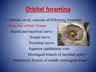
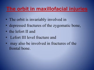
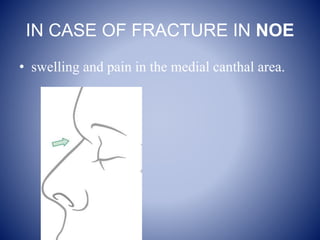
BY:-
Dr.SAUVIK SINGHA
PG-I yr STUDENT
Dept of oral and maxillofacial surgery
HDC&H
CONTENTS
 Introduction
 Osteology of orbit
 Contents of orbit
Innervation of the orbit
Vascular supply
The orbit in maxillofacial injuries
Surgical approach to the orbit
Safe distance in orbital dissection
conclusion
INTRODUCTION
• The orbits are pyramidal bony cavities lodging
the eyeballs, situated one on each side of the
root of the nose.
• They provide socket for rotatory movements of
the eye ball.
• The long axis of the each orbit passes
backward and medially.
VISUAL AXIS AND ORBITAL AXIS
• Axis passing through centre
of anterior and posterior
poles of the eyeball is known
as visual axis.
• It makes an angle of 20-25
degree with the orbital axis.
• Orbital axis is the line
passing through the optic
canal and centre of base of
orbit (anteriorly)
OSTEOLOGY OF ORBIT
Osteology of the orbit can be discussed under
following headings:-
 orbital margins
 orbital roof
 orbital floor
 medial wall
 lateral wall
ORBITAL MARGINS
quadrangular in shape
bounded by the following four margins:-
 Supraorbital margins-formed by frontal bone.
 Infraorbital margin-formed by zygomatic bone laterally
and the maxilla medially.
 Medial orbital margin- formed by frontal bone above and
by frontal process of maxilla
 Lateral orbital margins:-It is formed mostly by the frontal
process of zygomatic bone and zygomatic process of frontal
bone.
Bones Forming Orbit
• Seven bones make up the
bony orbit:
– Frontal
– Zygomatic
– Maxillary
– Ethmoidal
– Sphenoid
– Lacrimal
– Palatine
Orbital Roof
• orbital plate of
the frontal bone
• the lesser wing
of the sphenoid
bone.
Orbital Floor
– Maxillary
– Palatine
– Orbital plate of
the zygomatic
bone
Medial Orbital Wall
• Frontal process of
the maxillary
• Lacrimal
• Orbital plate of
the ethmoidal
• Lesser wing of
the sphenoid
• weakest
Lateral Orbital Wall
– Zygomatic
– Greater wing of the
sphenoid
• Strongest
• (Whitnall’s tubercle)
Orbital foramina
• Orbital cavity consists of following foramina:
• Superior orbital fissure:
lateral part-lacrimal nerve
frontal nerve
Trochlear nerve
Superior ophthalmic vein
Meningeal branch of lacrimal artery
Anastmotic branch of middle meningeal artery.
• Middle part:-occulomotor nerve
Nasocilliary nerve
Abducents nerve
Medial part:-inferior ophthalmic vein
Sympathetic nerves from the plexus around the
internal carotid artery.
Inferior orbital fissure:-zygomatic nerve
Infraorbital vessel
Maxillary nerve
Emissary vein
• Supra orbital foramen:-supraorbital nerves and
vessel
• Infra orbital foramen:-infraorbital nerve and
vessel
• Optic canal :- optic nerve
Ophthalmic artery
Central retinal vein
Content of orbit
Eyeball
Fascia: orbital and bulbar
Extraocular muscles
Vessels: ophthalmic artery ,ophthalmic veins and
lymphatics
Nerve: optic, occulomotor ,trochlear and abducents,
branches of ophthalmic and maxillary nerves.
Lacrimal glands
Orbital fat.
Orbital fascia
periorbita
It forms the periosteum of the bony orbit
Anteriorly, it is continuous with the
periosteum lining the bone around the
orbital margins
Posteriorly, it is continuous with
duramater and with the sheath of the
optic nerve.
At upper and lower margin this fascia
forms the flap like continuations called
orbital septum
Tenon’s capsule
• It forms the thin , loose membranous sheath
around the eye ball extending from optic nerve
to the sclerocorneal junction
• It is separated from the sclera by the episcleral
space which is traversed by the delicate
fibrous band.this allows the free movement of
the eyeball within the sheath.
The lower part of tenon’s capsule is thickened
and is named the suspensory ligament of eye
or suspensory ligament of lockwood
It is formed by the union of the margins of the
sheath of inferior rectus and inferior oblique
muscles with the medial and lateral check
ligaments.
Extra ocular muscle
Extra ocular
muscle
Voluntary
muscles
Four recti Two obliqui
The levator
palpebrae
superioris
Involuntary
muscles
The superior
tarsal muscle
Inferior tarsal
muscle
The orbitalis
Origin of voluntary muscle
• The four recti arise from a common annular
ring. Annulus of zinn
• This ring is attached to the orbital surface of
the apex of the orbit.
• It encloses optic canal and middle part of
superior orbital fissure.
• Superior oblique arises from the body of
sphenoid superomedial to optic canal
• Inferior oblique from the orbital surface of the
maxilla , lateral to the lacrimal groove.
• The levator palpebrae superioris arise from
orbital surface of lesser wing of sphenoid.
Insertion of voluntary muscles
• Recti are inserted into the sclera
• The tendon of the superior oblique passes
through a fibrocartilagenous pulley attached to
the trochlear fossa of the frontal bone.
• It is inserted into the sclera behind the equator
of the eyeball, between the superior rectus and
the lateral rectus.
• Inferior oblique is inserted close to the
superior oblique,a little below and posterior
INVOLUNTARY MUSCLES
• The superior tarsal muscle is the deeper portion of the
levator palpebrae superioris. It is inserted on the upper
margin of superior tarsus.
• It elevates the upper eye lids
• The inferior tarsal muscle extends from the fascial
sheath of the inferior rectus and inferior oblique to the
lower margin of inferior tarsus.it possibly depresses the
lower eyelid
• The orbitalis bridges the inferior orbital fissure. Its
action is uncertain.
Innervation of the orbit
The innervation of the orbit can be divided into
component:-
• extraoccular muscle
• General sensations are carried by occulomotor
nerve and trigeminal nerve.
• Autonomic supply..occulomotor nerve and
lacrimal nerve
• Vision –optic nerve
Nerve supply of extraocular
Muscles
• The four recti and two
oblique muscles
• All are supplied by
oculomotor nerve III
except superior
oblique (Trochlear N)
and lateral rectus
(Abducens)
Vascular supply
• An anastomosing network of the vessels
derived from the internal and external carotid
artery system supplies the orbit.
• Major arterial supply is ophthalmic artery,the
first major branch of the internal carotid artery.
• It enters the orbit through the optic canal.
• The ocular branches include:
central artery of retina
ant and post cilliary artery
• The orbital branches are :
lacrimal artery
Muscular artery
Extra orbital branch include
Posterior and anterior ethmoidal artery
Supra orbital artery
Medial palpebral
Supra trochlear branch
Dorsal nasal branch.
The orbit in maxillofacial injuries
• The orbit is invariably involved in
• depressed fractures of the zygomatic bone,
• the lefort II and
• Lefort III level fracture and
• may also be involved in fractures of the
frontal bone.
Racoon eyes.
• Subconjunctival Ecchymosis–
• Flame shaped hemorrhage with posterior limit not seen .
( Suspect # of the orbital walls )
Pupillary reaction
A light is used to assess pupillary reaction
• It may also be involved in extensive nasal
complex injuries which are described as
nasoethmoidal fracture
• A fracture of the orbit without involvement of
the rim is termed as “blow out” fractures and
most frequently involves the orbital floor.
IN CASE OF FRACTURE IN NOE
• swelling and pain in the medial canthal area.
Intercanthal distance
• in Caucasians more than 35 mm intercanthal distance is considered
abnormal.
• illustration demonstrates widening on the left with the medial canthus
positioned lateral to the position of the lateral nasal alar margin
Acuity testing
Visual field testing
Testing of ocular motility
Examine the patient to check the extraocular
muscle (EOM) are functioning properly.
If the extra ocular muscles (EOM) are not functioning properly the
surgeon should make sure that there is no entrapment of the soft
tissues. It is recommended to perform the forced duction test (holding
the limbus)under sedation, local, or general anesthesia
LEFORT INJURIES
Maxillary fractures are classified into lefort I,
lefort II and lefort III.(Ren’e lefort 1901).
These fracture lines run bilaterally.
LEFORT I INJURY
It separates the palate and tooth bearing segment
, bilaterally from the mid face.
This is also called low level fracture or guerin
fracture.the fracture line extends from the
nasal septum to the lateral pyriform rims ,
• the fracture line extends from the nasal septum
to the lateral pyriform rims ,travels
horizontally above the tooth apices,runs below
the zygomatic buttress,and crosses the lower
third of the pterygoid lamina.
• This class of injury doesn’t involves orbit
Lefort II injury
LEFORT III INJURY
• Also called suprazygomatic fractures.
• Fracture runs near frontonasal suture , then to
the full depth of ethmoidbone,including
cribiform plate is fractured,then along the
medial wall of orbit fracturing the orbital plate
of ethmoid under optic foramen,upto
infraorbital fissure,fracturing the roots of
pterygoid lamina.
Implication of medial wall fracture
• medial wall prone to fracture,
• This portion of the orbit separates the orbital
content from the ethmoidal air cell .so small
medial orbital wall# often presents with
orbital emphysema .This trapped air within
the orbit has the potential to cause vascular
compromise to intra orbital content.
• The medial wall and the rim provides the site
of insertion for the medial canthal tendon.So
the # of the medial wall may cause detachment
of the tendon leading to telecanthus.
• The anterior and the posterior limb of the
medial canthal ligament encase the
nasolacrimal sac and severe damage in this
area may lead to tear outflow obstruction.
• Anterior and posterior ethmoidal artery are
also the important surgical landmark.
• The arteries mark the level of cribiform plate
and the relationship of the anterior cranial
fossa to the orbit.
• The ethmoid artery mark the superior limit for
osteotomies during medial maxillectomy.
• The distance of the orbital rim to the anterior
ethmoid artery is approximately 20-25mm.
• The distance between the anterior and
posterior ethmoid arteries is 12 mm(8-19mm)
• The optic ring averages 6mm from post
ethmoid artery.
• Knowledge of these distance safely guides the
surgeon along the medial orbital wall.
BLOW OUT FRACTURES
• It refers to the fracture of floor of the orbit
without the fracture of orbital rim.
• It is accompanied by displacement of the
orbital content into the maxillary sinus.
• When there is direct trauma to the orbit with
an object larger than the globe size(cricket ball
injury) it causes increase in pressure within the
orbit,resulting in floor fracture and herniation
of fat into the maxillary sinus.
• Orbital content may also herniate through the
thin lamina papyracea of the ethmoid bone on
the medial wall of the orbit.
• Any significant increase in orbital volume
occuring as a result of fracture of the walls of
the orbit will lead to displacement of the
tissues through the defect causing enophthmos
.
• The most common indication for an emergent
repair of orbital floor fracture is extraoccular
muscle entrapment.
• Most commonly involved muscle is inferior
rectus.
• Diagnosis can be confirmed by CT scan
Blow in fractures
A blow in fracture is an inwardly displaced
fracture of the orbital rim or wall resulting in
decreased orbital volume.
If the # segment is not too much displaced
,usually it does not require any treatment. But
the fracture of the lateral orbital wall are
treated surgically.
Subciliary incision
Surgical approach to orbit
EXTENDED LOWER EYELID
APPROACH
Transconjuctival approach
Supraorbital Eyebrow
Approach
Upper Eyelid Approach
Surgical complications
• Various complications which may occur
during and after orbital surgeries are:
• Vision loss due to orbital fracture
• Retrobulbar hematoma
• Occulocardiac reflex
Vision loss due to orbital fractures
• Serious injury to the optic nerve is an uncommon
, usually permanent complication of the orbital
fracture.
• Ocassionaly it is due to the reversible changes
such as edema ,contusion or compression of nerve
.
• In such cases emergency optic nerve
decompression is advocated but only if the case is
unresponsive to the medical management..
Retro bulbar hematoma
• It is an uncommon ,vision threatening
complication of orbit and eyelid surgery
It can be: post traumatic
Post anaesthetic
Post surgical
Occulocardiac reflex
• It is vagally mediated bradycardia associated with
stimulation of the orbital content including the
globe and extraoccular muscle
• The reflex is mediated by the nerve connection
between the ophthalmic branch of the trigeminal
cranial nerve via ciliary ganglion and the vagus
nerve.
• The afferent synapse with the visceral motor
nucleus of the vagus nerve located in the reticular
formation of the brain stem
Safe distance in orbital dissection
• Knowledge of safe distance of dissection in
orbital surgeries is mandatory for preservation
of vital structures.
Orbital floor exploration:- the orbital floor
dissection can be extended posteriorly for the
safe distance of 30 mm
Care has to be taken for infra orbital artery
which exist from the inferior orbital fissure
approx 14 mm posterior to the rim.
• Medial orbitomy:-ant &post ethmoidal
foramina are located 24 and 36 mm posterior
to anterior lacrimal crest along the fronto
ethmoidal suture. So the cauterisation of these
vessel under direct visualisation should be
done.
• Lateral orbitomy:- zygomatico facial nerve and
artery traverses infero lateral aspect of the
orbit.
• Orbital roof exploration:- an exploration
distance of 30mm from superior orbital rim or
anterior lacrimal crest can be safe.
Orbital reconstruction
• This is done to recreate the floor of the orbit
which was lost due to trauma.
• This procedure is performed using a lower eye lid
approach.
• Some materials that can be used are autologous
bone cartilage
methyl methacrylate
silicon polymer
metal sheets
and mesh.
Superior orbital Fissure Syndrome
TON
• Traumatic optic neuropathy (TON) refers to an acute
injury of the optic nerve secondary to trauma. The optic
nerve axons may be damaged either directly or
indirectly and the visual loss may be partial or
complete.
• An indirect injury to the optic nerve typically occurs
from the transmission of forces to the optic canal from
blunt head trauma.
• This is in contrast to direct TON, which results from an
anatomical disruption of the optic nerve fibers from
penetrating orbital trauma, bone fragments within the
optic canal, or nerve sheath hematomas.
usually the result of impaired function of the extraocular muscles
(EOMs), where both eyes are still functional but they cannot turn to
target the desired object
diplopia
Dacryocystitis
• Greek dákryon (tear),cysta (sac), and -itis
(inflammation).
• It causes pain, redness, and swelling over the inner
aspect of the lower eyelid and epiphora.
• . It is most commonly caused by Staphylococcus aureus
and Streptococcus pneumoniae.
• The most common complication is corneal ulceration,
frequently in association with S. pneumoniae.
• The mainstays of treatment are oral antibiotics, warm
compresses, and relief of nasolacrimal duct obstruction
by dacryocystorhinostomy
conclusion
• Proper management of the orbital fracture can
be complex mainly due to the variability of the
injuries ,the proximity of the eye and brain,
therefore careful attention should be given to a
thorough preoperative assessment , proper
surgical plan & technique with a clear
understanding of relevant anatomy.
Reference
THANK YOU

Orbit final

  • 1.
    BY:- Dr.SAUVIK SINGHA PG-I yrSTUDENT Dept of oral and maxillofacial surgery HDC&H
  • 2.
    CONTENTS  Introduction  Osteologyof orbit  Contents of orbit Innervation of the orbit Vascular supply The orbit in maxillofacial injuries Surgical approach to the orbit Safe distance in orbital dissection conclusion
  • 3.
    INTRODUCTION • The orbitsare pyramidal bony cavities lodging the eyeballs, situated one on each side of the root of the nose. • They provide socket for rotatory movements of the eye ball. • The long axis of the each orbit passes backward and medially.
  • 4.
    VISUAL AXIS ANDORBITAL AXIS • Axis passing through centre of anterior and posterior poles of the eyeball is known as visual axis. • It makes an angle of 20-25 degree with the orbital axis. • Orbital axis is the line passing through the optic canal and centre of base of orbit (anteriorly)
  • 5.
    OSTEOLOGY OF ORBIT Osteologyof the orbit can be discussed under following headings:-  orbital margins  orbital roof  orbital floor  medial wall  lateral wall
  • 6.
    ORBITAL MARGINS quadrangular inshape bounded by the following four margins:-  Supraorbital margins-formed by frontal bone.  Infraorbital margin-formed by zygomatic bone laterally and the maxilla medially.  Medial orbital margin- formed by frontal bone above and by frontal process of maxilla  Lateral orbital margins:-It is formed mostly by the frontal process of zygomatic bone and zygomatic process of frontal bone.
  • 7.
    Bones Forming Orbit •Seven bones make up the bony orbit: – Frontal – Zygomatic – Maxillary – Ethmoidal – Sphenoid – Lacrimal – Palatine
  • 8.
    Orbital Roof • orbitalplate of the frontal bone • the lesser wing of the sphenoid bone.
  • 9.
    Orbital Floor – Maxillary –Palatine – Orbital plate of the zygomatic bone
  • 10.
    Medial Orbital Wall •Frontal process of the maxillary • Lacrimal • Orbital plate of the ethmoidal • Lesser wing of the sphenoid • weakest
  • 11.
    Lateral Orbital Wall –Zygomatic – Greater wing of the sphenoid • Strongest • (Whitnall’s tubercle)
  • 12.
    Orbital foramina • Orbitalcavity consists of following foramina: • Superior orbital fissure: lateral part-lacrimal nerve frontal nerve Trochlear nerve Superior ophthalmic vein Meningeal branch of lacrimal artery Anastmotic branch of middle meningeal artery.
  • 13.
    • Middle part:-occulomotornerve Nasocilliary nerve Abducents nerve Medial part:-inferior ophthalmic vein Sympathetic nerves from the plexus around the internal carotid artery. Inferior orbital fissure:-zygomatic nerve Infraorbital vessel Maxillary nerve Emissary vein
  • 14.
    • Supra orbitalforamen:-supraorbital nerves and vessel • Infra orbital foramen:-infraorbital nerve and vessel • Optic canal :- optic nerve Ophthalmic artery Central retinal vein
  • 15.
    Content of orbit Eyeball Fascia:orbital and bulbar Extraocular muscles Vessels: ophthalmic artery ,ophthalmic veins and lymphatics Nerve: optic, occulomotor ,trochlear and abducents, branches of ophthalmic and maxillary nerves. Lacrimal glands Orbital fat.
  • 16.
    Orbital fascia periorbita It formsthe periosteum of the bony orbit Anteriorly, it is continuous with the periosteum lining the bone around the orbital margins Posteriorly, it is continuous with duramater and with the sheath of the optic nerve. At upper and lower margin this fascia forms the flap like continuations called orbital septum
  • 17.
    Tenon’s capsule • Itforms the thin , loose membranous sheath around the eye ball extending from optic nerve to the sclerocorneal junction • It is separated from the sclera by the episcleral space which is traversed by the delicate fibrous band.this allows the free movement of the eyeball within the sheath.
  • 18.
    The lower partof tenon’s capsule is thickened and is named the suspensory ligament of eye or suspensory ligament of lockwood It is formed by the union of the margins of the sheath of inferior rectus and inferior oblique muscles with the medial and lateral check ligaments.
  • 19.
    Extra ocular muscle Extraocular muscle Voluntary muscles Four recti Two obliqui The levator palpebrae superioris Involuntary muscles The superior tarsal muscle Inferior tarsal muscle The orbitalis
  • 20.
    Origin of voluntarymuscle • The four recti arise from a common annular ring. Annulus of zinn • This ring is attached to the orbital surface of the apex of the orbit. • It encloses optic canal and middle part of superior orbital fissure.
  • 21.
    • Superior obliquearises from the body of sphenoid superomedial to optic canal • Inferior oblique from the orbital surface of the maxilla , lateral to the lacrimal groove. • The levator palpebrae superioris arise from orbital surface of lesser wing of sphenoid.
  • 22.
    Insertion of voluntarymuscles • Recti are inserted into the sclera • The tendon of the superior oblique passes through a fibrocartilagenous pulley attached to the trochlear fossa of the frontal bone. • It is inserted into the sclera behind the equator of the eyeball, between the superior rectus and the lateral rectus. • Inferior oblique is inserted close to the superior oblique,a little below and posterior
  • 23.
    INVOLUNTARY MUSCLES • Thesuperior tarsal muscle is the deeper portion of the levator palpebrae superioris. It is inserted on the upper margin of superior tarsus. • It elevates the upper eye lids • The inferior tarsal muscle extends from the fascial sheath of the inferior rectus and inferior oblique to the lower margin of inferior tarsus.it possibly depresses the lower eyelid • The orbitalis bridges the inferior orbital fissure. Its action is uncertain.
  • 24.
    Innervation of theorbit The innervation of the orbit can be divided into component:- • extraoccular muscle • General sensations are carried by occulomotor nerve and trigeminal nerve. • Autonomic supply..occulomotor nerve and lacrimal nerve • Vision –optic nerve
  • 25.
    Nerve supply ofextraocular Muscles • The four recti and two oblique muscles • All are supplied by oculomotor nerve III except superior oblique (Trochlear N) and lateral rectus (Abducens)
  • 26.
    Vascular supply • Ananastomosing network of the vessels derived from the internal and external carotid artery system supplies the orbit. • Major arterial supply is ophthalmic artery,the first major branch of the internal carotid artery. • It enters the orbit through the optic canal.
  • 27.
    • The ocularbranches include: central artery of retina ant and post cilliary artery • The orbital branches are : lacrimal artery Muscular artery Extra orbital branch include Posterior and anterior ethmoidal artery Supra orbital artery Medial palpebral Supra trochlear branch Dorsal nasal branch.
  • 28.
    The orbit inmaxillofacial injuries • The orbit is invariably involved in • depressed fractures of the zygomatic bone, • the lefort II and • Lefort III level fracture and • may also be involved in fractures of the frontal bone.
  • 29.
  • 30.
    • Subconjunctival Ecchymosis– •Flame shaped hemorrhage with posterior limit not seen . ( Suspect # of the orbital walls )
  • 31.
    Pupillary reaction A lightis used to assess pupillary reaction
  • 32.
    • It mayalso be involved in extensive nasal complex injuries which are described as nasoethmoidal fracture • A fracture of the orbit without involvement of the rim is termed as “blow out” fractures and most frequently involves the orbital floor.
  • 33.
    IN CASE OFFRACTURE IN NOE • swelling and pain in the medial canthal area.
  • 34.
    Intercanthal distance • inCaucasians more than 35 mm intercanthal distance is considered abnormal. • illustration demonstrates widening on the left with the medial canthus positioned lateral to the position of the lateral nasal alar margin
  • 35.
  • 36.
  • 37.
  • 38.
  • 39.
    Examine the patientto check the extraocular muscle (EOM) are functioning properly.
  • 40.
    If the extraocular muscles (EOM) are not functioning properly the surgeon should make sure that there is no entrapment of the soft tissues. It is recommended to perform the forced duction test (holding the limbus)under sedation, local, or general anesthesia
  • 41.
    LEFORT INJURIES Maxillary fracturesare classified into lefort I, lefort II and lefort III.(Ren’e lefort 1901). These fracture lines run bilaterally. LEFORT I INJURY It separates the palate and tooth bearing segment , bilaterally from the mid face. This is also called low level fracture or guerin fracture.the fracture line extends from the nasal septum to the lateral pyriform rims ,
  • 42.
    • the fractureline extends from the nasal septum to the lateral pyriform rims ,travels horizontally above the tooth apices,runs below the zygomatic buttress,and crosses the lower third of the pterygoid lamina. • This class of injury doesn’t involves orbit
  • 43.
  • 44.
    LEFORT III INJURY •Also called suprazygomatic fractures. • Fracture runs near frontonasal suture , then to the full depth of ethmoidbone,including cribiform plate is fractured,then along the medial wall of orbit fracturing the orbital plate of ethmoid under optic foramen,upto infraorbital fissure,fracturing the roots of pterygoid lamina.
  • 45.
    Implication of medialwall fracture • medial wall prone to fracture, • This portion of the orbit separates the orbital content from the ethmoidal air cell .so small medial orbital wall# often presents with orbital emphysema .This trapped air within the orbit has the potential to cause vascular compromise to intra orbital content.
  • 46.
    • The medialwall and the rim provides the site of insertion for the medial canthal tendon.So the # of the medial wall may cause detachment of the tendon leading to telecanthus. • The anterior and the posterior limb of the medial canthal ligament encase the nasolacrimal sac and severe damage in this area may lead to tear outflow obstruction.
  • 47.
    • Anterior andposterior ethmoidal artery are also the important surgical landmark. • The arteries mark the level of cribiform plate and the relationship of the anterior cranial fossa to the orbit. • The ethmoid artery mark the superior limit for osteotomies during medial maxillectomy.
  • 48.
    • The distanceof the orbital rim to the anterior ethmoid artery is approximately 20-25mm. • The distance between the anterior and posterior ethmoid arteries is 12 mm(8-19mm) • The optic ring averages 6mm from post ethmoid artery. • Knowledge of these distance safely guides the surgeon along the medial orbital wall.
  • 49.
    BLOW OUT FRACTURES •It refers to the fracture of floor of the orbit without the fracture of orbital rim. • It is accompanied by displacement of the orbital content into the maxillary sinus. • When there is direct trauma to the orbit with an object larger than the globe size(cricket ball injury) it causes increase in pressure within the orbit,resulting in floor fracture and herniation of fat into the maxillary sinus.
  • 50.
    • Orbital contentmay also herniate through the thin lamina papyracea of the ethmoid bone on the medial wall of the orbit. • Any significant increase in orbital volume occuring as a result of fracture of the walls of the orbit will lead to displacement of the tissues through the defect causing enophthmos .
  • 51.
    • The mostcommon indication for an emergent repair of orbital floor fracture is extraoccular muscle entrapment. • Most commonly involved muscle is inferior rectus. • Diagnosis can be confirmed by CT scan
  • 55.
    Blow in fractures Ablow in fracture is an inwardly displaced fracture of the orbital rim or wall resulting in decreased orbital volume. If the # segment is not too much displaced ,usually it does not require any treatment. But the fracture of the lateral orbital wall are treated surgically.
  • 56.
  • 57.
  • 58.
  • 59.
  • 60.
  • 61.
    Surgical complications • Variouscomplications which may occur during and after orbital surgeries are: • Vision loss due to orbital fracture • Retrobulbar hematoma • Occulocardiac reflex
  • 62.
    Vision loss dueto orbital fractures • Serious injury to the optic nerve is an uncommon , usually permanent complication of the orbital fracture. • Ocassionaly it is due to the reversible changes such as edema ,contusion or compression of nerve . • In such cases emergency optic nerve decompression is advocated but only if the case is unresponsive to the medical management..
  • 63.
    Retro bulbar hematoma •It is an uncommon ,vision threatening complication of orbit and eyelid surgery It can be: post traumatic Post anaesthetic Post surgical
  • 64.
    Occulocardiac reflex • Itis vagally mediated bradycardia associated with stimulation of the orbital content including the globe and extraoccular muscle • The reflex is mediated by the nerve connection between the ophthalmic branch of the trigeminal cranial nerve via ciliary ganglion and the vagus nerve. • The afferent synapse with the visceral motor nucleus of the vagus nerve located in the reticular formation of the brain stem
  • 65.
    Safe distance inorbital dissection • Knowledge of safe distance of dissection in orbital surgeries is mandatory for preservation of vital structures. Orbital floor exploration:- the orbital floor dissection can be extended posteriorly for the safe distance of 30 mm Care has to be taken for infra orbital artery which exist from the inferior orbital fissure approx 14 mm posterior to the rim.
  • 66.
    • Medial orbitomy:-ant&post ethmoidal foramina are located 24 and 36 mm posterior to anterior lacrimal crest along the fronto ethmoidal suture. So the cauterisation of these vessel under direct visualisation should be done. • Lateral orbitomy:- zygomatico facial nerve and artery traverses infero lateral aspect of the orbit.
  • 67.
    • Orbital roofexploration:- an exploration distance of 30mm from superior orbital rim or anterior lacrimal crest can be safe.
  • 68.
    Orbital reconstruction • Thisis done to recreate the floor of the orbit which was lost due to trauma. • This procedure is performed using a lower eye lid approach. • Some materials that can be used are autologous bone cartilage methyl methacrylate silicon polymer metal sheets and mesh.
  • 69.
  • 70.
    TON • Traumatic opticneuropathy (TON) refers to an acute injury of the optic nerve secondary to trauma. The optic nerve axons may be damaged either directly or indirectly and the visual loss may be partial or complete. • An indirect injury to the optic nerve typically occurs from the transmission of forces to the optic canal from blunt head trauma. • This is in contrast to direct TON, which results from an anatomical disruption of the optic nerve fibers from penetrating orbital trauma, bone fragments within the optic canal, or nerve sheath hematomas.
  • 72.
    usually the resultof impaired function of the extraocular muscles (EOMs), where both eyes are still functional but they cannot turn to target the desired object diplopia
  • 73.
    Dacryocystitis • Greek dákryon(tear),cysta (sac), and -itis (inflammation). • It causes pain, redness, and swelling over the inner aspect of the lower eyelid and epiphora. • . It is most commonly caused by Staphylococcus aureus and Streptococcus pneumoniae. • The most common complication is corneal ulceration, frequently in association with S. pneumoniae. • The mainstays of treatment are oral antibiotics, warm compresses, and relief of nasolacrimal duct obstruction by dacryocystorhinostomy
  • 77.
    conclusion • Proper managementof the orbital fracture can be complex mainly due to the variability of the injuries ,the proximity of the eye and brain, therefore careful attention should be given to a thorough preoperative assessment , proper surgical plan & technique with a clear understanding of relevant anatomy.
  • 78.
  • 79.