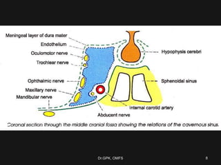
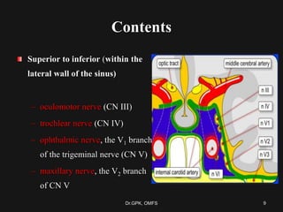
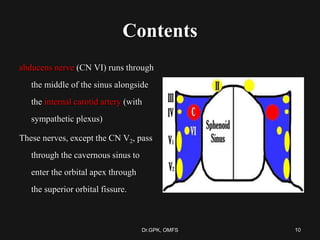
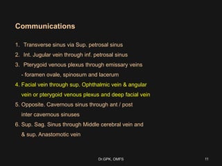
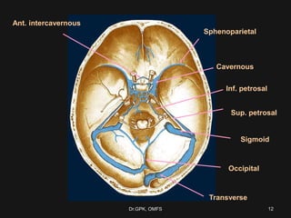
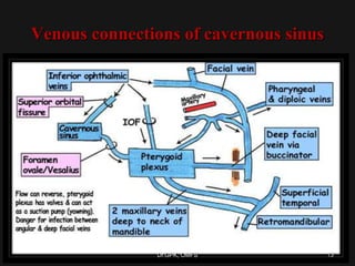
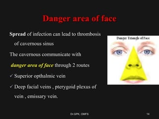
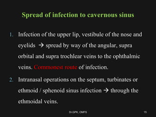
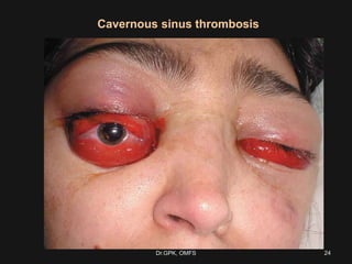
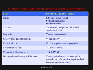
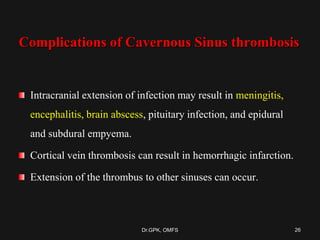
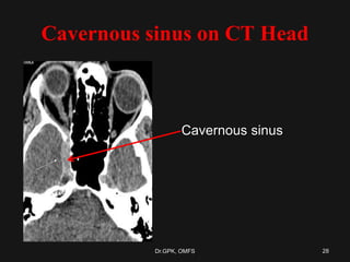
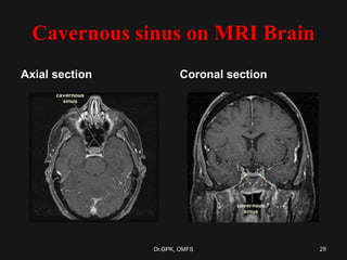
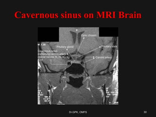
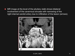
Cavernous Sinus Thrombosis
Dr.G.P.Kumar
Dept of OMFS
Dr.GPK, OMFS 1
Learning outcome
Knowledge about the anatomy of cavernous
sinus (C1)
Understand the significance of cavernous sinus
(C2)
Understand the importance of cavernous sinus
thrombosis (C2)
Knowledge about Medical management of
cavernous sinus (C1)
Dr.GPK, OMFS 2
Dural Venous Sinuses
The Dural venous sinuses are spaces between
the periosteal and meningeal layers of dura mater, which are
lined by endothelial cells.
They collect venous blood from the veins that drain the brain and
bony skull, and ultimately drain into the internal jugular vein.
Dr.GPK, OMFS 3
Classification
Paired venous sinuses
Cavernous sinuses
Petrosal sinuses
Transverse sinuses
Sigmoid sinuses
Spheno parietal sinuses
Unpaired venous sinuses
Superior sagittal sinus
Inferior sagittal sinus
Straight sinus
Occipital sinus
Basilar plexus of sinuses
Dr.GPK, OMFS 4
Cavernous sinus Anatomy
Large venous space situated in the middle cranial fossa, on either side of
body of the sphenoid bone.
Each sinus is about 2 cm long and 1 cm wide.
Interior is divided into a number of spaces or caverns by trabeculae.
5Dr.GPK, OMFS
6Dr.GPK, OMFS
Cavernous sinus Anatomy
Boundaries
Anterior - extends into medial
end of superior orbital fissure.
Posterior – up to apex of
petrous temporal bone.
Medial – Pituitary above and
sphenoid below
Lateral – temporal lobe and
uncus
Superior – optic chiasma
Inferior - endosteal dura mater,
greater wing of sphenoid
7Dr.GPK, OMFS
8Dr.GPK, OMFS
Contents
Superior to inferior (within the
lateral wall of the sinus)
– oculomotor nerve (CN III)
– trochlear nerve (CN IV)
– ophthalmic nerve, the V1 branch
of the trigeminal nerve (CN V)
– maxillary nerve, the V2 branch
of CN V
9Dr.GPK, OMFS
Contents
abducens nerve (CN VI) runs through
the middle of the sinus alongside
the internal carotid artery (with
sympathetic plexus)
These nerves, except the CN V2, pass
through the cavernous sinus to
enter the orbital apex through
the superior orbital fissure.
10Dr.GPK, OMFS
Communications
1. Transverse sinus via Sup. petrosal sinus
2. Int. Jugular vein through inf. petrosal sinus
3. Pterygoid venous plexus through emissary veins
- foramen ovale, spinosum and lacerum
4. Facial vein through sup. Ophthalmic vein & angular
vein or pterygoid venous plexus and deep facial vein
5. Opposite. Cavernous sinus through ant / post
inter cavernous sinuses
6. Sup. Sag. Sinus through Middle cerebral vein and
& sup. Anastomotic vein
11Dr.GPK, OMFS
Sphenoparietal
Cavernous
Ant. intercavernous
Sup. petrosal
Inf. petrosal
Transverse
Sigmoid
Occipital
12Dr.GPK, OMFS
Venous connections of cavernous sinus
13Dr.GPK, OMFS
Danger area of face
Spread of infection can lead to thrombosis
of cavernous sinus
The cavernous communicate with
danger area of face through 2 routes
 Superior opthalmic vein
 Deep facial veins , pterygoid plexus of
vein , emissary vein.
14Dr.GPK, OMFS
Spread of infection to cavernous sinus
1. Infection of the upper lip, vestibule of the nose and
eyelids  spread by way of the angular, supra
orbital and supra trochlear veins to the ophthalmic
veins. Commonest route of infection.
2. Intranasal operations on the septum, turbinates or
ethmoid / sphenoid sinus infection  through the
ethmoidal veins.
15Dr.GPK, OMFS
Spread of infection to cavernous sinus
3. Operations on the tonsil, peritonsillar abscess, surgery or
osteomyelitis of the maxilla, dental extraction and deep
cervical abscess  spread by pterygoid plexus or by direct
extension to the internal jugular vein.
4. Involvement of the middle ear and mastoid with lateral sinus
phlebitis or thrombosis  retrograde spread through the
petrosal sinuses to the cavernous sinus.
16Dr.GPK, OMFS
Etiology of CST
Septic CST
Infectious
Aseptic CST
Trauma
Post surgery
Rhinoplasty
Cataract extraction
Basal skull (including maxillary)
Tooth extraction
Hematologic
Polycythemia rubra vera
Acute lymphocytic leukemia
Malignancy
Nasopharyngeal tumor
Other
Ulcerative colitis
Dehydration
Heroin
17Dr.GPK, OMFS
Septic cavernous sinus thrombosis
Most commonly results from contiguous spread of infection from
the nose (50%),
sphenoidal or ethmoidal sinuses (30%) and
dental infections (10%).
Staphylococcus aureus is the most common - found in 70% of the cases.
Streptococcus is the second leading cause.
Gram-negative rods and anaerobes may also lead to cavernous sinus thrombosis.
Rarely fungal (Aspergillus fumigatus and mucormycosis).
18Dr.GPK, OMFS
Dental Infections
The upper and lower jaws and their teeth are drained by the superior
and inferior alveolar veins which are tributaries (directly or indirectly)
to the pterygoid venous plexus.
This plexus lies in the infra temporal fossa and communicates with
the cavernous sinus via an emissary vein passing through the
foramen ovale.
Infection could therefore spread from the jaws to the pterygoid
plexus and then into the cavernous sinus via this emissary vein.
Dr.GPK, OMFS 19
Cavernous Sinus thrombosis
Characterized by multiple cranial neuropathies
Clinical feature :
 General feature of infection – fever , rigors ,malaise, and severe frontal &
periorbital pain.
 U/L exophthalmoses & tender eye ball
 Edema of eyelid & chemosis of conjuctiva
Oculomotor feature :
 External ophthalmoplegia
 Ptosis
 Slight exophthalmos
 dilated pupil with loss of accommodation reflex
20Dr.GPK, OMFS
Cavernous Sinus thrombosis
Impairment of ocular motor nerves, Horner’s syndrome and
sensory loss of the first or second divisions of the trigeminal
nerve in various combination
The pupil may be involved or spared or may appear
spared with concomitant oculo sympathetic involvement.
21Dr.GPK, OMFS
Clinical features
Infections from face, orbit,
sphenoid sinus can cause
thrombosis.
III,IV,V1,V2,VI can be
involved resulting in various
diplopias
Rupture of ICA can cause
pulsatile exophthalmos
Caput medusae in cavernous
sinus thrombosis
Dr.GPK, OMFS 22
Cavernous sinus thrombosis
23Dr.GPK, OMFS
Cavernous sinus thrombosis
24Dr.GPK, OMFS
Occular manifestation of cavernous sinus thrombosis
SIGN INVOLVED STRUCTURES
Ptosis Edema of upper eye lid
Sympathetic plexus
III cranial nerve
Chemosis Thrombosis of superior and inferior
ophthalamic vein
Proptosis Venous engorgement
Sensory loss/ Periorbital pain V cranial nerve
Corneal ulcers Corneal exposure due to proptosis
Lateral rectus palsy VI cranial nerve
Complete ophthalmoplegia CN II, IV, VI
Decreased visual acuity or blindness Central retinal artery/ vein occlusion
secondary to ICA arteritis, septic emboli,
ischemic optic neuropathy
25Dr.GPK, OMFS
Complications of Cavernous Sinus thrombosis
Intracranial extension of infection may result in meningitis,
encephalitis, brain abscess, pituitary infection, and epidural
and subdural empyema.
Cortical vein thrombosis can result in hemorrhagic infarction.
Extension of the thrombus to other sinuses can occur.
26Dr.GPK, OMFS
Imaging of cavernous sinus
27Dr.GPK, OMFS
Cavernous sinus on CT Head
Cavernous sinus
28Dr.GPK, OMFS
Cavernous sinus on MRI Brain
Axial section Coronal section
29Dr.GPK, OMFS
Cavernous sinus on MRI Brain
30Dr.GPK, OMFS
MR image at the level of the pituitary stalk shows bilateral
involvement of the cavernous sinuses with narrowing of the
right internal carotid artery due to infiltration of the lesion (arrows).
Dr.GPK, OMFS 31
TREATMENT OF
CAVERNOUS SINUS
THROMBOSIS
32Dr.GPK, OMFS
TREATMENT OF CAVERNOUS SINUS THROMBOSIS
Septic cavernous sinus thrombosis –
The mainstay of therapy is early and aggressive antibiotic
administration.
Although S aureus is the usual cause, broad-spectrum coverage for gram-
positive, gram-negative, and anaerobic organisms should be instituted
pending the outcome of cultures.
Empiric antibiotic therapy should include a penicillinase-resistant penicillin
plus a third generation cephalosporin.
Vancomycin may be added for MRSA.
IV antibiotics are recommended for a minimum of 3-4 weeks.
33Dr.GPK, OMFS
TREATMENT OF CAVERNOUS SINUS THROMBOSIS
The indication of anticoagulation is still debated because of possible
bleeding complications and an eventual suppressive role of the thrombus on
the extension of the infectious thrombophlebitis.
Limited evidence suggests anticoagulant drugs are probably safe and may
be beneficial for people with sinus thrombosis.
Anticoagulant is contraindicated in the presence of intra cerebral
hemorrhage or other bleeding diathesis.
34Dr.GPK, OMFS
Fungal infection
 Intracranial extension is the most dreaded complication of fungal sinusitis with high
mortality rates.
 Aspergillus is the most common.
Mucormycosis also occurs but rare
Uncontrolled diabetics are more susceptible to mucormycosis .
The clinical signs of Mucormycosis are commonly opthalmoplegia, proptosis,
blindness, palatal ulcer, coma and stupor.
 Mode of spread =
Hematogenous spread
Direct extension
35Dr.GPK, OMFS
Fungal infection - treatment
Line of management-included debridement, clearing of disease from the
sinuses and antifungal therapy with systemic Amphotericin B.
Posaconazole (Noxafil) a triazole, is currently considered a second-line
drug for treatment of mucormycosis and the typical dose is 400 mg twice
daily (total of 800 mg/d).
Patients on posaconazole should avoid antacids, especially proton pump
inhibitors.
Posaconazole has also been used as sequential therapy after the initial
administration and control of the disease with amphotericin B.
Voriconazole is the preferred agent against aspergillus.
36Dr.GPK, OMFS
Prognosis
 100% mortality prior to effective antimicrobials
 Typically, death is due to sepsis or central nervous system (CNS)
infection.
 With aggressive management, the mortality rate is now less than
30%.
 Morbidity, however, remains high, and complete recovery is rare.
 Roughly one sixth of patients are left with some degree of visual
impairment, and one half (50 %) have cranial nerve deficits.
37Dr.GPK, OMFS
REFERENCES
1. Oral & maxillofacial surgery - Neelima Malick
2. Principles of Oral & maxillofacial surgery - Peterson
3. Maxillofacial infection - Topazian
Dr.GPK, OMFS 38
Dr.GPK, OMFS 39

Cavenous sinus thrombosis

  • 1.
  • 2.
    Learning outcome Knowledge aboutthe anatomy of cavernous sinus (C1) Understand the significance of cavernous sinus (C2) Understand the importance of cavernous sinus thrombosis (C2) Knowledge about Medical management of cavernous sinus (C1) Dr.GPK, OMFS 2
  • 3.
    Dural Venous Sinuses TheDural venous sinuses are spaces between the periosteal and meningeal layers of dura mater, which are lined by endothelial cells. They collect venous blood from the veins that drain the brain and bony skull, and ultimately drain into the internal jugular vein. Dr.GPK, OMFS 3
  • 4.
    Classification Paired venous sinuses Cavernoussinuses Petrosal sinuses Transverse sinuses Sigmoid sinuses Spheno parietal sinuses Unpaired venous sinuses Superior sagittal sinus Inferior sagittal sinus Straight sinus Occipital sinus Basilar plexus of sinuses Dr.GPK, OMFS 4
  • 5.
    Cavernous sinus Anatomy Largevenous space situated in the middle cranial fossa, on either side of body of the sphenoid bone. Each sinus is about 2 cm long and 1 cm wide. Interior is divided into a number of spaces or caverns by trabeculae. 5Dr.GPK, OMFS
  • 6.
  • 7.
    Cavernous sinus Anatomy Boundaries Anterior- extends into medial end of superior orbital fissure. Posterior – up to apex of petrous temporal bone. Medial – Pituitary above and sphenoid below Lateral – temporal lobe and uncus Superior – optic chiasma Inferior - endosteal dura mater, greater wing of sphenoid 7Dr.GPK, OMFS
  • 8.
  • 9.
    Contents Superior to inferior(within the lateral wall of the sinus) – oculomotor nerve (CN III) – trochlear nerve (CN IV) – ophthalmic nerve, the V1 branch of the trigeminal nerve (CN V) – maxillary nerve, the V2 branch of CN V 9Dr.GPK, OMFS
  • 10.
    Contents abducens nerve (CNVI) runs through the middle of the sinus alongside the internal carotid artery (with sympathetic plexus) These nerves, except the CN V2, pass through the cavernous sinus to enter the orbital apex through the superior orbital fissure. 10Dr.GPK, OMFS
  • 11.
    Communications 1. Transverse sinusvia Sup. petrosal sinus 2. Int. Jugular vein through inf. petrosal sinus 3. Pterygoid venous plexus through emissary veins - foramen ovale, spinosum and lacerum 4. Facial vein through sup. Ophthalmic vein & angular vein or pterygoid venous plexus and deep facial vein 5. Opposite. Cavernous sinus through ant / post inter cavernous sinuses 6. Sup. Sag. Sinus through Middle cerebral vein and & sup. Anastomotic vein 11Dr.GPK, OMFS
  • 12.
    Sphenoparietal Cavernous Ant. intercavernous Sup. petrosal Inf.petrosal Transverse Sigmoid Occipital 12Dr.GPK, OMFS
  • 13.
    Venous connections ofcavernous sinus 13Dr.GPK, OMFS
  • 14.
    Danger area offace Spread of infection can lead to thrombosis of cavernous sinus The cavernous communicate with danger area of face through 2 routes  Superior opthalmic vein  Deep facial veins , pterygoid plexus of vein , emissary vein. 14Dr.GPK, OMFS
  • 15.
    Spread of infectionto cavernous sinus 1. Infection of the upper lip, vestibule of the nose and eyelids  spread by way of the angular, supra orbital and supra trochlear veins to the ophthalmic veins. Commonest route of infection. 2. Intranasal operations on the septum, turbinates or ethmoid / sphenoid sinus infection  through the ethmoidal veins. 15Dr.GPK, OMFS
  • 16.
    Spread of infectionto cavernous sinus 3. Operations on the tonsil, peritonsillar abscess, surgery or osteomyelitis of the maxilla, dental extraction and deep cervical abscess  spread by pterygoid plexus or by direct extension to the internal jugular vein. 4. Involvement of the middle ear and mastoid with lateral sinus phlebitis or thrombosis  retrograde spread through the petrosal sinuses to the cavernous sinus. 16Dr.GPK, OMFS
  • 17.
    Etiology of CST SepticCST Infectious Aseptic CST Trauma Post surgery Rhinoplasty Cataract extraction Basal skull (including maxillary) Tooth extraction Hematologic Polycythemia rubra vera Acute lymphocytic leukemia Malignancy Nasopharyngeal tumor Other Ulcerative colitis Dehydration Heroin 17Dr.GPK, OMFS
  • 18.
    Septic cavernous sinusthrombosis Most commonly results from contiguous spread of infection from the nose (50%), sphenoidal or ethmoidal sinuses (30%) and dental infections (10%). Staphylococcus aureus is the most common - found in 70% of the cases. Streptococcus is the second leading cause. Gram-negative rods and anaerobes may also lead to cavernous sinus thrombosis. Rarely fungal (Aspergillus fumigatus and mucormycosis). 18Dr.GPK, OMFS
  • 19.
    Dental Infections The upperand lower jaws and their teeth are drained by the superior and inferior alveolar veins which are tributaries (directly or indirectly) to the pterygoid venous plexus. This plexus lies in the infra temporal fossa and communicates with the cavernous sinus via an emissary vein passing through the foramen ovale. Infection could therefore spread from the jaws to the pterygoid plexus and then into the cavernous sinus via this emissary vein. Dr.GPK, OMFS 19
  • 20.
    Cavernous Sinus thrombosis Characterizedby multiple cranial neuropathies Clinical feature :  General feature of infection – fever , rigors ,malaise, and severe frontal & periorbital pain.  U/L exophthalmoses & tender eye ball  Edema of eyelid & chemosis of conjuctiva Oculomotor feature :  External ophthalmoplegia  Ptosis  Slight exophthalmos  dilated pupil with loss of accommodation reflex 20Dr.GPK, OMFS
  • 21.
    Cavernous Sinus thrombosis Impairmentof ocular motor nerves, Horner’s syndrome and sensory loss of the first or second divisions of the trigeminal nerve in various combination The pupil may be involved or spared or may appear spared with concomitant oculo sympathetic involvement. 21Dr.GPK, OMFS
  • 22.
    Clinical features Infections fromface, orbit, sphenoid sinus can cause thrombosis. III,IV,V1,V2,VI can be involved resulting in various diplopias Rupture of ICA can cause pulsatile exophthalmos Caput medusae in cavernous sinus thrombosis Dr.GPK, OMFS 22
  • 23.
  • 24.
  • 25.
    Occular manifestation ofcavernous sinus thrombosis SIGN INVOLVED STRUCTURES Ptosis Edema of upper eye lid Sympathetic plexus III cranial nerve Chemosis Thrombosis of superior and inferior ophthalamic vein Proptosis Venous engorgement Sensory loss/ Periorbital pain V cranial nerve Corneal ulcers Corneal exposure due to proptosis Lateral rectus palsy VI cranial nerve Complete ophthalmoplegia CN II, IV, VI Decreased visual acuity or blindness Central retinal artery/ vein occlusion secondary to ICA arteritis, septic emboli, ischemic optic neuropathy 25Dr.GPK, OMFS
  • 26.
    Complications of CavernousSinus thrombosis Intracranial extension of infection may result in meningitis, encephalitis, brain abscess, pituitary infection, and epidural and subdural empyema. Cortical vein thrombosis can result in hemorrhagic infarction. Extension of the thrombus to other sinuses can occur. 26Dr.GPK, OMFS
  • 27.
    Imaging of cavernoussinus 27Dr.GPK, OMFS
  • 28.
    Cavernous sinus onCT Head Cavernous sinus 28Dr.GPK, OMFS
  • 29.
    Cavernous sinus onMRI Brain Axial section Coronal section 29Dr.GPK, OMFS
  • 30.
    Cavernous sinus onMRI Brain 30Dr.GPK, OMFS
  • 31.
    MR image atthe level of the pituitary stalk shows bilateral involvement of the cavernous sinuses with narrowing of the right internal carotid artery due to infiltration of the lesion (arrows). Dr.GPK, OMFS 31
  • 32.
  • 33.
    TREATMENT OF CAVERNOUSSINUS THROMBOSIS Septic cavernous sinus thrombosis – The mainstay of therapy is early and aggressive antibiotic administration. Although S aureus is the usual cause, broad-spectrum coverage for gram- positive, gram-negative, and anaerobic organisms should be instituted pending the outcome of cultures. Empiric antibiotic therapy should include a penicillinase-resistant penicillin plus a third generation cephalosporin. Vancomycin may be added for MRSA. IV antibiotics are recommended for a minimum of 3-4 weeks. 33Dr.GPK, OMFS
  • 34.
    TREATMENT OF CAVERNOUSSINUS THROMBOSIS The indication of anticoagulation is still debated because of possible bleeding complications and an eventual suppressive role of the thrombus on the extension of the infectious thrombophlebitis. Limited evidence suggests anticoagulant drugs are probably safe and may be beneficial for people with sinus thrombosis. Anticoagulant is contraindicated in the presence of intra cerebral hemorrhage or other bleeding diathesis. 34Dr.GPK, OMFS
  • 35.
    Fungal infection  Intracranialextension is the most dreaded complication of fungal sinusitis with high mortality rates.  Aspergillus is the most common. Mucormycosis also occurs but rare Uncontrolled diabetics are more susceptible to mucormycosis . The clinical signs of Mucormycosis are commonly opthalmoplegia, proptosis, blindness, palatal ulcer, coma and stupor.  Mode of spread = Hematogenous spread Direct extension 35Dr.GPK, OMFS
  • 36.
    Fungal infection -treatment Line of management-included debridement, clearing of disease from the sinuses and antifungal therapy with systemic Amphotericin B. Posaconazole (Noxafil) a triazole, is currently considered a second-line drug for treatment of mucormycosis and the typical dose is 400 mg twice daily (total of 800 mg/d). Patients on posaconazole should avoid antacids, especially proton pump inhibitors. Posaconazole has also been used as sequential therapy after the initial administration and control of the disease with amphotericin B. Voriconazole is the preferred agent against aspergillus. 36Dr.GPK, OMFS
  • 37.
    Prognosis  100% mortalityprior to effective antimicrobials  Typically, death is due to sepsis or central nervous system (CNS) infection.  With aggressive management, the mortality rate is now less than 30%.  Morbidity, however, remains high, and complete recovery is rare.  Roughly one sixth of patients are left with some degree of visual impairment, and one half (50 %) have cranial nerve deficits. 37Dr.GPK, OMFS
  • 38.
    REFERENCES 1. Oral &maxillofacial surgery - Neelima Malick 2. Principles of Oral & maxillofacial surgery - Peterson 3. Maxillofacial infection - Topazian Dr.GPK, OMFS 38
  • 39.