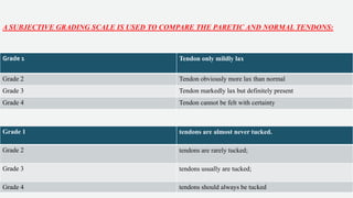
FORCED DUCTION
TEST
DR ANISHA RATHOD
MS,FPOS(PEDIATRICOPHTHALMOLOGY
AND STRABISMUS)
• THE AUTHOR HAS NO FINANCIAL INTEREST INTHE SUBJECT MATTER BEING PRESENTED
FINANCIAL DISCLOSURE
• The forced duction test was first described for evaluating Duane syndrome in the 1900s
• Critical for the diagnosis of muscle palsies or restrictions.
• Not useful if ocular rotations are full.
• It may be performed when the patient is under general anesthesia or in the office utilizing
topical anesthesia.
METHOD
• Properly performed the test is short and relatively painless.
• A topical anesthetic such as tetracaine is applied, either as eye drops or with a cotton-tipped applicator
• Patient is instructed to cover one eye and with the other, look in the direction of the muscle suspected of having
limited rotation.
• The patient is asked to look as far as possible into the field of gaze being tested
• Frequently a tendency for an eye with limited ocular rotation to “fall off” or migrate back toward the primary
position
.
• Results of the forced duction test may be misleading because the examiner is able to rotate the eye passively
• Holding the lids apart with one hand, the examiner uses forceps for globe fixation.
• The examiner should grasp as close to the limbus as possible opposite the side of gaze limitation.
• This area corresponds to that where Tenon's capsule and conjunctiva are fused in one layer.
• This limits stretching of the conjunctiva and gives the examiner a firmer grasp in rotating the globe
• Knapp stressed that pushing the eye into the orbit -give a false-negative result as full rotation may be simulated.
• For vertical rectus muscles, Jampolsky" believes that the test should be performed in 23 degrees of abduction
• Retroplacement of the globe may distinguish -inferior and superior restrictions to upgaze limitation.
• In an upgaze deficiency such as Brown syndrome, retroplacement of the globe during upward rotation
exaggerates a restriction caused by the superior oblique muscle.
• A “knife's edge” sensation may be present when performing the traction test in Brown syndrome.
• Stimulating the vagal response may cause syncopal attacks.
INTERPRETATION OF RESULTS
Absolute Restriction.
• The first type, an absolute restriction (e.g. Graves ophthalmopathy or Brown syndrome)
• An immediate increase in resistance is felt when forceps are applied with just a small amount of passive forced
duction movement.
• The examiner cannot rotate the globe past a certain point despite exerting maximum force.
Uniform Restriction.
• In a uniform type of restriction the examiner feels a linearly progressive increase in resistance to passive traction
across the arc of rotation.
• Examples of this type include restrictions secondary to scar tissue or muscle contracture.
Leash Phenomenon.
• In leash phenomenon there is a range of normal resistance followed by a sudden increase toward the end of the
field of gaze as the end of the “leash”is reached.
• Cases of moderate contracture and fibrosis.
• Long standing extraocular muscle palsy, indicating that agonist muscle weakness -secondary mechanical restriction
of the antagonist.
• A direct leash is due to scar tissue or stiffness of the muscle opposite the gaze limitation.
• A reverse leash is due to scar tissue or muscle tightness on the same side as the gaze limitation.
Common Pitfalls in Diagnosis
• Apprehensive patients may squeeze their eyes and guard muscle movement (forceps grasping the globe.)
• If the patient does not adequately look into the field being examined, the antagonist muscle remains innervated.
• A resistance will be felt and misinterpreted as a restriction.
• Conjunctival tearing
• Hemorrhage.
PHARMACOLOGIC EFFECTS.
• Short-acting depolarizing agents such as succinylcholine cause a sustained contraction of the extraocular
muscles that persists for 15 to 20 minutes after administration.
• (Examiner should wait 20 minutes before doing the test.)
• Passive length-tension curves convert to straight parallel lines after intravenous administration of this
agent.
• Alternatively, nondepolarizing agents such as pancuronium may be used without affecting forced duction
testing results.
ERRORS IN TECHNIQUE.
• Failure to follow natural arc of muscle movement/pushing the globe in its socket can yield false-negative results
(shortening arc of contact.)
• Proptosing the globe too much, on the other hand, may give a false-positive result.
• Overstretching of tissues during surgery -false sense of relieved restriction.
POSTERIOR RESTRICTIONS.
Restrictions may occur far more posteriorly
FALSE-NEGATIVE TEST.
• Failure to recognize the presence of a restriction when one exists constitutes a false-negative test.
• This commonly occurs in cases of mild restriction
COEXISTING PARESIS AND RESTRICTION.
• Kushner reported the association of rectus muscle paresis and restriction in orbital floor fractures.
• One may diagnose limited rotations due to a restriction and overlook an associated muscle palsy.
• Force generation testing and saccadic velocity analysis provide useful adjunctive information.
• This problem is encountered in:
• Long-standing paresis with secondary contracture of the antagonist
• Thyroid ophthalmopathy
CO-CONTRACTION SYNDROMES AND ABERRANT INNERVATIONS.
• Tongue documented her experience with FDT in patients with Duane syndrome.
• Esotropic type with DRS in the left eye,
• FDT on the left LRmuscle free on attempted abduction+paradoxically positive in adduction.
• The resistance to rotation is relieved when the patient is asked to fixate in abduction (left gaze)while the globe
is rotated into adduction.
• Restriction in adduction is due to aberrant firing of the LR muscle in right gaze,
• Relieving restriction on abduction is due to the absence of LR firing in left gaze
FORCE GENERATION TESTING
• PRINCIPLE-By applying a counteracting force or resistance to a series of isometrically contracting
muscles, the strength of the muscle or muscle groups is estimated.
• Scott first described -patients with lateral rectus paralysis.
OFFICE FORCE GENERATION TESTING
• Patient needs to be reassured and informed about the goals and expectations of the procedure.
• Topical anesthetic is applied in the usual manner.
• The forceps are positioned slightly anterior to the rectus muscle .
• Potential complications include conjunctival haemorrhage and corneal abrasion
• The position of the forceps should be changed to the limbus opposite the duction effort to reduce the risk
of corneal abrasion.
INTERPRETATION OF RESULTS
• Paresis means a weakened muscle, but the paretic muscle still is able to generate some movement or contraction.
• Complete palsy means absence of innervation and the inability to generate any significant force.
• Muscle contracts normally in its field of gaze-the observer is unable to rotate the globe in the opposite direction.
• Muscle is paretic-the examiner can rotate the globe but notes resistance.
• In a completely palsied muscle- the observer is able to rotate the globe with no resistance felt in the opposite
direction.
• The results can be classified as normal, weak, or absent force generation.
OBLIQUE TRACTION TESTING
• Intraoperative traction testing
• The technique for assessing laxity of the SO tendon is based on the method of “exaggerated forced ductions” of the
obliques, described by Guyton for evaluating patients with SO overaction.
Step 1: With the surgeon seated above the head of the supine, anesthetized patient, two toothed forceps are used to
grasp the limbus diagonally, that is, at the 2 and 8-o'clock positions on the left eye and the 4- and 10-o'clock positions
on the right eye.
Step 2: The eye is rotated up into an elevated, adducted position in the superior nasal quadrant while simultaneously
at apex putting the tendon at maximum stretch
Step 3: Once the tendon is put on stretch, the eye is moved back and forth (temporally and nasally) while maintaining
the tendon taut.
This back and forth movement allows the surgeon to “feel” the tendon as a band
across which the globe is rocked.
Step 4: Steps 1 to 3 are repeated on the fellow eye.
A SUBJECTIVE GRADING SCALE IS USED TO COMPARE THE PARETIC AND NORMAL TENDONS:
Grade 1 Tendon only mildly lax
Grade 2 Tendon obviously more lax than normal
Grade 3 Tendon markedly lax but definitely present
Grade 4 Tendon cannot be felt with certainty
Grade 1 tendons are almost never tucked.
Grade 2 tendons are rarely tucked;
Grade 3 tendons usually are tucked;
Grade 4 tendons should always be tucked
THANK YOU

Forced duction test

  • 1.
    FORCED DUCTION TEST DR ANISHARATHOD MS,FPOS(PEDIATRICOPHTHALMOLOGY AND STRABISMUS)
  • 2.
    • THE AUTHORHAS NO FINANCIAL INTEREST INTHE SUBJECT MATTER BEING PRESENTED FINANCIAL DISCLOSURE
  • 3.
    • The forcedduction test was first described for evaluating Duane syndrome in the 1900s • Critical for the diagnosis of muscle palsies or restrictions. • Not useful if ocular rotations are full. • It may be performed when the patient is under general anesthesia or in the office utilizing topical anesthesia.
  • 4.
    METHOD • Properly performedthe test is short and relatively painless. • A topical anesthetic such as tetracaine is applied, either as eye drops or with a cotton-tipped applicator • Patient is instructed to cover one eye and with the other, look in the direction of the muscle suspected of having limited rotation. • The patient is asked to look as far as possible into the field of gaze being tested • Frequently a tendency for an eye with limited ocular rotation to “fall off” or migrate back toward the primary position
  • 5.
    . • Results ofthe forced duction test may be misleading because the examiner is able to rotate the eye passively • Holding the lids apart with one hand, the examiner uses forceps for globe fixation. • The examiner should grasp as close to the limbus as possible opposite the side of gaze limitation. • This area corresponds to that where Tenon's capsule and conjunctiva are fused in one layer. • This limits stretching of the conjunctiva and gives the examiner a firmer grasp in rotating the globe
  • 6.
    • Knapp stressedthat pushing the eye into the orbit -give a false-negative result as full rotation may be simulated. • For vertical rectus muscles, Jampolsky" believes that the test should be performed in 23 degrees of abduction • Retroplacement of the globe may distinguish -inferior and superior restrictions to upgaze limitation. • In an upgaze deficiency such as Brown syndrome, retroplacement of the globe during upward rotation exaggerates a restriction caused by the superior oblique muscle. • A “knife's edge” sensation may be present when performing the traction test in Brown syndrome. • Stimulating the vagal response may cause syncopal attacks.
  • 7.
    INTERPRETATION OF RESULTS AbsoluteRestriction. • The first type, an absolute restriction (e.g. Graves ophthalmopathy or Brown syndrome) • An immediate increase in resistance is felt when forceps are applied with just a small amount of passive forced duction movement. • The examiner cannot rotate the globe past a certain point despite exerting maximum force. Uniform Restriction. • In a uniform type of restriction the examiner feels a linearly progressive increase in resistance to passive traction across the arc of rotation. • Examples of this type include restrictions secondary to scar tissue or muscle contracture.
  • 8.
    Leash Phenomenon. • Inleash phenomenon there is a range of normal resistance followed by a sudden increase toward the end of the field of gaze as the end of the “leash”is reached. • Cases of moderate contracture and fibrosis. • Long standing extraocular muscle palsy, indicating that agonist muscle weakness -secondary mechanical restriction of the antagonist. • A direct leash is due to scar tissue or stiffness of the muscle opposite the gaze limitation. • A reverse leash is due to scar tissue or muscle tightness on the same side as the gaze limitation.
  • 9.
    Common Pitfalls inDiagnosis • Apprehensive patients may squeeze their eyes and guard muscle movement (forceps grasping the globe.) • If the patient does not adequately look into the field being examined, the antagonist muscle remains innervated. • A resistance will be felt and misinterpreted as a restriction. • Conjunctival tearing • Hemorrhage.
  • 10.
    PHARMACOLOGIC EFFECTS. • Short-actingdepolarizing agents such as succinylcholine cause a sustained contraction of the extraocular muscles that persists for 15 to 20 minutes after administration. • (Examiner should wait 20 minutes before doing the test.) • Passive length-tension curves convert to straight parallel lines after intravenous administration of this agent. • Alternatively, nondepolarizing agents such as pancuronium may be used without affecting forced duction testing results.
  • 11.
    ERRORS IN TECHNIQUE. •Failure to follow natural arc of muscle movement/pushing the globe in its socket can yield false-negative results (shortening arc of contact.) • Proptosing the globe too much, on the other hand, may give a false-positive result. • Overstretching of tissues during surgery -false sense of relieved restriction. POSTERIOR RESTRICTIONS. Restrictions may occur far more posteriorly FALSE-NEGATIVE TEST. • Failure to recognize the presence of a restriction when one exists constitutes a false-negative test. • This commonly occurs in cases of mild restriction
  • 12.
    COEXISTING PARESIS ANDRESTRICTION. • Kushner reported the association of rectus muscle paresis and restriction in orbital floor fractures. • One may diagnose limited rotations due to a restriction and overlook an associated muscle palsy. • Force generation testing and saccadic velocity analysis provide useful adjunctive information. • This problem is encountered in: • Long-standing paresis with secondary contracture of the antagonist • Thyroid ophthalmopathy
  • 13.
    CO-CONTRACTION SYNDROMES ANDABERRANT INNERVATIONS. • Tongue documented her experience with FDT in patients with Duane syndrome. • Esotropic type with DRS in the left eye, • FDT on the left LRmuscle free on attempted abduction+paradoxically positive in adduction. • The resistance to rotation is relieved when the patient is asked to fixate in abduction (left gaze)while the globe is rotated into adduction. • Restriction in adduction is due to aberrant firing of the LR muscle in right gaze, • Relieving restriction on abduction is due to the absence of LR firing in left gaze
  • 14.
    FORCE GENERATION TESTING •PRINCIPLE-By applying a counteracting force or resistance to a series of isometrically contracting muscles, the strength of the muscle or muscle groups is estimated. • Scott first described -patients with lateral rectus paralysis. OFFICE FORCE GENERATION TESTING • Patient needs to be reassured and informed about the goals and expectations of the procedure. • Topical anesthetic is applied in the usual manner. • The forceps are positioned slightly anterior to the rectus muscle . • Potential complications include conjunctival haemorrhage and corneal abrasion • The position of the forceps should be changed to the limbus opposite the duction effort to reduce the risk of corneal abrasion.
  • 15.
    INTERPRETATION OF RESULTS •Paresis means a weakened muscle, but the paretic muscle still is able to generate some movement or contraction. • Complete palsy means absence of innervation and the inability to generate any significant force. • Muscle contracts normally in its field of gaze-the observer is unable to rotate the globe in the opposite direction. • Muscle is paretic-the examiner can rotate the globe but notes resistance. • In a completely palsied muscle- the observer is able to rotate the globe with no resistance felt in the opposite direction. • The results can be classified as normal, weak, or absent force generation.
  • 16.
    OBLIQUE TRACTION TESTING •Intraoperative traction testing • The technique for assessing laxity of the SO tendon is based on the method of “exaggerated forced ductions” of the obliques, described by Guyton for evaluating patients with SO overaction.
  • 17.
    Step 1: Withthe surgeon seated above the head of the supine, anesthetized patient, two toothed forceps are used to grasp the limbus diagonally, that is, at the 2 and 8-o'clock positions on the left eye and the 4- and 10-o'clock positions on the right eye. Step 2: The eye is rotated up into an elevated, adducted position in the superior nasal quadrant while simultaneously at apex putting the tendon at maximum stretch Step 3: Once the tendon is put on stretch, the eye is moved back and forth (temporally and nasally) while maintaining the tendon taut. This back and forth movement allows the surgeon to “feel” the tendon as a band across which the globe is rocked. Step 4: Steps 1 to 3 are repeated on the fellow eye.
  • 18.
    A SUBJECTIVE GRADINGSCALE IS USED TO COMPARE THE PARETIC AND NORMAL TENDONS: Grade 1 Tendon only mildly lax Grade 2 Tendon obviously more lax than normal Grade 3 Tendon markedly lax but definitely present Grade 4 Tendon cannot be felt with certainty Grade 1 tendons are almost never tucked. Grade 2 tendons are rarely tucked; Grade 3 tendons usually are tucked; Grade 4 tendons should always be tucked
  • 19.