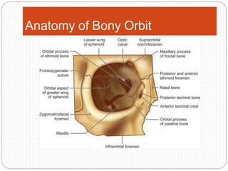
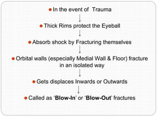
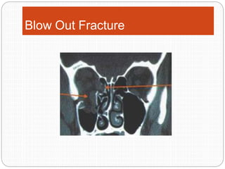
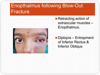
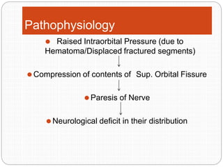
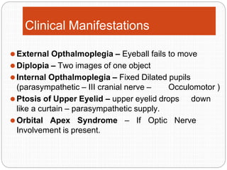
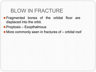
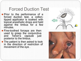
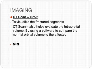
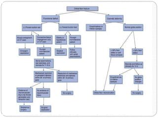
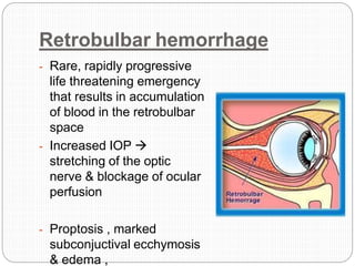
Orbital Fracture
Anatomy of Orbit
⚫ Orbit – conical cavity
⚫Base – Anteriorly
⚫Apex – directed at Optic Foramen
⚫Orbital Volume – 30cc ; Globe – 7cc
⚫By Age 5 years orbital growth is 85% complete , Finalised
between 7 years of age
⚫Bones – 7 (Maxillary, Zygomatic, Frontal, Ethmoid, Larimal,
Palatine, and Sphenoid )
⚫Four Walls – roof, lateral, medial, floor
⚫Medial wall & floor – thin ;
⚫Lateral wall & Roof – Stronger.
⚫Floor weakened – due to – Infraorbtal Canal passing through it
⚫Medial wall – Formed by – Lamina Papyracea of
Ethmoidal bone & Lacrimal bone
⚫Vital Contents – Eyeball (Non Compressible)
⚫6 EXTRAOCULAR MUSCLES –
⚫Originate from Common Tendinous fibrous ring
⚫Annulus of Zinn – Fibrous ring – Common origin of 4
Rectus Muscle
⚫OPTIC NERVE – direct extension of Gray Matter
⚫Very sensitie to Compression and once damaged fails to
regenerate
SUPERIOR ORBITAL FISSURE
⚫Transmits – 3rd , 4th & 6th Cranial Nerves
⚫Opthalmic Division of 5th Cranial Nerve
Cranial Nerves and its Supply
⚫Occulomotor (C.N.-III)- Supplies –
⚫Superior Rectus
⚫Medial Rectus
⚫Inferior Rectus
⚫Inferior Obliqus
⚫Abducent Nerve – Lateral Rectus (LR6 )
⚫Trochlear Nerve – Superior Obliqus (SO4)
⚫Theoretically, the mechanical load of the orbital
floor is approximately 0.0005 N/mm2 (30 g orbital
content onto 600 mm2 of orbital floor area), as
indicated by several cadaver investigations.
CLASSIFICATION
⚫ORBITAL WALL FRACTURES –
1. Blow Out Fracture
2. Pure Blow Out Fracture
3. Impure Blow Out Fracture
4. Blow In Fracture
⚫ISOLATED ORBITAL WALL FRACTURE :
5. Roof
6. Floor
7. Medial wall
8. Lateral wall
⚫ISOLATED FRACTURES OF THE ORBITAL RIM
1. SUPERIOR RIM
2. INFERIOR RIM
3. MEDIAL RIM
4. LATERAL RIM
⚫COMPLEX COMMINUTED FRACTURES :
⚫Nasoethmoidal and fronto-naso-orbital fractures
Anatomy of Bony Orbit
Orbital Fissures/Canals and Their
Contents
Pathophysiology of Orbital Fractures
⚫In the event of Trauma
⚫Thick Rims protect the Eyeball
⚫Absorb shock by Fracturing themselves
⚫Orbital walls (especially Medial Wall & Floor) fracture
in an isolated way
⚫Gets displaces Inwards or Outwards
⚫Called as ‘Blow-In’ or ‘Blow-Out’ fractures
⚫PURE Blow Out OR Blow In –
⚫Orbital Walls fracture in Isolation
⚫IMPURE Blow Out or Blow In Fracture –
⚫Walls + Rim
Blow Out Fracture
Blow Out Fracture
⚫Term coined by – Smith and Regan – 1957
⚫First described by MacKenzie in Paris in 1844
PATHOPHYSIOLOGY
⚫Buckling Theory –
⚫This theory states that if a force was to strike any part of
the orbital rim, it will cause walls to undergo a rippling
effect & the force striking the rim will transfer to the
weaker portion especially the floor & cause them to
distort & eventually fracture
Pathophysiology of blow out fracture
of the orbit
⚫ Hydraulic Theory (Pfeiffer in 1943) – he said that it is
evident that the force of the blow received by the eyeball was
transmitted by it to the walls of the orbit with fracture of the
delicate portions.
- Therefore direct injury to the globe forcing it into the orbit was
necessary.
⚫ Medial Wall & Floor –Thin & Fragile
Clinical Features
⚫ Circumorbital Edema -
⚫ Subconjunctival Bleeding – due to fracture subperiosteal bleeding 
escapes in subconjunctival plane.
⚫ Enopthalmous  Increase in size of Orbit  Eyeball sinks
⚫ Periorbital Fat Herniates through fractured walls
⚫ ‘Hanging-drop’ Appearance
⚫ Unilateral Epistaxis – bleeding into antrum
⚫ Numbness in area of distribution of Infraorbital Nerve
⚫ Diplopia or Vertical gage – Inferior Rectus or Inferior Obliqus gets
entrapped in fracture  inability of eyeball to move in vertical direction.
Enopthalmus following Blow-Out
Fracture
⚫Retracting action of
extraocular muscles –
Enopthalmus.
⚫Diplopia – Entrapment
of Inferior Rectus &
Inferior Obliqus
Pathophysiology
⚫ Raised Intraorbital Pressure (due to
Hematoma/Displaced fractured segments)
⚫Compression of contents of Sup. Orbital Fissure
⚫Paresis of Nerve
⚫Neurological deficit in their distribution
⚫Occulomotor supplies – SR , MR, IR , IO
⚫Abducent – LR
⚫Trochlear – SO
⚫Due to paresis of these nerves  all these
extraocular muscles undergo paralysis  eyeball
fails to move  ‘External Opthalmoplegia’
⚫As affected eye does not move  whereas
contralateral normal eyeball moves  focal axis
gets disturbed  two images - Diplopia
Clinical Manifestations
⚫External Opthalmoplegia – Eyeball fails to move
⚫Diplopia – Two images of one object
⚫Internal Opthalmoplegia – Fixed Dilated pupils
(parasympathetic – III cranial nerve – Occulomotor )
⚫Ptosis of Upper Eyelid – upper eyelid drops down
like a curtain – parasympathetic supply.
⚫Orbital Apex Syndrome – If Optic Nerve
Involvement is present.
BLOW IN FRACTURE
⚫Fragmented bones of the orbital floor are
displaced into the orbit.
⚫Proptosis – Exopthalmous
⚫More commonly seen in fractures of – orbital roof
CLINICAL EXAMINATION
⚫Initial Opthalmological evaluation –
1. Periorbital Examination
2. visual acuity – SNELLEN CHART
3. ocular motility – FORCED DUCTION TEST
4. Pupillary responses ,- pupillary size, shape&
symmetry, light reactivity,
5. Visual fields – HESS CHART
6. Fundoscopic examination
- TONOMETRY – to assess Intraocular pressure
(Normal 10-20mmHg)
7. Hertel Exopthalmometer – measure exopthalmous
Forced Duction Test
⚫ Prior to the performance of a
forced duction test, a cotton-
tipped applicator is soaked with
topical anesthetic drops and held
against the limbus for a few
minutes.
⚫ Fine-toothed forceps are then
used to grasp the conjunctiva
and Tenon’s capsule just
posterior to the limbus.
⚫ The patient is then asked to look
in the direction of restriction of
movement of the eye .
Tonometer
Snellen chart
IMAGING
⚫CT Scan – Orbit
- To visualize the fractured segments
- CT Scan – also helps evaluate the Intraorbital
volume. By using a software to compare the
normal orbital volume to the affected
- MRI
Management
Indications for surgical
managment
⚫Unresolving soft tissue entrapment with disabling
diplopia
⚫Enopthalmous greater than 2mm
⚫CT scan evidence of a large fracture
Incisions
•Existing lacerations
•LowerEyelid –
• 1)Subciliary
• 2) Subtarsal
• 3) Infraorbital
approaches
•Transconjuctival
Approach – Lower Eyelid
Pre-formed Orbital Implant
ADVANTAGES :
•Radiopacity
•Smooth Surface
•Minimal or no countouring necessary
DISADVANTAGE :
•Cost
Bone Graft
Disadvantages :
•Additional Donor site needed
•Possible contour and dimensional changes due to
remodeling
•Difficult to shape according to patients anatomy
Porous Polyethylene Sheet
(PPE)
Disadvantages :
• Not Radiopaque (Not visble on Post Operative
Radiographs
•Lack of Rigidity when very thin wafer of PPE is used.
When a more thick rigid wafer is used there is a risk
of causing dystopia
POLYETHYLENE AND TITANIUM
MESH
⚫By combining titanium mesh
with porous polyethylene –
⚫Material becomes radioopaque
⚫More rigid than porous PPE.
⚫ADVANTAGE :
⚫Stability
⚫Contouring
⚫Adequate in large three wall
fractures
⚫Radiopacity
⚫No Donor Site Needed
⚫DISADVANTAGES : Less
Drainage from orbit than with
RESORBABLE MATERIALS
⚫ Thermoplastic and Non
Thermoplastic Materials
⚫ ADVANTAGES :
⚫ Availability Handling/
⚫ Contourability (only for
thermoplastics)
⚫ Smooth surface and smooth
edges
⚫ Disadvantages:
- No radiopacity
- Degradation of material with
possible contour loss Sterile
infection / inflammatory response
- Difficult to shape according to
patients anatomy (only for non-
thermoplastics)
COMPLICATIONS
⚫ EARLY COMPLICATIONS :
⚫ 1. Hemorrhagic or orbital hematoma – treated by
-lateral cathotomy immediately, lateral canthal
Tendonlysis ,
Lateral canthotomy – indicated when –
- Decreased visual acuity
- Introcular pressure more than 40mmHg
- Proptosis
- Opthalmoplegia
Retrobulbar hemorrhage
- Rare, rapidly progressive
life threatening emergency
that results in accumulation
of blood in the retrobulbar
space
- Increased IOP 
stretching of the optic
nerve & blockage of ocular
perfusion
- Proptosis , marked
subconjuctival ecchymosis
& edema ,
⚫ Symptoms seen are – pain , decreasing visual acuity,
diplopia
⚫ Treatment includes
⚫ iv mannitol – (used to treat raised intracranial
pressure)
⚫ Acteazolamide – carbonic anhydrase inhibitor ;
diuresis in PCT of kidney – excretion of NA, K, Cl –
lowering BP, IOP
⚫ Megadose Steroid Therapy – 100mg Dexa as an i.v.
bolus with 40mg 6 hourly in severe unresponsive
cases ( Anderson et al 1982)
3. Blindness
⚫OCULOCARDIAC REFLEX/ TRIGEMINOCARDIAC/
TRIGEMINOVAGAL REFLEX –
- The oculocardiac reflex pathway begins with the
- afferent fibres of the long & short ciliary nerves that
travel with
- the opthalmic division of the trigeminal nerve to
- the gasserion ganglion via
- the sensory nucleus of the trigeminal nerve.
- In the floor of the 4th ventricle short internuncial
fibres in the reticular formation connect them with the
efferent pathway from the motor nucleus of the
vagus nerve to the depressor nerve ending in the
mucle tissue of the heart.
⚫CLINICAL FEATURES –
- Bradycardia
- Faintness
- Further stimulation can lead to cardiac
dysrhythmias
- Atrioventricular blocks
- Asystole
- Bradycardia has been attributed to Trigeminal
derived vagal reflex
LATE COMPLICATIONS
⚫Altered vision
⚫Diplopia
⚫Ectropion – lower eyelid turns outward
⚫ Epiphora – overflow of tears onto the face –
insufficient tear film drainage from eyed in that
tears will drain down the face rather than through
nasolacrimal system
⚫Enopthalmous
⚫THANK YOU

Orbital Fractures

  • 1.
  • 2.
    Anatomy of Orbit ⚫Orbit – conical cavity ⚫Base – Anteriorly ⚫Apex – directed at Optic Foramen ⚫Orbital Volume – 30cc ; Globe – 7cc ⚫By Age 5 years orbital growth is 85% complete , Finalised between 7 years of age ⚫Bones – 7 (Maxillary, Zygomatic, Frontal, Ethmoid, Larimal, Palatine, and Sphenoid ) ⚫Four Walls – roof, lateral, medial, floor ⚫Medial wall & floor – thin ; ⚫Lateral wall & Roof – Stronger. ⚫Floor weakened – due to – Infraorbtal Canal passing through it
  • 3.
    ⚫Medial wall –Formed by – Lamina Papyracea of Ethmoidal bone & Lacrimal bone ⚫Vital Contents – Eyeball (Non Compressible) ⚫6 EXTRAOCULAR MUSCLES – ⚫Originate from Common Tendinous fibrous ring ⚫Annulus of Zinn – Fibrous ring – Common origin of 4 Rectus Muscle ⚫OPTIC NERVE – direct extension of Gray Matter ⚫Very sensitie to Compression and once damaged fails to regenerate SUPERIOR ORBITAL FISSURE ⚫Transmits – 3rd , 4th & 6th Cranial Nerves ⚫Opthalmic Division of 5th Cranial Nerve
  • 4.
    Cranial Nerves andits Supply ⚫Occulomotor (C.N.-III)- Supplies – ⚫Superior Rectus ⚫Medial Rectus ⚫Inferior Rectus ⚫Inferior Obliqus ⚫Abducent Nerve – Lateral Rectus (LR6 ) ⚫Trochlear Nerve – Superior Obliqus (SO4)
  • 5.
    ⚫Theoretically, the mechanicalload of the orbital floor is approximately 0.0005 N/mm2 (30 g orbital content onto 600 mm2 of orbital floor area), as indicated by several cadaver investigations.
  • 6.
    CLASSIFICATION ⚫ORBITAL WALL FRACTURES– 1. Blow Out Fracture 2. Pure Blow Out Fracture 3. Impure Blow Out Fracture 4. Blow In Fracture ⚫ISOLATED ORBITAL WALL FRACTURE : 5. Roof 6. Floor 7. Medial wall 8. Lateral wall
  • 7.
    ⚫ISOLATED FRACTURES OFTHE ORBITAL RIM 1. SUPERIOR RIM 2. INFERIOR RIM 3. MEDIAL RIM 4. LATERAL RIM ⚫COMPLEX COMMINUTED FRACTURES : ⚫Nasoethmoidal and fronto-naso-orbital fractures
  • 8.
  • 9.
  • 11.
  • 12.
    ⚫In the eventof Trauma ⚫Thick Rims protect the Eyeball ⚫Absorb shock by Fracturing themselves ⚫Orbital walls (especially Medial Wall & Floor) fracture in an isolated way ⚫Gets displaces Inwards or Outwards ⚫Called as ‘Blow-In’ or ‘Blow-Out’ fractures
  • 13.
    ⚫PURE Blow OutOR Blow In – ⚫Orbital Walls fracture in Isolation ⚫IMPURE Blow Out or Blow In Fracture – ⚫Walls + Rim
  • 14.
  • 15.
    Blow Out Fracture ⚫Termcoined by – Smith and Regan – 1957 ⚫First described by MacKenzie in Paris in 1844 PATHOPHYSIOLOGY ⚫Buckling Theory – ⚫This theory states that if a force was to strike any part of the orbital rim, it will cause walls to undergo a rippling effect & the force striking the rim will transfer to the weaker portion especially the floor & cause them to distort & eventually fracture
  • 16.
    Pathophysiology of blowout fracture of the orbit ⚫ Hydraulic Theory (Pfeiffer in 1943) – he said that it is evident that the force of the blow received by the eyeball was transmitted by it to the walls of the orbit with fracture of the delicate portions. - Therefore direct injury to the globe forcing it into the orbit was necessary. ⚫ Medial Wall & Floor –Thin & Fragile
  • 17.
    Clinical Features ⚫ CircumorbitalEdema - ⚫ Subconjunctival Bleeding – due to fracture subperiosteal bleeding  escapes in subconjunctival plane. ⚫ Enopthalmous  Increase in size of Orbit  Eyeball sinks ⚫ Periorbital Fat Herniates through fractured walls ⚫ ‘Hanging-drop’ Appearance ⚫ Unilateral Epistaxis – bleeding into antrum ⚫ Numbness in area of distribution of Infraorbital Nerve ⚫ Diplopia or Vertical gage – Inferior Rectus or Inferior Obliqus gets entrapped in fracture  inability of eyeball to move in vertical direction.
  • 18.
    Enopthalmus following Blow-Out Fracture ⚫Retractingaction of extraocular muscles – Enopthalmus. ⚫Diplopia – Entrapment of Inferior Rectus & Inferior Obliqus
  • 19.
    Pathophysiology ⚫ Raised IntraorbitalPressure (due to Hematoma/Displaced fractured segments) ⚫Compression of contents of Sup. Orbital Fissure ⚫Paresis of Nerve ⚫Neurological deficit in their distribution
  • 20.
    ⚫Occulomotor supplies –SR , MR, IR , IO ⚫Abducent – LR ⚫Trochlear – SO ⚫Due to paresis of these nerves  all these extraocular muscles undergo paralysis  eyeball fails to move  ‘External Opthalmoplegia’ ⚫As affected eye does not move  whereas contralateral normal eyeball moves  focal axis gets disturbed  two images - Diplopia
  • 21.
    Clinical Manifestations ⚫External Opthalmoplegia– Eyeball fails to move ⚫Diplopia – Two images of one object ⚫Internal Opthalmoplegia – Fixed Dilated pupils (parasympathetic – III cranial nerve – Occulomotor ) ⚫Ptosis of Upper Eyelid – upper eyelid drops down like a curtain – parasympathetic supply. ⚫Orbital Apex Syndrome – If Optic Nerve Involvement is present.
  • 22.
    BLOW IN FRACTURE ⚫Fragmentedbones of the orbital floor are displaced into the orbit. ⚫Proptosis – Exopthalmous ⚫More commonly seen in fractures of – orbital roof
  • 23.
    CLINICAL EXAMINATION ⚫Initial Opthalmologicalevaluation – 1. Periorbital Examination 2. visual acuity – SNELLEN CHART 3. ocular motility – FORCED DUCTION TEST 4. Pupillary responses ,- pupillary size, shape& symmetry, light reactivity, 5. Visual fields – HESS CHART 6. Fundoscopic examination - TONOMETRY – to assess Intraocular pressure (Normal 10-20mmHg) 7. Hertel Exopthalmometer – measure exopthalmous
  • 24.
    Forced Duction Test ⚫Prior to the performance of a forced duction test, a cotton- tipped applicator is soaked with topical anesthetic drops and held against the limbus for a few minutes. ⚫ Fine-toothed forceps are then used to grasp the conjunctiva and Tenon’s capsule just posterior to the limbus. ⚫ The patient is then asked to look in the direction of restriction of movement of the eye .
  • 25.
  • 26.
    IMAGING ⚫CT Scan –Orbit - To visualize the fractured segments - CT Scan – also helps evaluate the Intraorbital volume. By using a software to compare the normal orbital volume to the affected - MRI
  • 27.
  • 29.
    Indications for surgical managment ⚫Unresolvingsoft tissue entrapment with disabling diplopia ⚫Enopthalmous greater than 2mm ⚫CT scan evidence of a large fracture
  • 30.
    Incisions •Existing lacerations •LowerEyelid – •1)Subciliary • 2) Subtarsal • 3) Infraorbital approaches •Transconjuctival Approach – Lower Eyelid
  • 31.
    Pre-formed Orbital Implant ADVANTAGES: •Radiopacity •Smooth Surface •Minimal or no countouring necessary DISADVANTAGE : •Cost
  • 32.
    Bone Graft Disadvantages : •AdditionalDonor site needed •Possible contour and dimensional changes due to remodeling •Difficult to shape according to patients anatomy
  • 33.
    Porous Polyethylene Sheet (PPE) Disadvantages: • Not Radiopaque (Not visble on Post Operative Radiographs •Lack of Rigidity when very thin wafer of PPE is used. When a more thick rigid wafer is used there is a risk of causing dystopia
  • 34.
    POLYETHYLENE AND TITANIUM MESH ⚫Bycombining titanium mesh with porous polyethylene – ⚫Material becomes radioopaque ⚫More rigid than porous PPE. ⚫ADVANTAGE : ⚫Stability ⚫Contouring ⚫Adequate in large three wall fractures ⚫Radiopacity ⚫No Donor Site Needed ⚫DISADVANTAGES : Less Drainage from orbit than with
  • 35.
    RESORBABLE MATERIALS ⚫ Thermoplasticand Non Thermoplastic Materials ⚫ ADVANTAGES : ⚫ Availability Handling/ ⚫ Contourability (only for thermoplastics) ⚫ Smooth surface and smooth edges ⚫ Disadvantages: - No radiopacity - Degradation of material with possible contour loss Sterile infection / inflammatory response - Difficult to shape according to patients anatomy (only for non- thermoplastics)
  • 36.
    COMPLICATIONS ⚫ EARLY COMPLICATIONS: ⚫ 1. Hemorrhagic or orbital hematoma – treated by -lateral cathotomy immediately, lateral canthal Tendonlysis , Lateral canthotomy – indicated when – - Decreased visual acuity - Introcular pressure more than 40mmHg - Proptosis - Opthalmoplegia
  • 37.
    Retrobulbar hemorrhage - Rare,rapidly progressive life threatening emergency that results in accumulation of blood in the retrobulbar space - Increased IOP  stretching of the optic nerve & blockage of ocular perfusion - Proptosis , marked subconjuctival ecchymosis & edema ,
  • 38.
    ⚫ Symptoms seenare – pain , decreasing visual acuity, diplopia ⚫ Treatment includes ⚫ iv mannitol – (used to treat raised intracranial pressure) ⚫ Acteazolamide – carbonic anhydrase inhibitor ; diuresis in PCT of kidney – excretion of NA, K, Cl – lowering BP, IOP ⚫ Megadose Steroid Therapy – 100mg Dexa as an i.v. bolus with 40mg 6 hourly in severe unresponsive cases ( Anderson et al 1982) 3. Blindness
  • 39.
    ⚫OCULOCARDIAC REFLEX/ TRIGEMINOCARDIAC/ TRIGEMINOVAGALREFLEX – - The oculocardiac reflex pathway begins with the - afferent fibres of the long & short ciliary nerves that travel with - the opthalmic division of the trigeminal nerve to - the gasserion ganglion via - the sensory nucleus of the trigeminal nerve. - In the floor of the 4th ventricle short internuncial fibres in the reticular formation connect them with the efferent pathway from the motor nucleus of the vagus nerve to the depressor nerve ending in the mucle tissue of the heart.
  • 40.
    ⚫CLINICAL FEATURES – -Bradycardia - Faintness - Further stimulation can lead to cardiac dysrhythmias - Atrioventricular blocks - Asystole - Bradycardia has been attributed to Trigeminal derived vagal reflex
  • 41.
    LATE COMPLICATIONS ⚫Altered vision ⚫Diplopia ⚫Ectropion– lower eyelid turns outward ⚫ Epiphora – overflow of tears onto the face – insufficient tear film drainage from eyed in that tears will drain down the face rather than through nasolacrimal system ⚫Enopthalmous
  • 42.