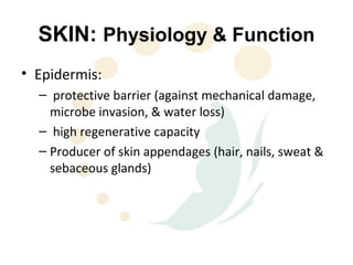
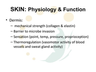
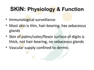
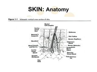
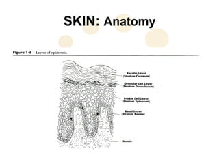
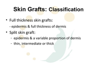
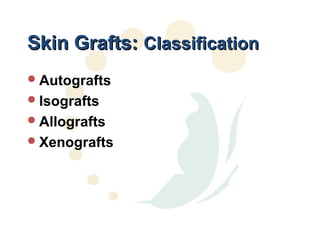
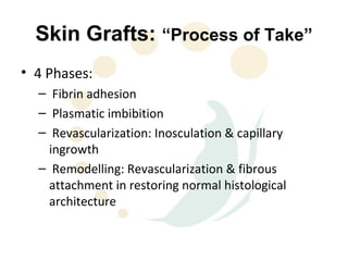
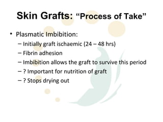
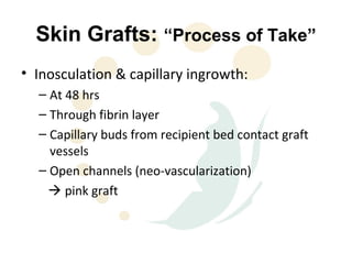
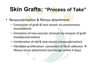
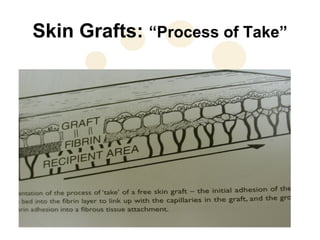
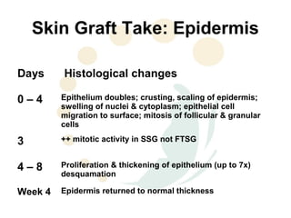
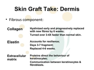
This document discusses the physiology and process of skin grafts. It describes the layers of skin - epidermis and dermis - and their functions. It explains the classification of different types of skin grafts including full thickness and split thickness grafts. The document then outlines the four phases of "take" that a skin graft undergoes as it revascularizes and attaches to the recipient bed. It provides details on the histological and structural changes that occur in both the epidermis and dermis as a graft heals over time. Factors that influence graft survival and potential causes of graft failure are also summarized.
Introduction
Burns
Clinically Relevant Anatomy Of Hand
Common Hand Problems In Burns
Surgical Management
Evidence based Physical Therapy Rehabilitation
Outcome Measures
Summary
References
Introduction
Burns
Clinically Relevant Anatomy Of Hand
Common Hand Problems In Burns
Surgical Management
Evidence based Physical Therapy Rehabilitation
Outcome Measures
Summary
References
Brief Anatomy of Skin and Skin GraftingRishi Gupta
Brief Anatomy of Skin and Skin Grafting.
Anatomy of Skin
History of skin grafting.
Recent Advances in Skin Grafting.
Dermal Substitutes.
Cell cultures in skin grafting.
A presentation
a. The anatomy of the skin
b. The types of skin grafts
c. Indications of a skin graft
d. Mechanism of a graft take
e. Causes of graft failure
f. How to perform skin grafting
3. SKIN: Physiology & Function
• Epidermis:
– protective barrier (against mechanical damage,
microbe invasion, & water loss)
– high regenerative capacity
– Producer of skin appendages (hair, nails, sweat &
sebaceous glands)
4. SKIN: Physiology & Function
• Dermis:
– mechanical strength (collagen & elastin)
– Barrier to microbe invasion
– Sensation (point, temp, pressure, proprioception)
– Thermoregulation (vasomotor activity of blood
vessels and sweat gland activity)
5. SKIN: Physiology & Function
• Immunological surveillance
• Most skin is thin, hair-bearing, has sebaceous
glands
• Skin of palms/soles/flexor surface of digits is
thick, not hair-bearing, no sebaceous glands
• Vascular supply confined to dermis
14. Skin Grafts: “Process of Take”
• Plasmatic Imbibition:
– Initially graft ischaemic (24 – 48 hrs)
– Fibrin adhesion
– Imbibition allows the graft to survive this period
– ? Important for nutrition of graft
– ? Stops drying out
15. Skin Grafts: “Process of Take”
• Inosculation & capillary ingrowth:
– At 48 hrs
– Through fibrin layer
– Capillary buds from recipient bed contact graft
vessels
– Open channels (neo-vascularization)
pink graft
16. Skin Grafts: “Process of Take”
• Revascularization & fibrous attachment:
– Connection of graft & host vessels via anastomoses
(inosculation)
– Formation of new vascular channels by invasion of graft
(neovascularisation)
– Combination of old & new vessels (revascularisation)
– Fibroblast proliferation: conversion of fibrin adhesion
fibrous tissue attachment (anchorage within 4 days)
18. Skin Graft Take: Epidermis
Days Histological changes
0–4 Epithelium doubles; crusting, scaling of epidermis;
swelling of nuclei & cytoplasm; epithelial cell
migration to surface; mitosis of follicular & granular
cells
3 ++ mitotic activity in SSG not FTSG
4–8 Proliferation & thickening of epithelium (up to 7x)
desquamation
Week 4 Epidermis returned to normal thickness
19. Skin Graft Take: Epidermis
Day Histochemical changes
4 Increased RNA in basal cells, indicating protein
synthesis
10 RNA returns to normal
20. Skin Graft Take: Dermis
• Fibrous component:
Collagen Hyalinized early and progressively replaced
with new fibres by 6 weeks;
Turned over 3-4X faster than normal skin.
Elastin Accounts for resilience;
Days 3-7 fragment;
Replaced 4-6 weeks.
Extracellular Proteins direct the behaviour of
matrix keratinocytes;
Communication between keratinocytes &
fibroblasts.
21. Skin Graft Take: Dermis
• Appendages:
- sweating dependent on no. of transplanted sweat glands &
degree of sympathetic reinnervation; will sweat like recipient
site in FTSG only
- sebaceous gland activity mostly in thicker grafts: SSG usually
dry & shiny
- hair grows from FTSG if well taken with no complications
22. Skin Graft Healing
• Initially white then pinkens with new blood
supply
• Lymphatic drainage by day 6
• Collagen replacement from day 7 to week 6
• Vascular remodelling for months
23. Skin Graft Healing
• Contraction:
- shrinks immediately due to elastic recoil: – FTSG
40%; medium SSG 20%; thin SSG 10%.
- secondary contracture as heals:
- FTSG remains same size after above shrinkage;
- SSG will contract as much as possible;
- more dermis = less contraction
- ? Due to myofibroblasts
24. Skin Graft Healing
• Reinnervation:
– from margins to bed;
– 4/52 to 2 years;
– Depends on graft thickness and bed;
– Uneventful healing leads to near normal 2PD;
– Cold sensitivity can be a problem.
25. Skin Graft Expansion
• Based on principle that wounds
reepithelialized from the periphery
• Expansion provides larger areas from which
epithelium can grow
• Larger areas can be covered with less skin
26. Skin Graft Expansion
• Meshing
- covers large area
- easier to contour
- fluid can drain through holes
- cosmetic results less than ideal
- various mesh ratio
27. Skin Graft Survival
• Meticulous technique
• Atraumatic graft handling
• Well vascularized bed
• Haemostasis
• Immobilization
• No proximal constricting bandages