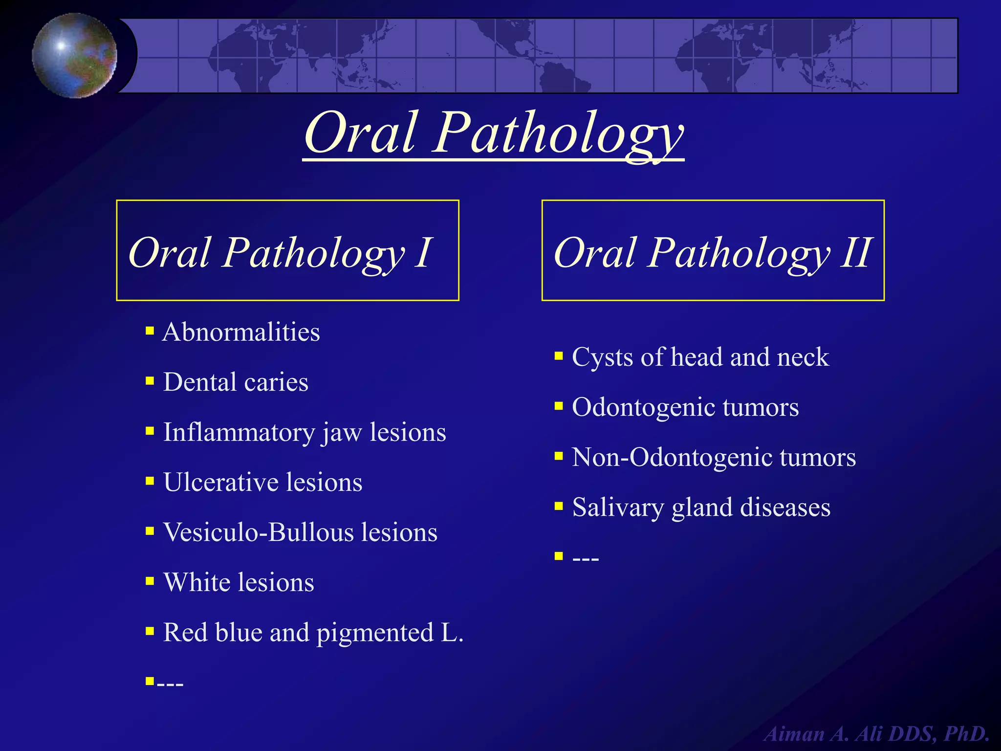
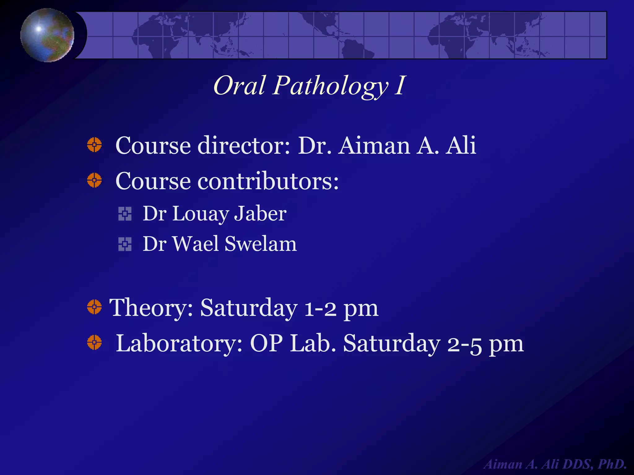
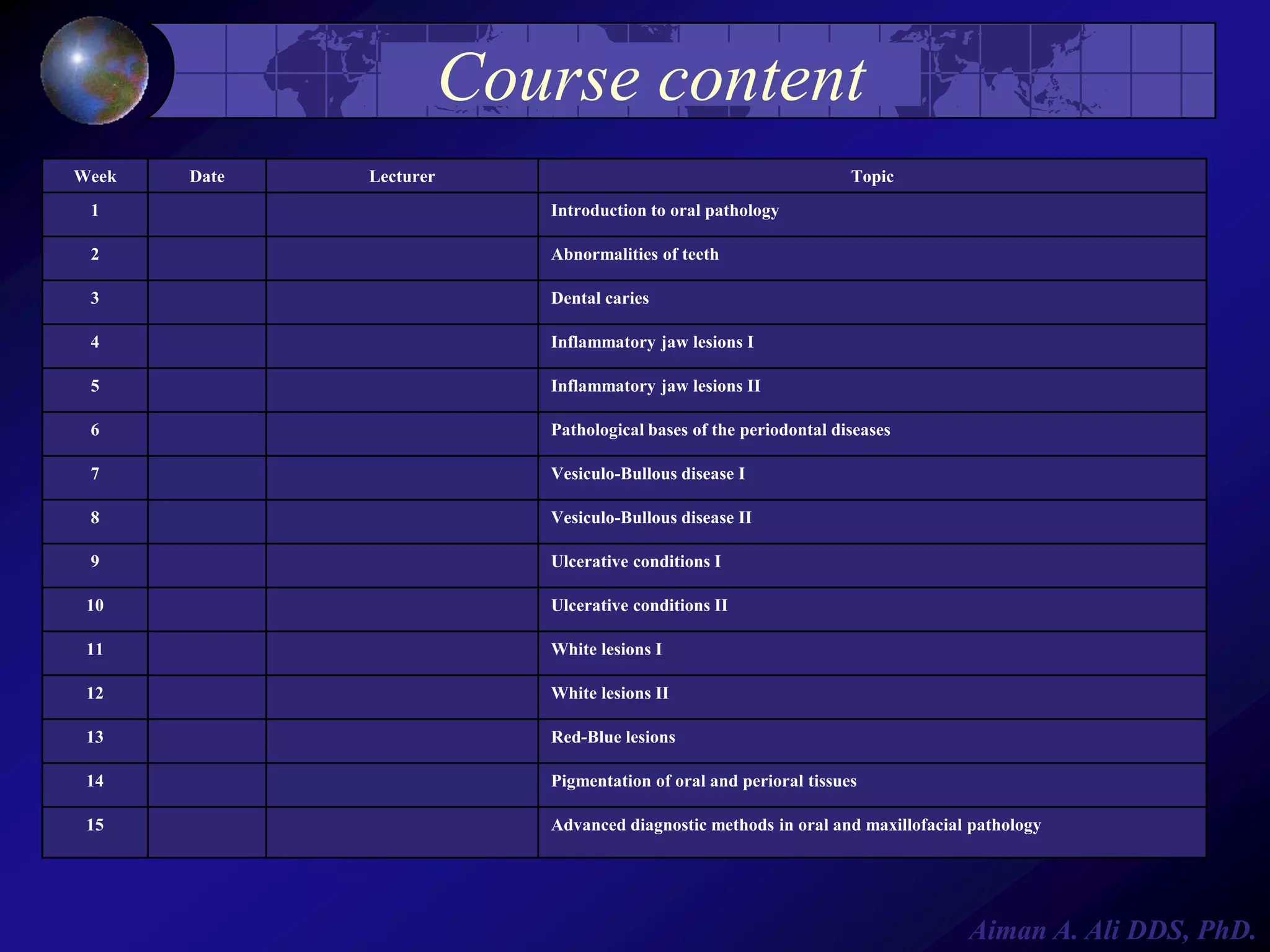
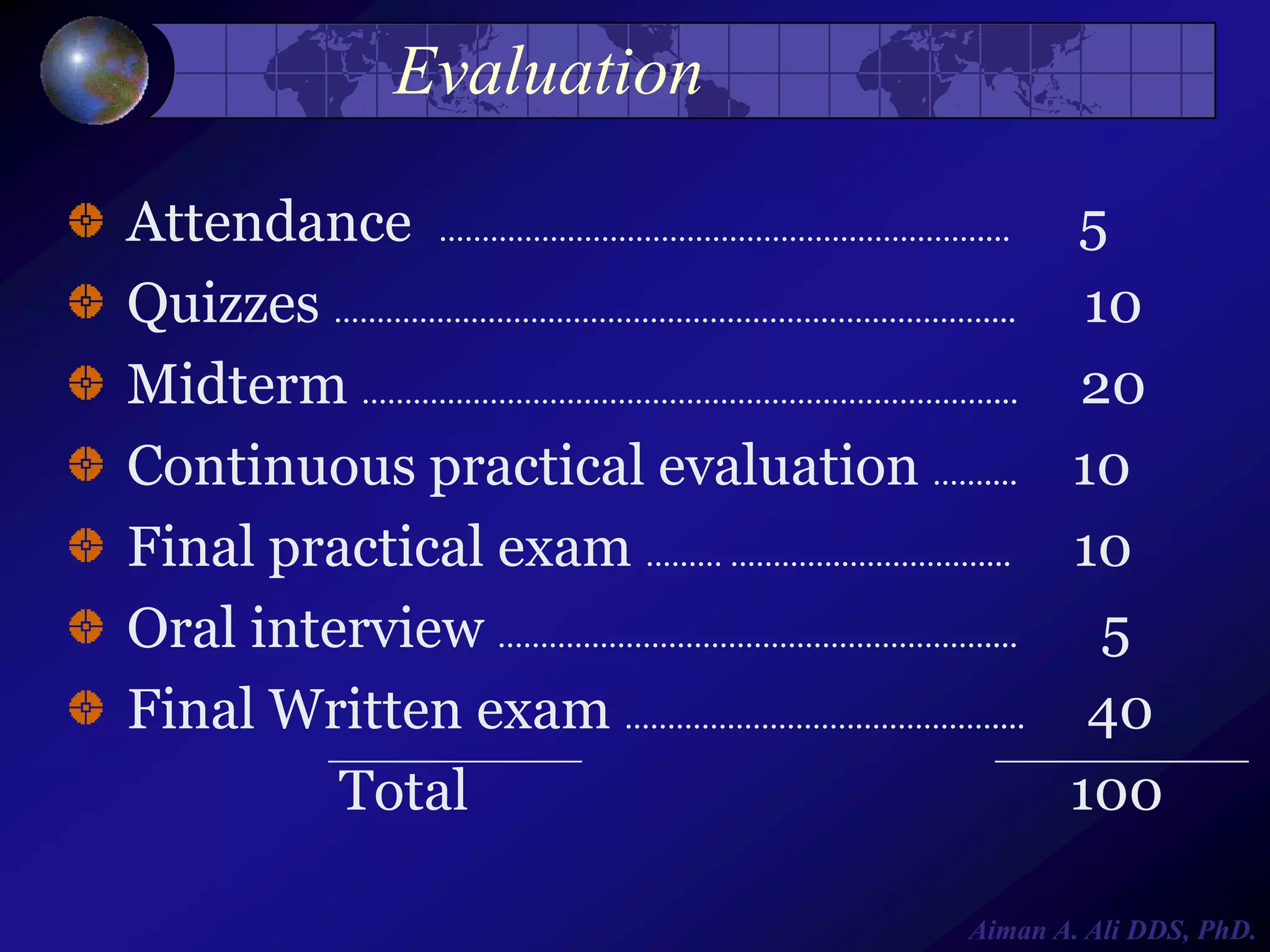
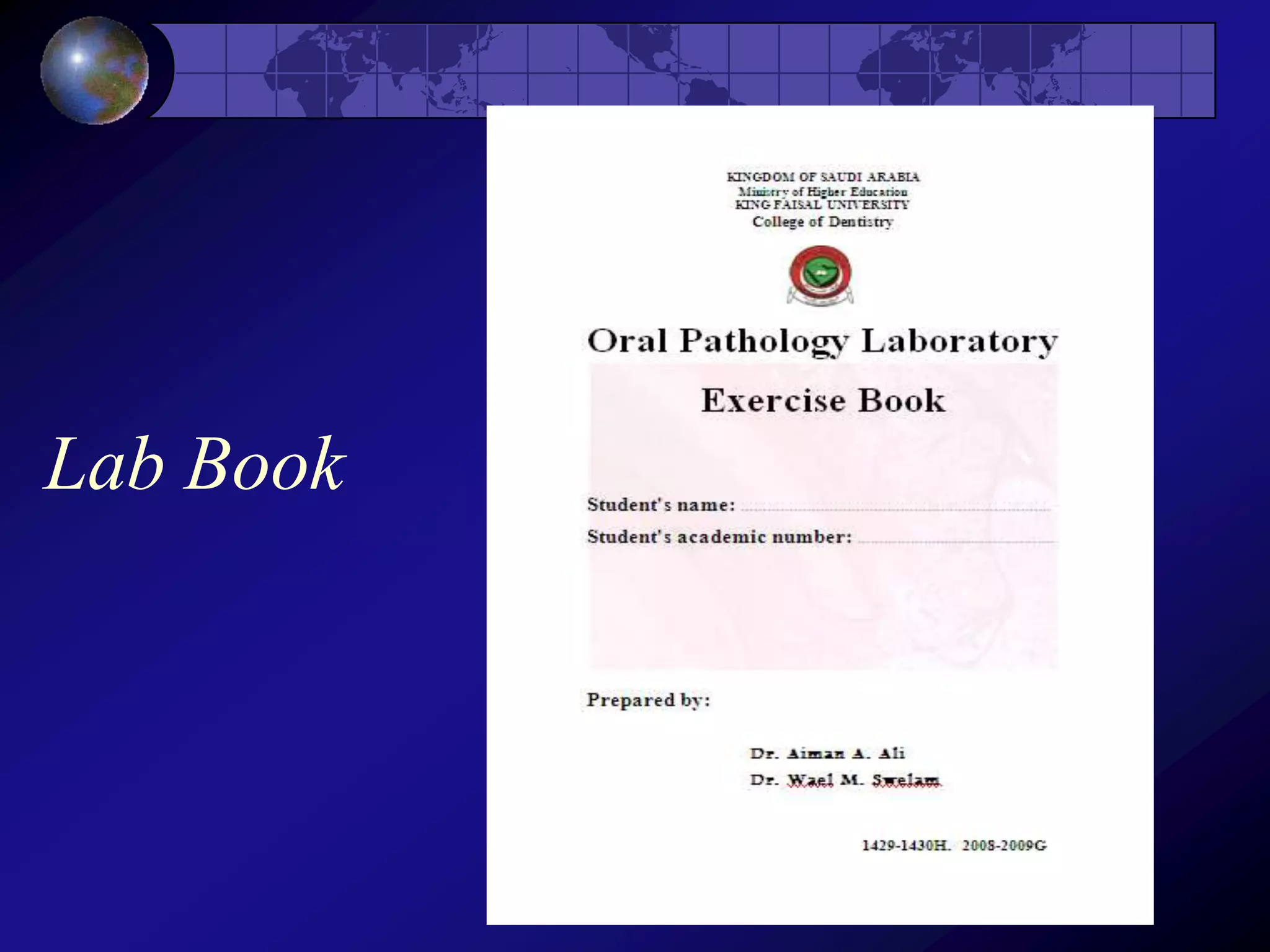
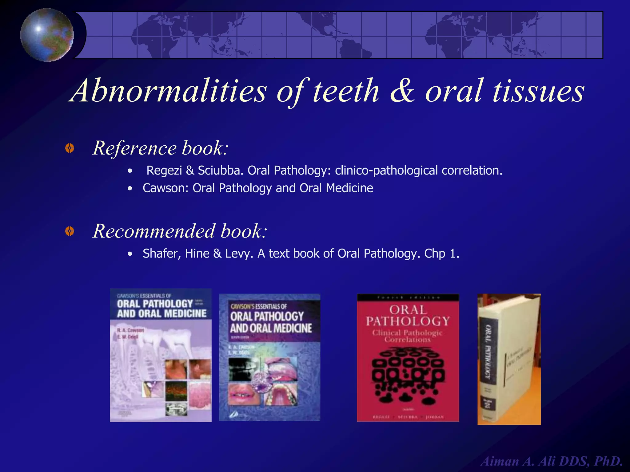
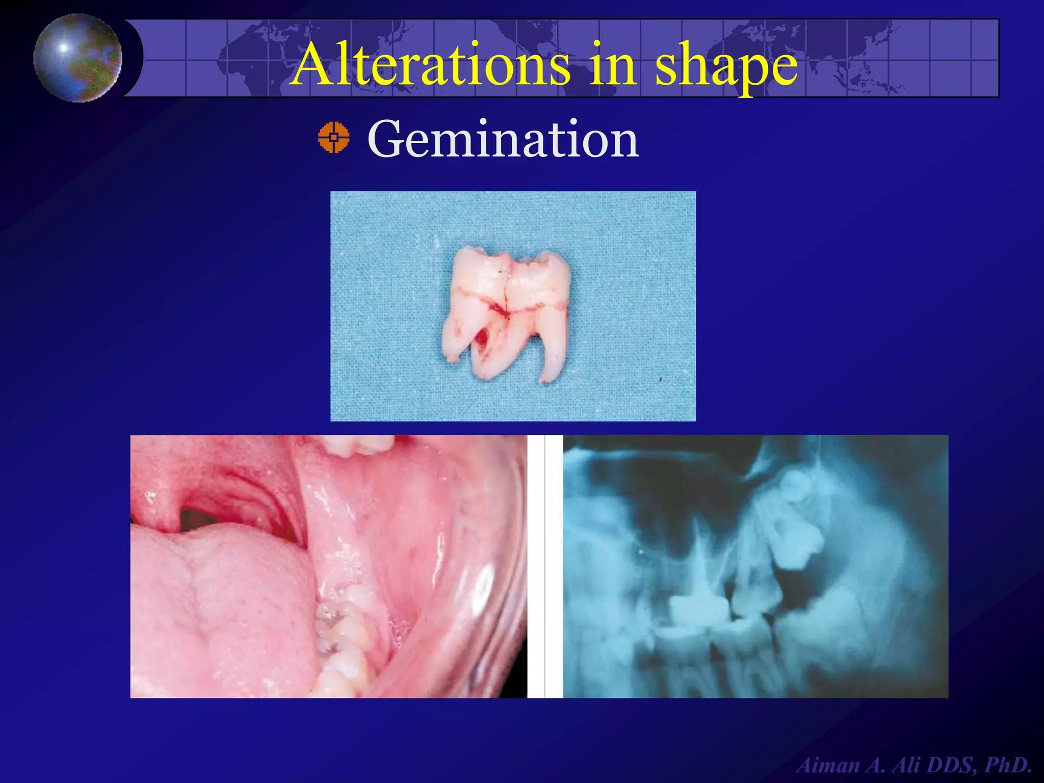
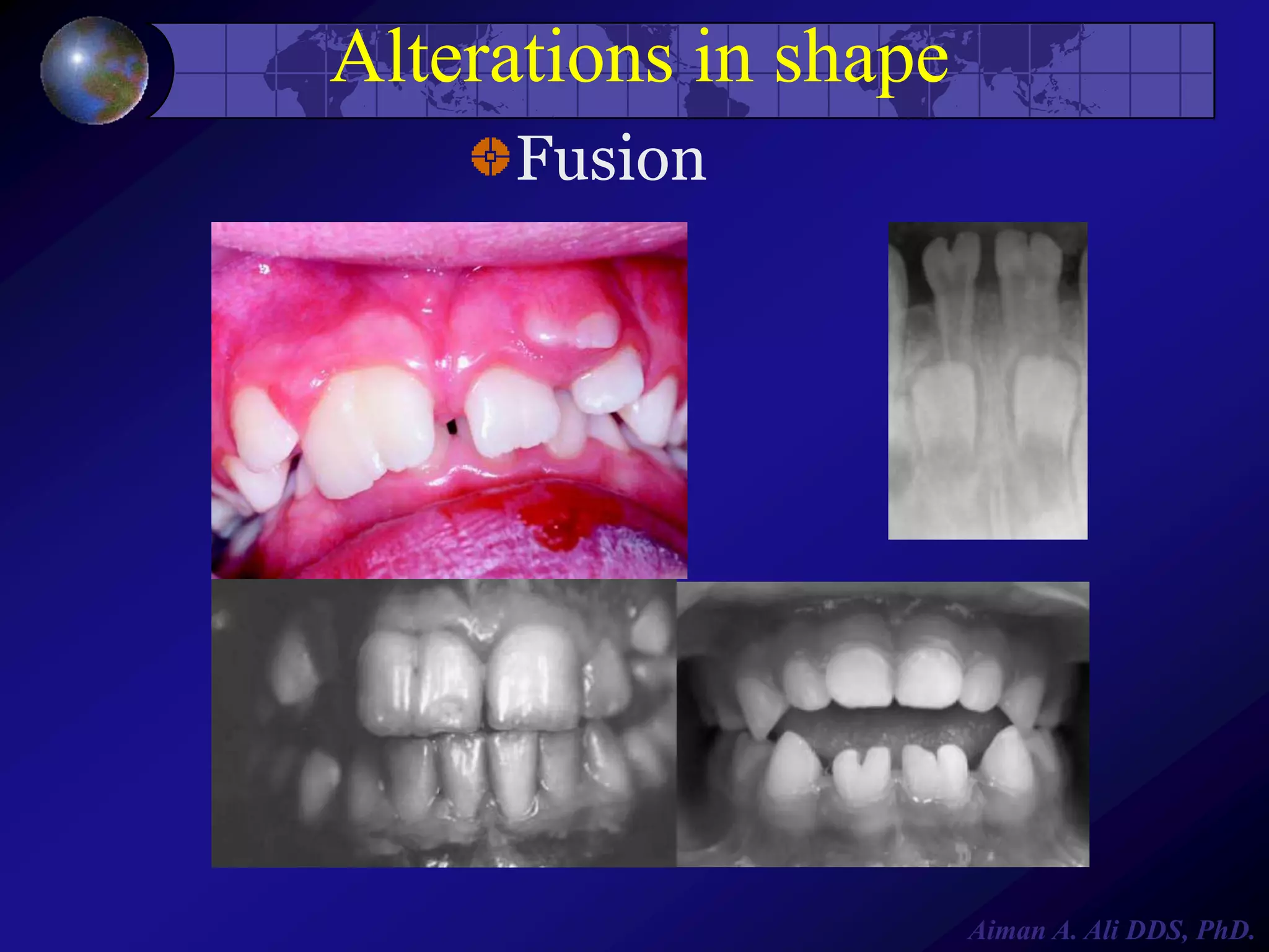
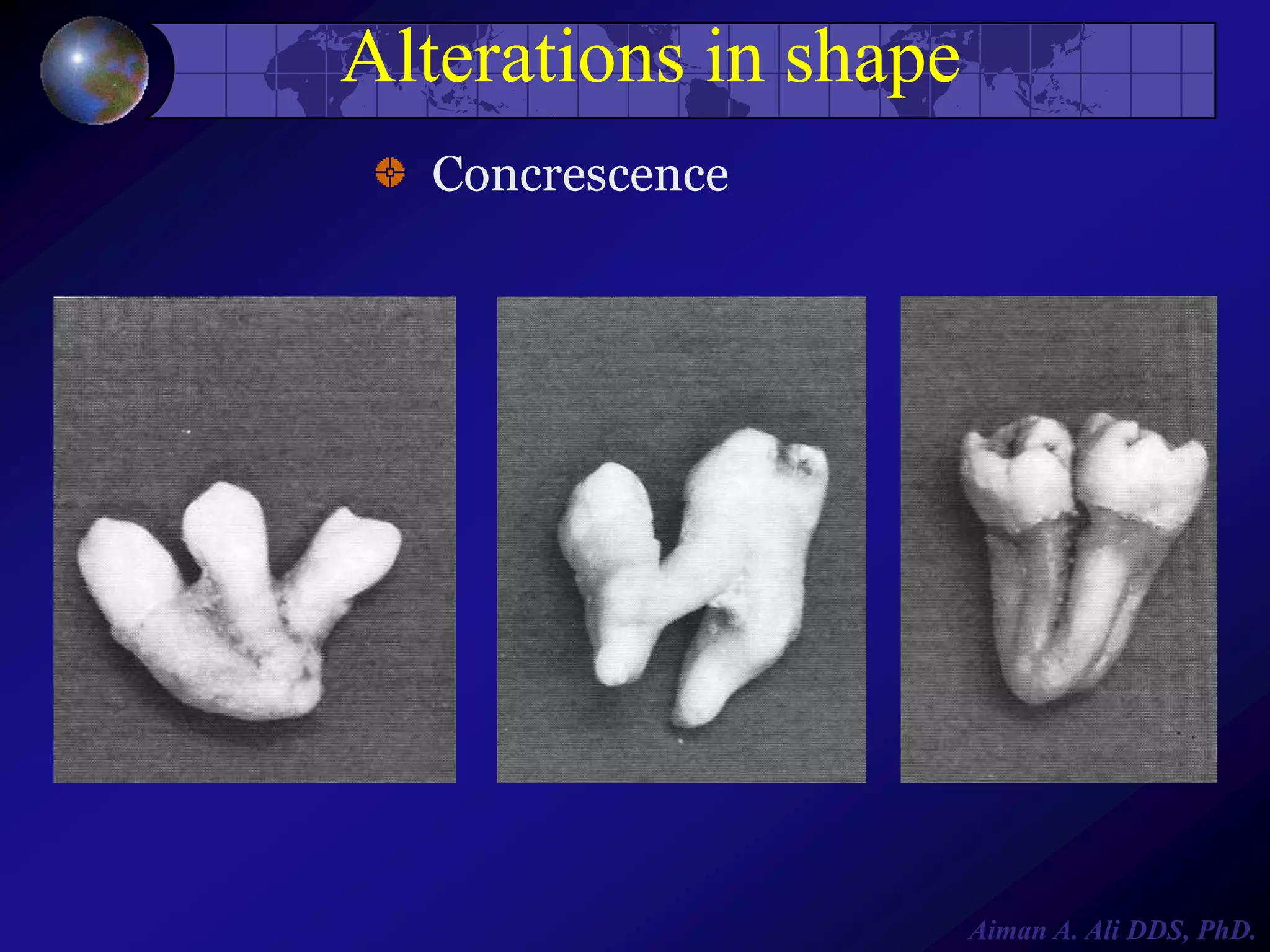
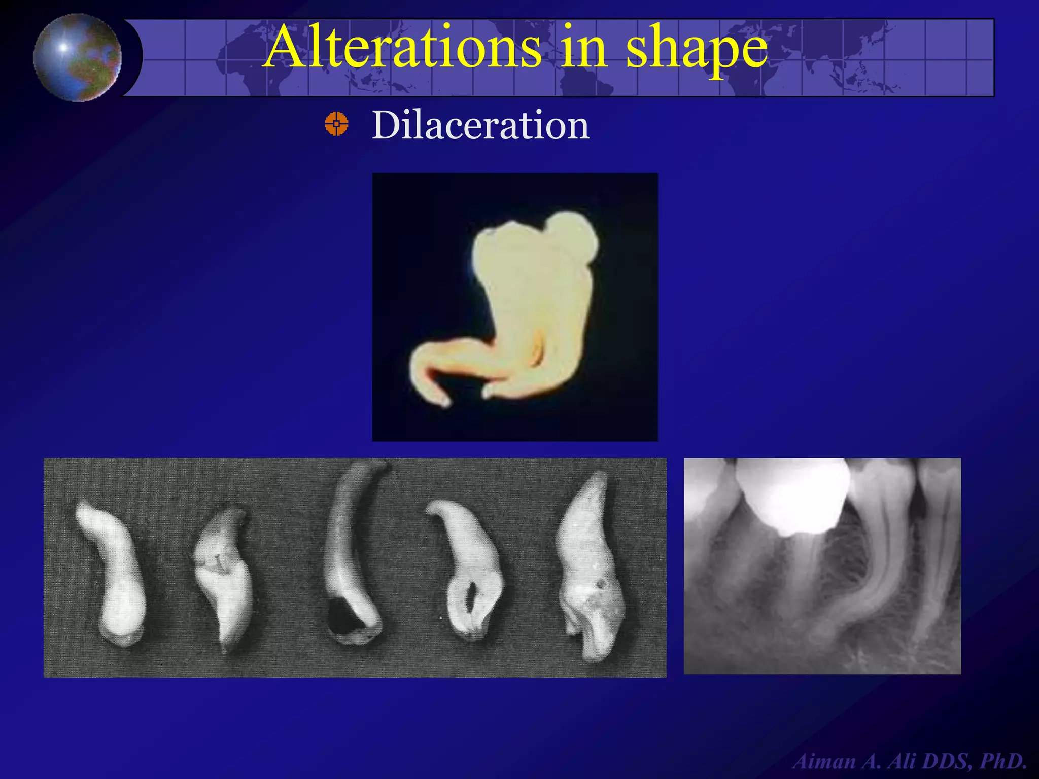
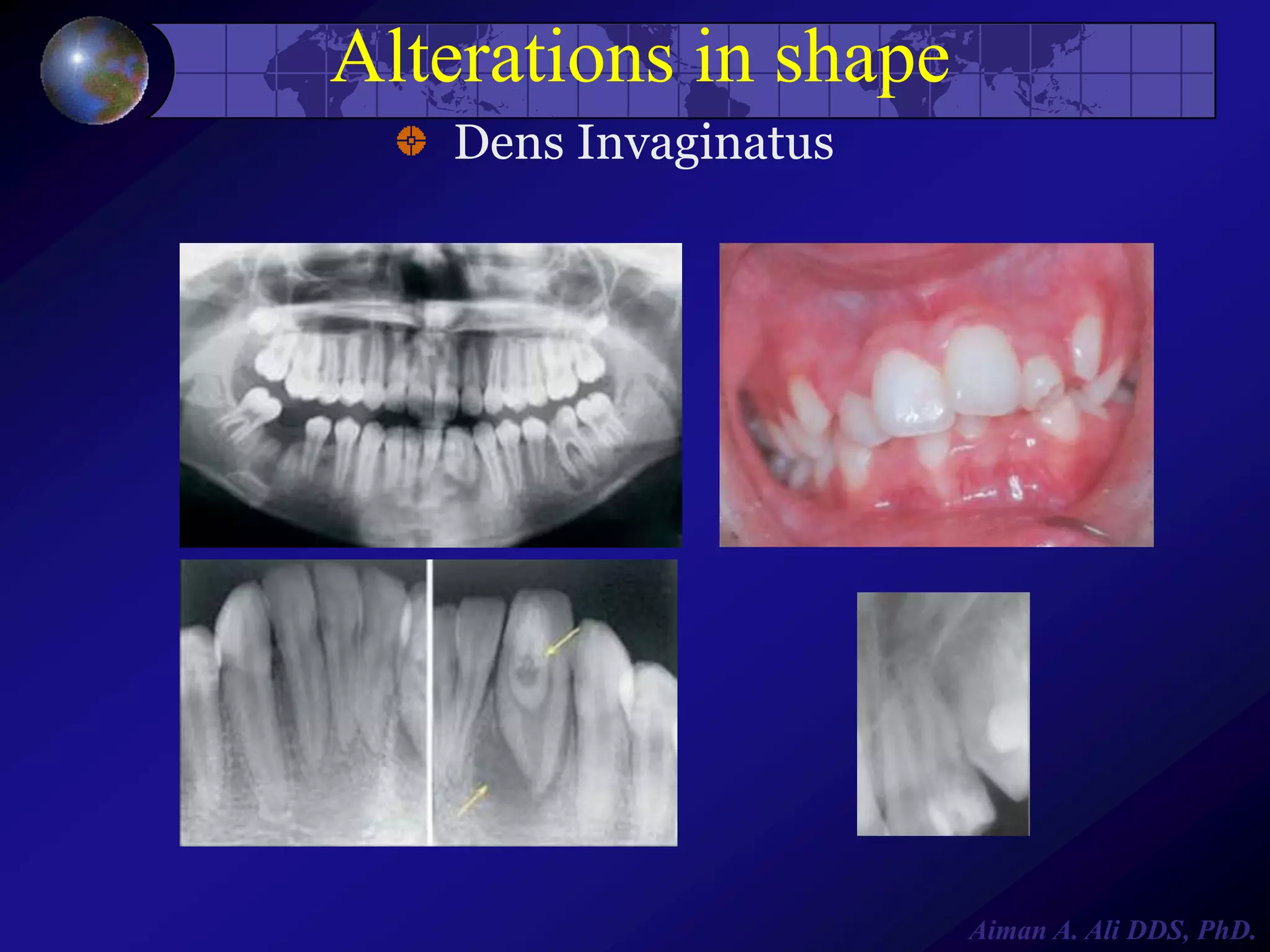
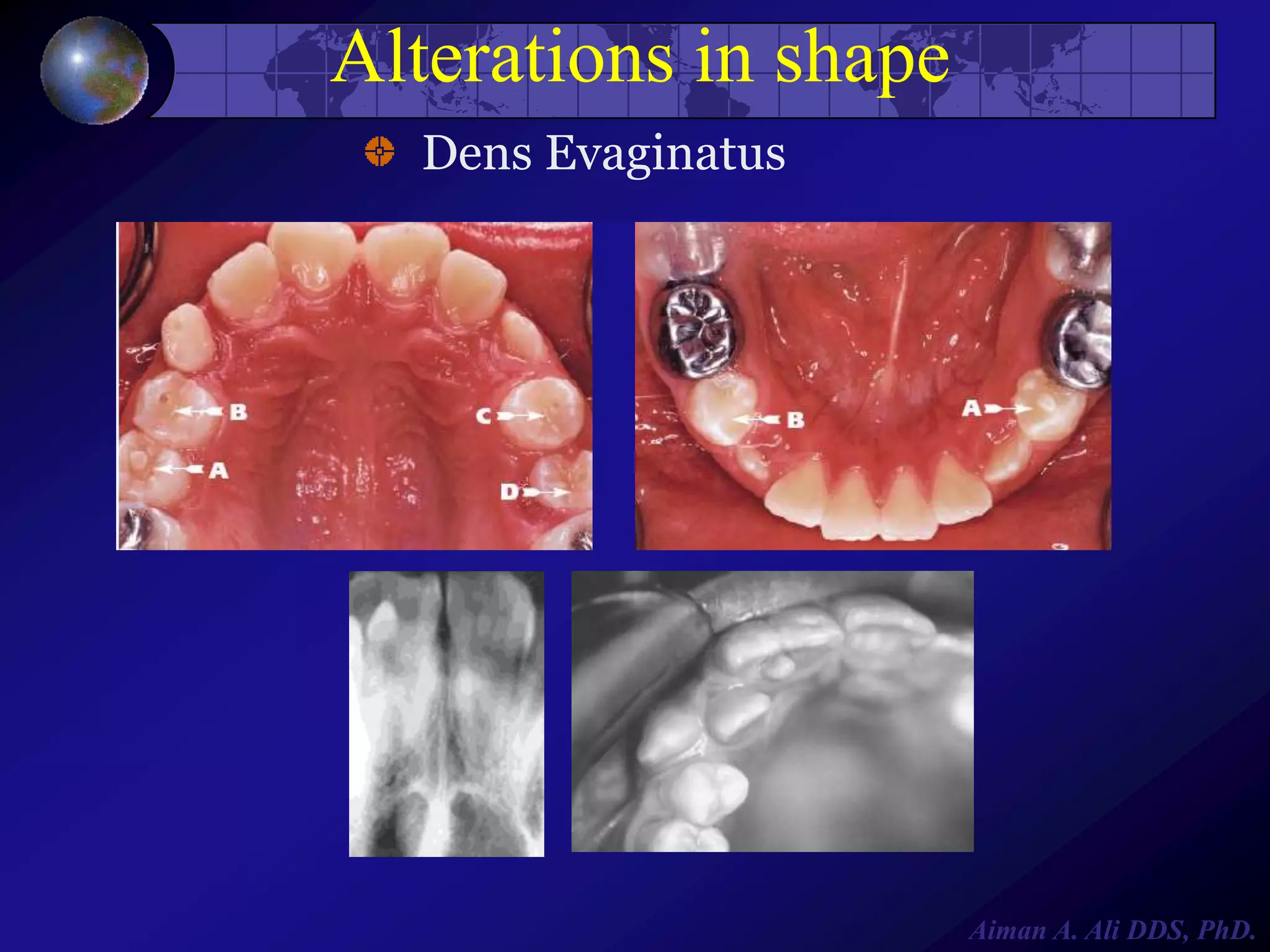
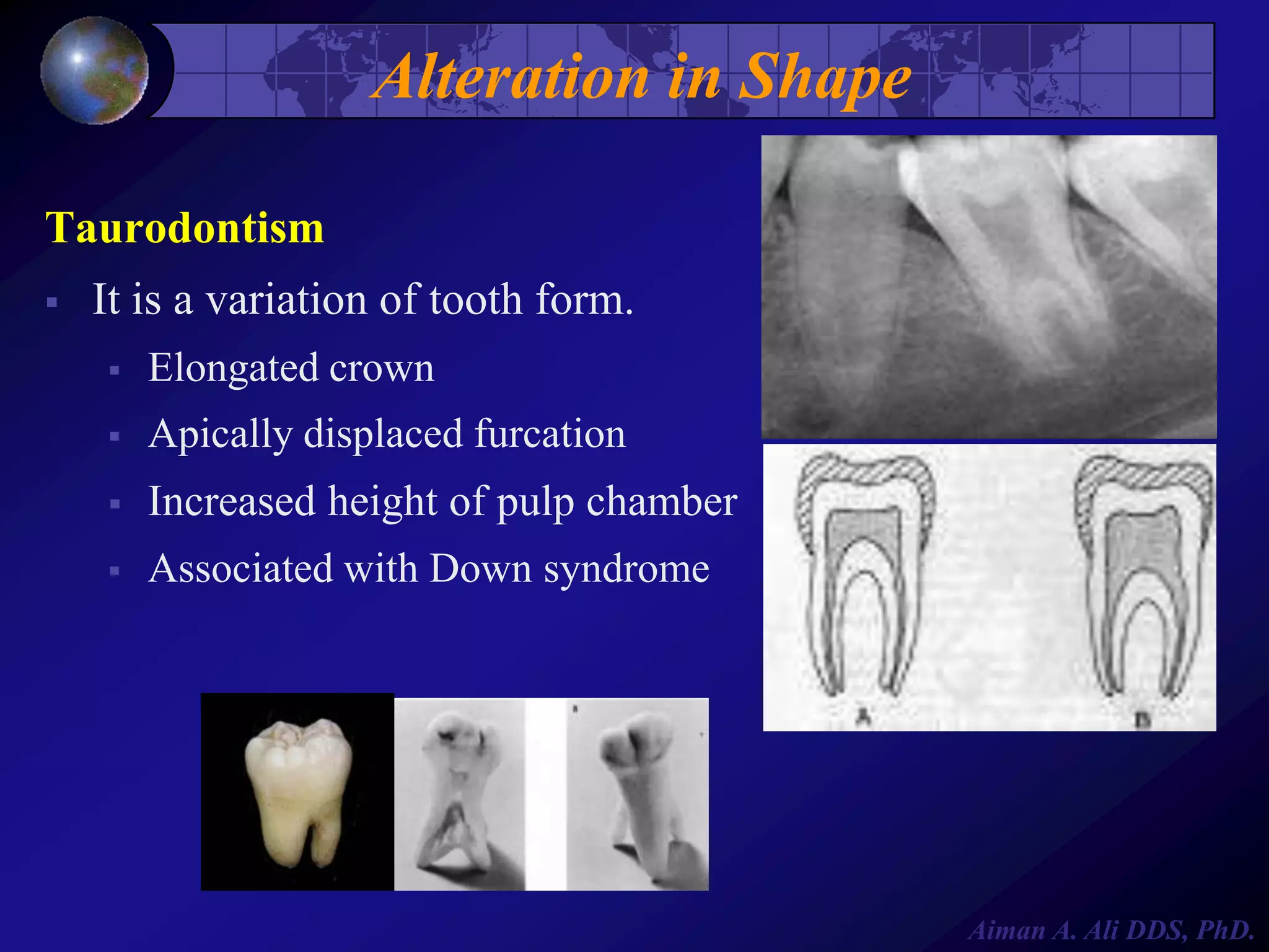
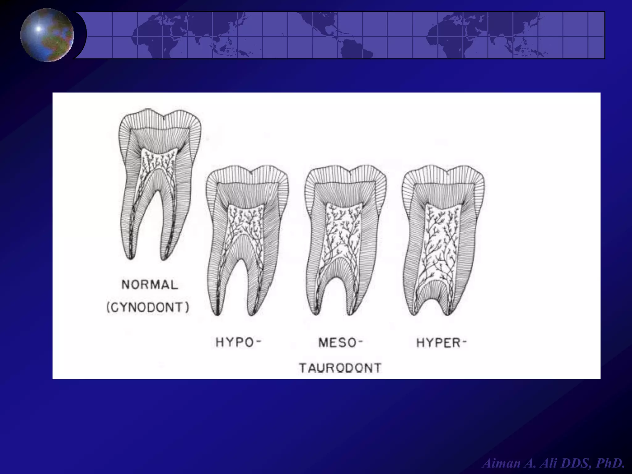
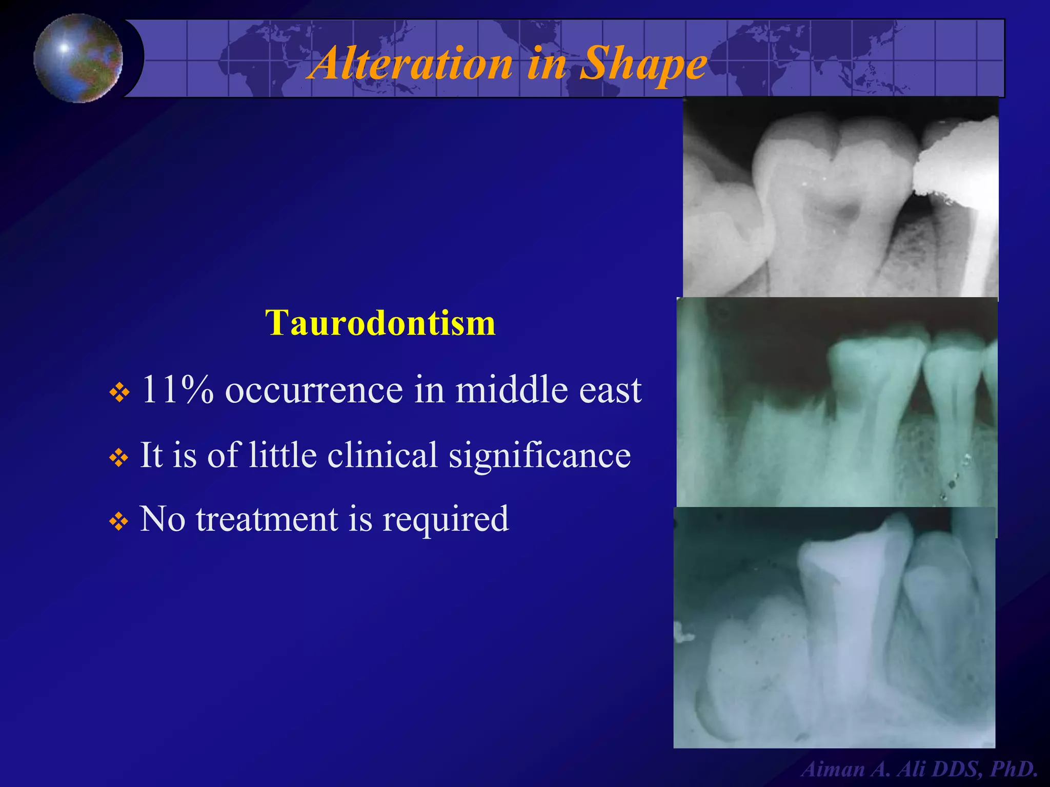
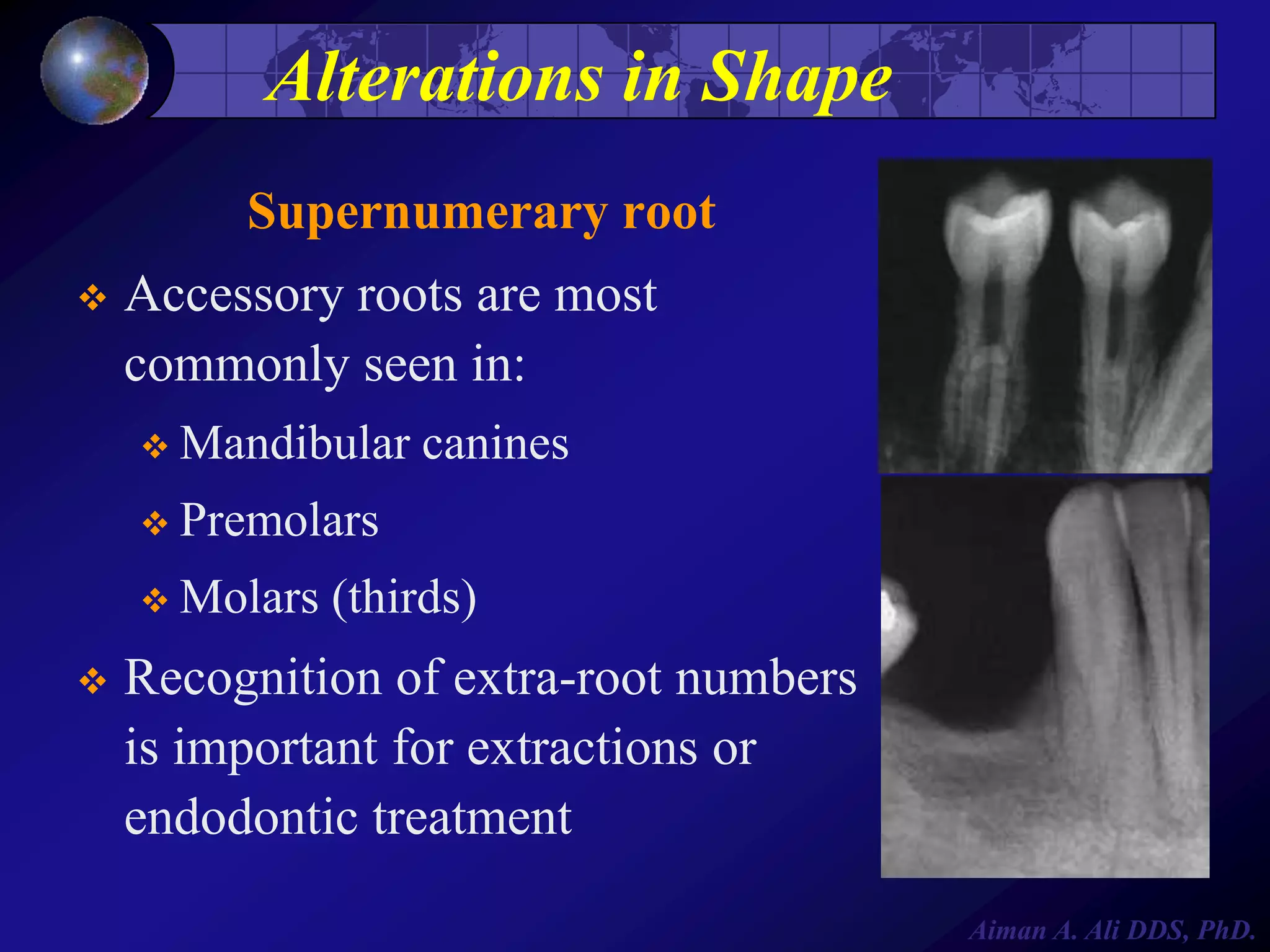
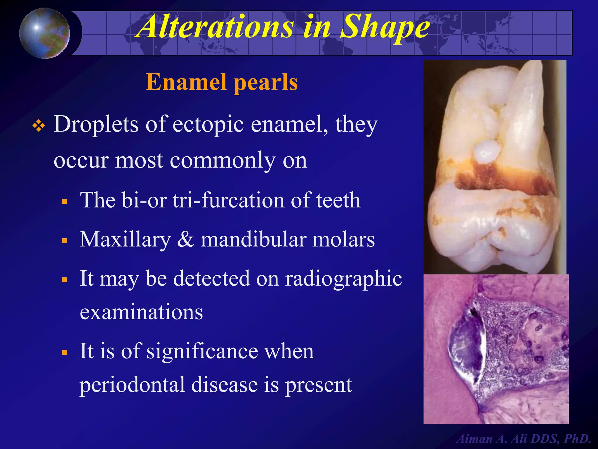
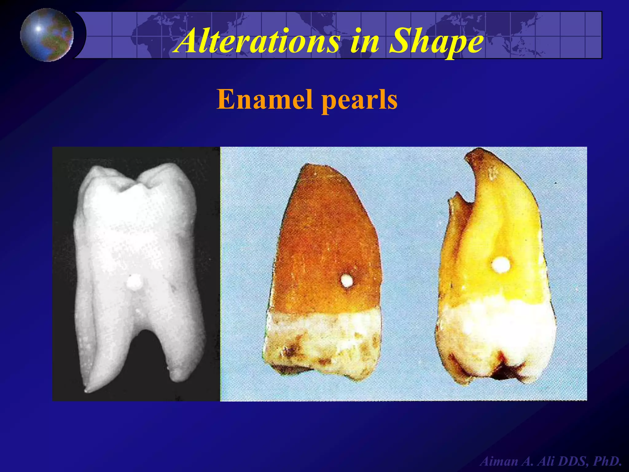
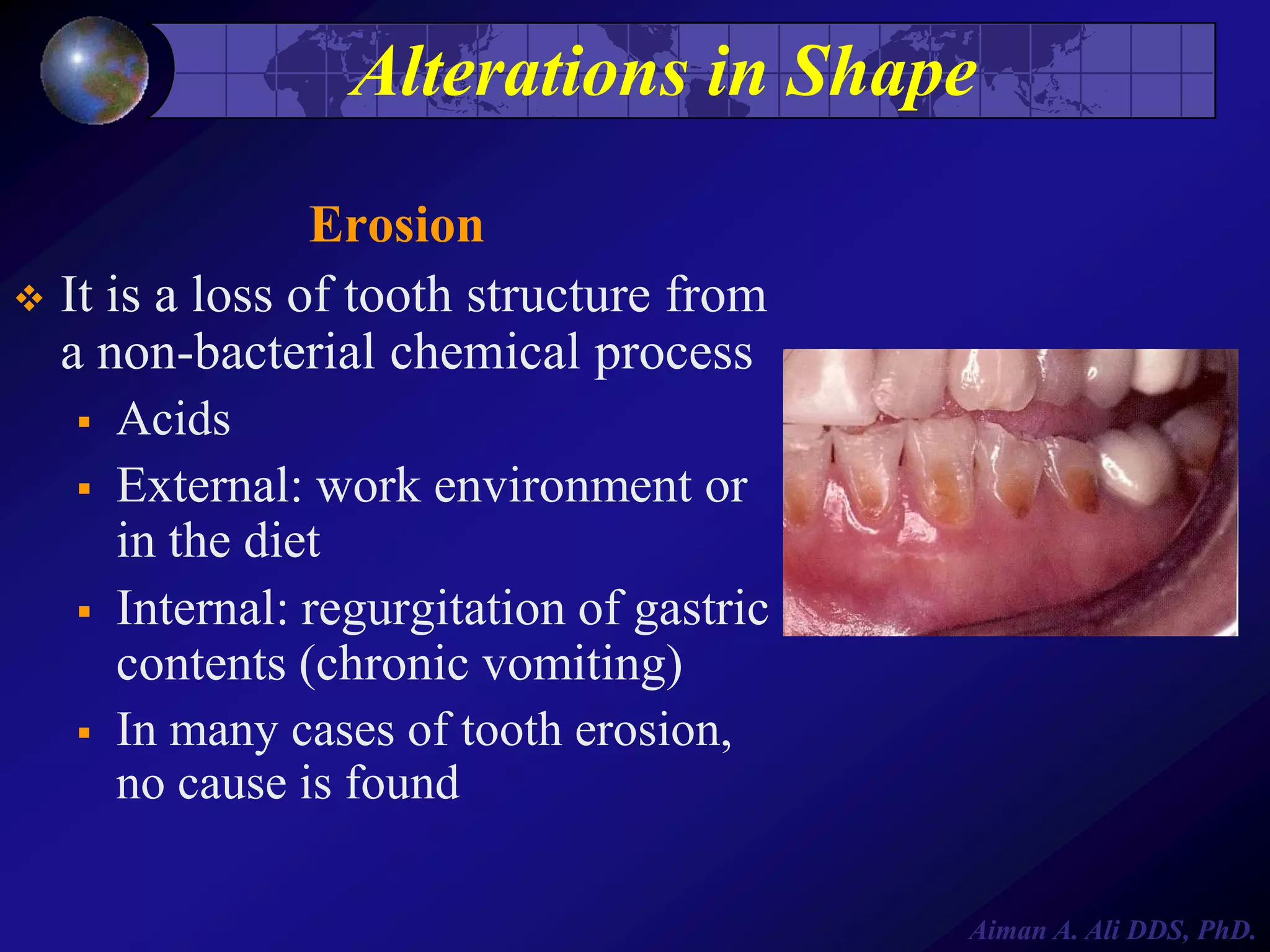
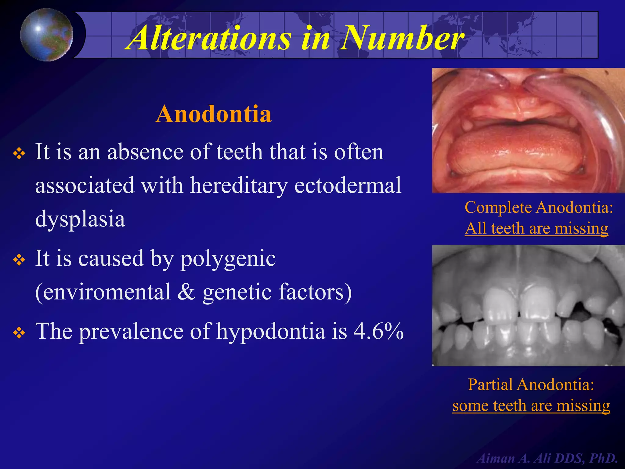
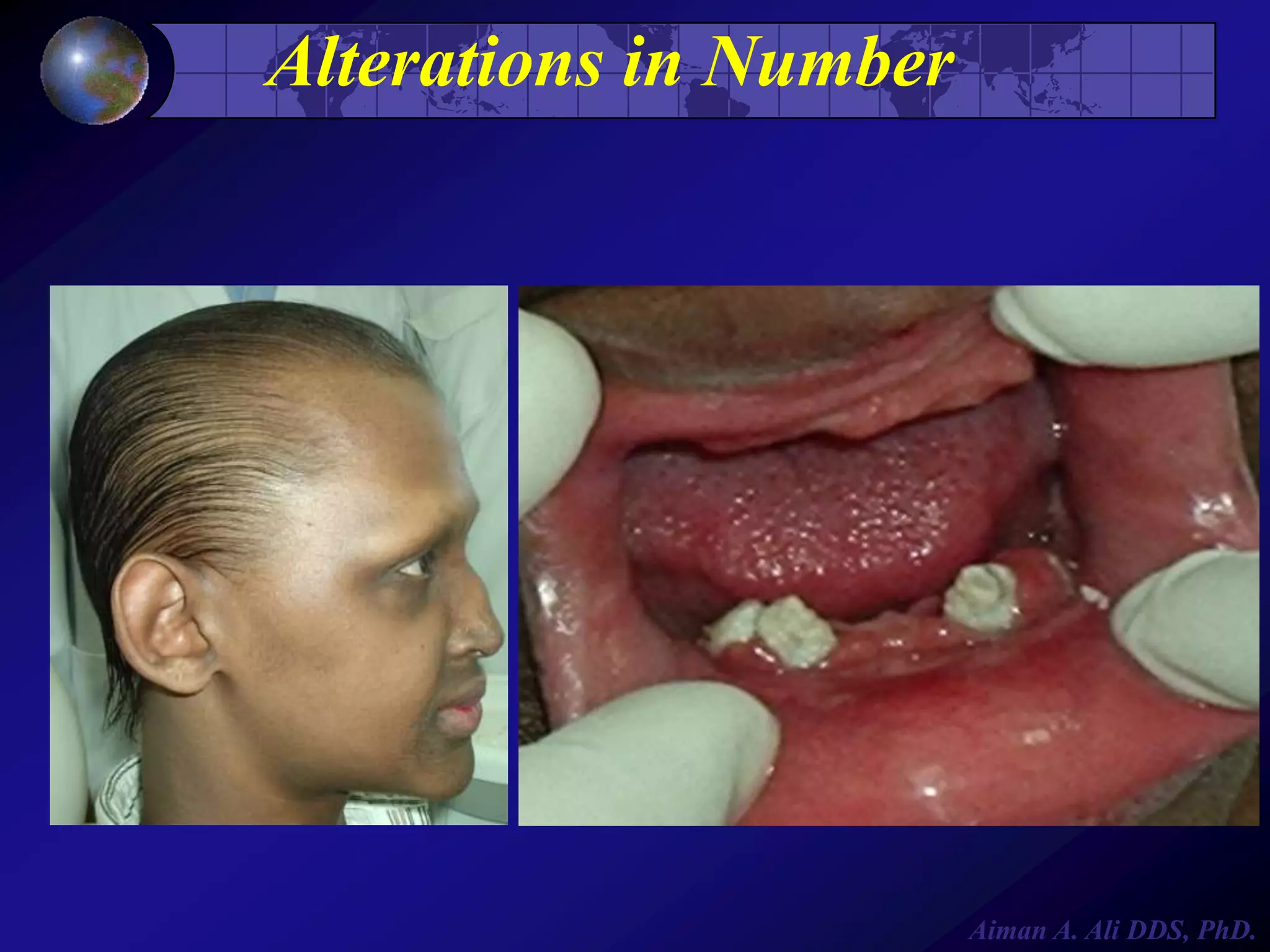
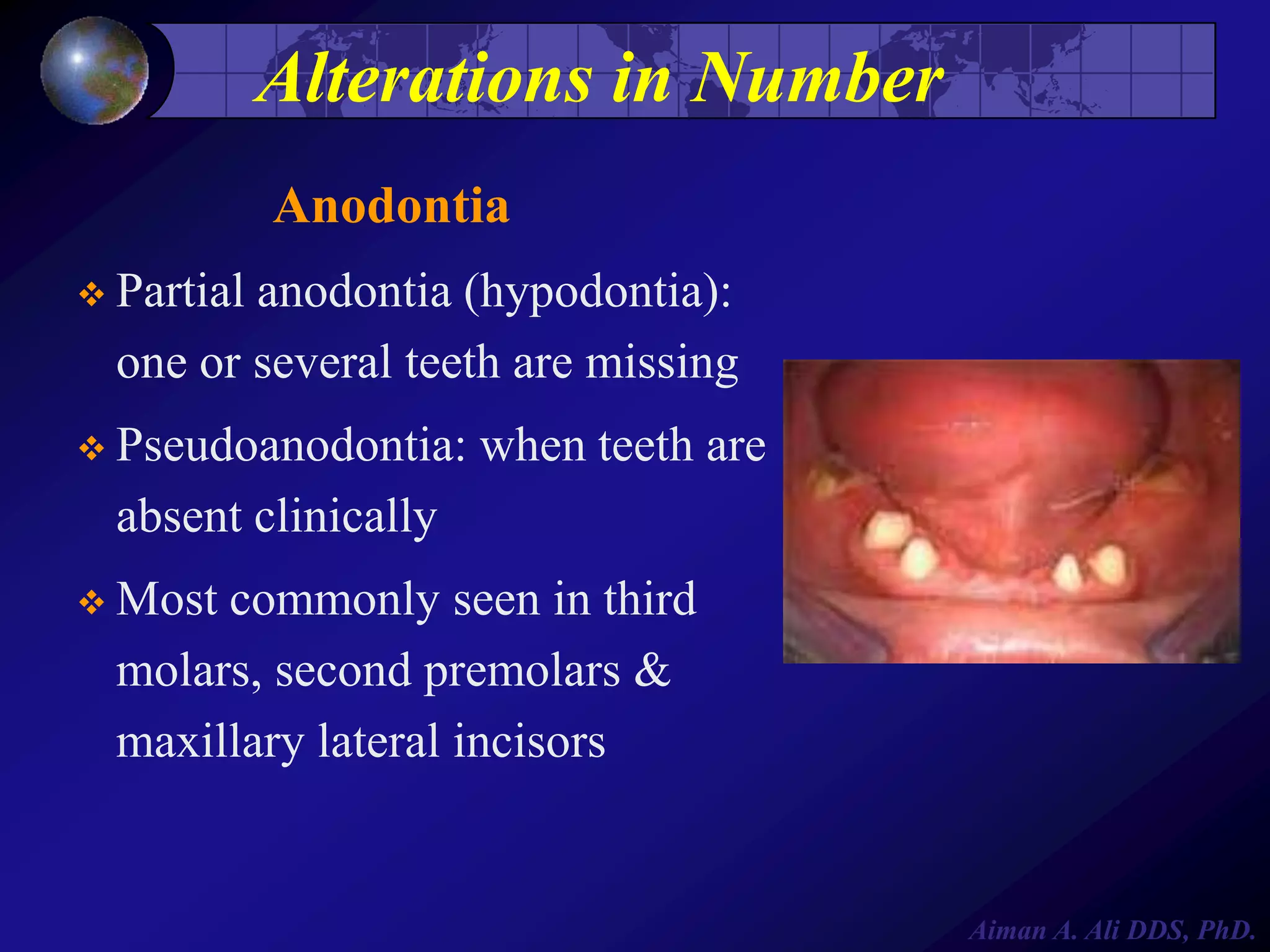
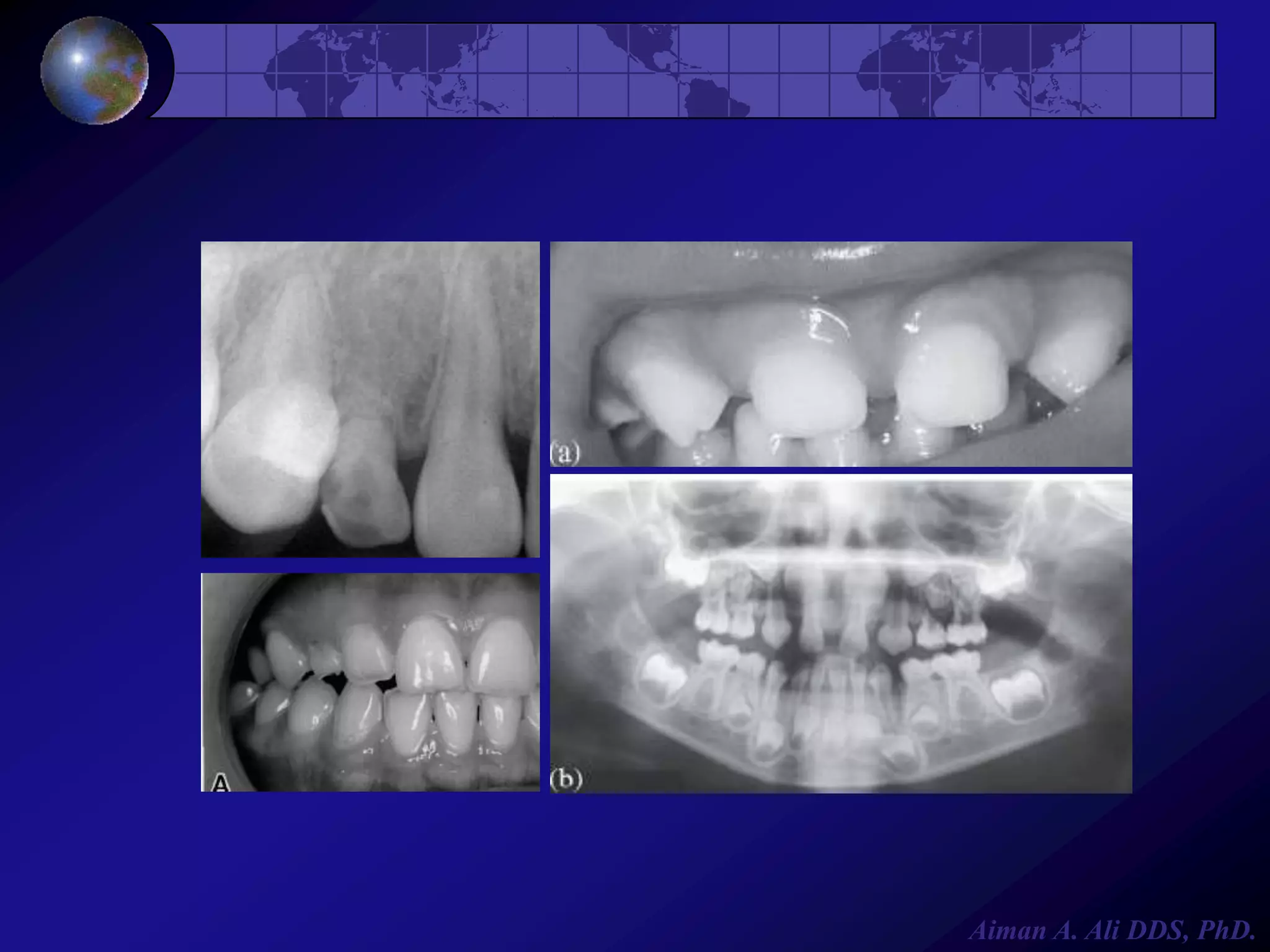
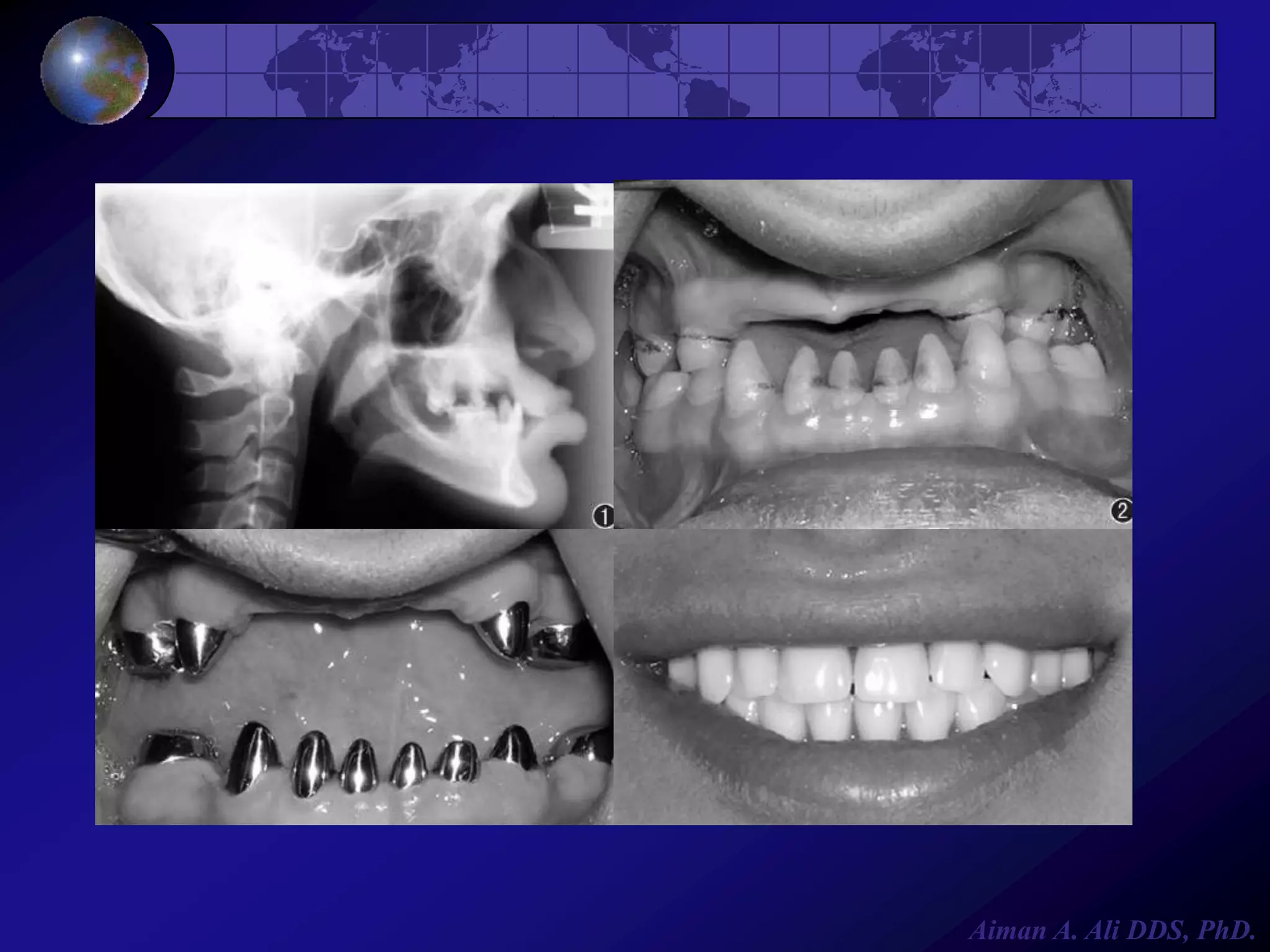
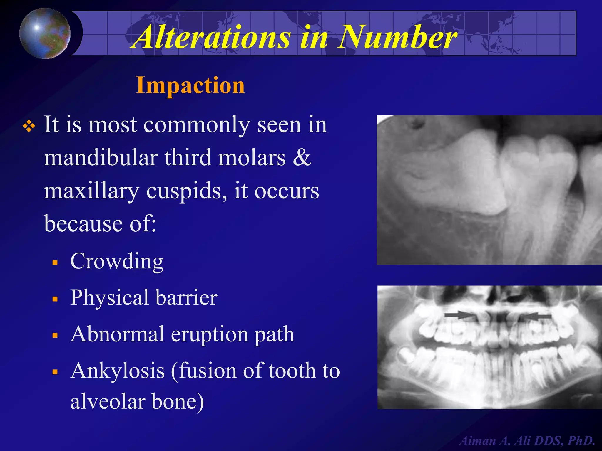
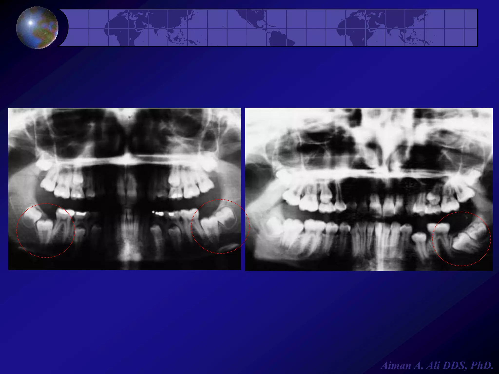
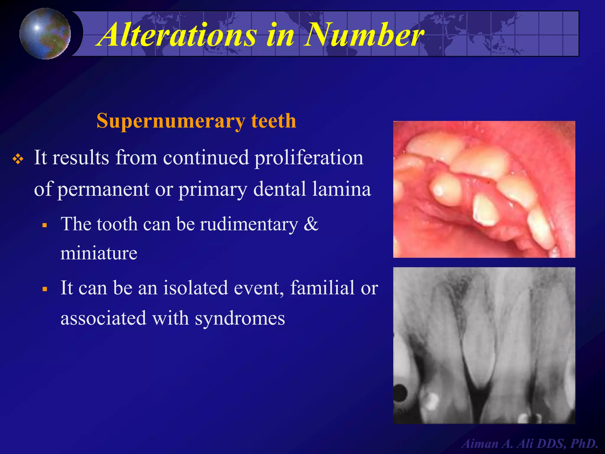
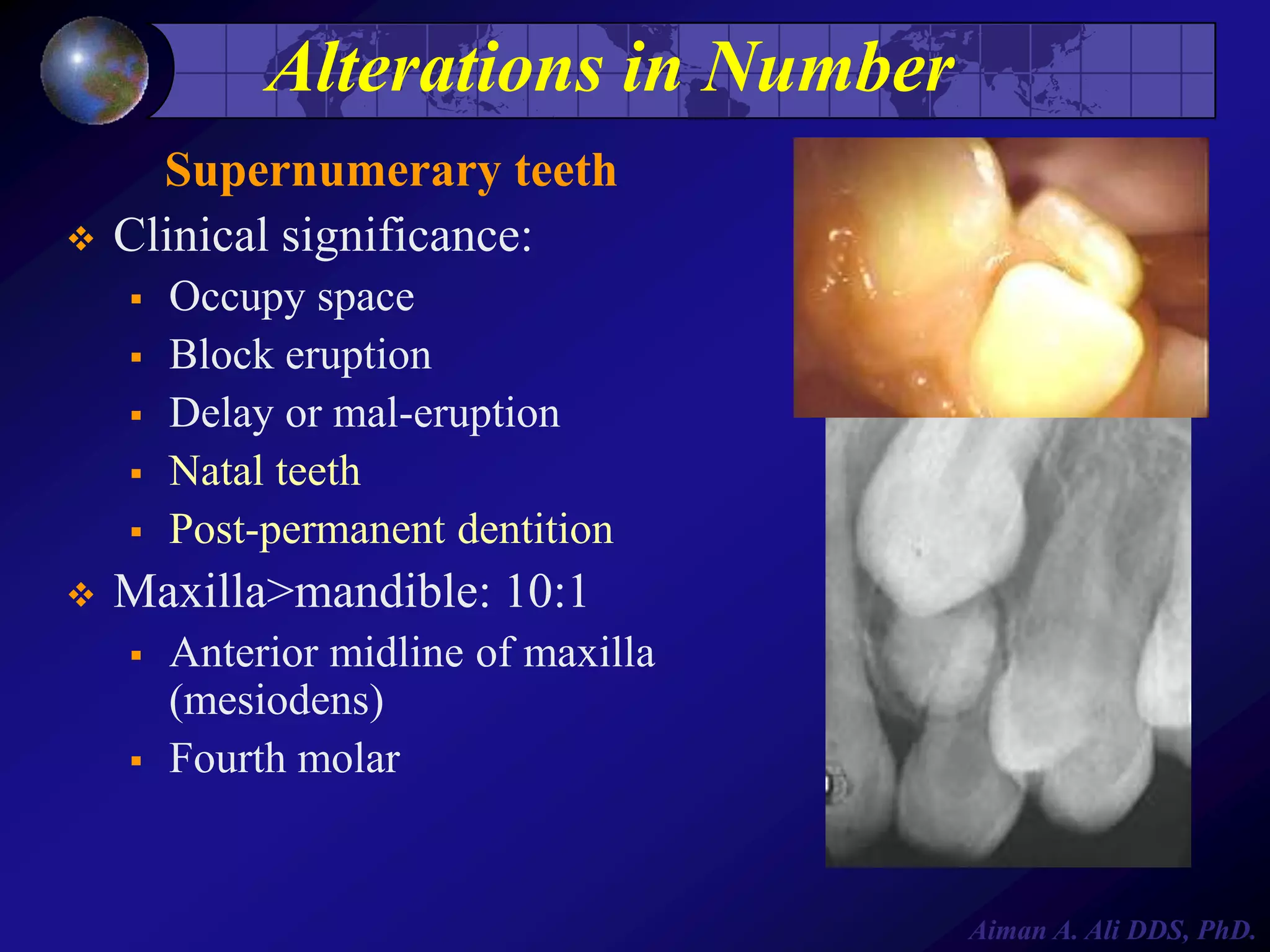
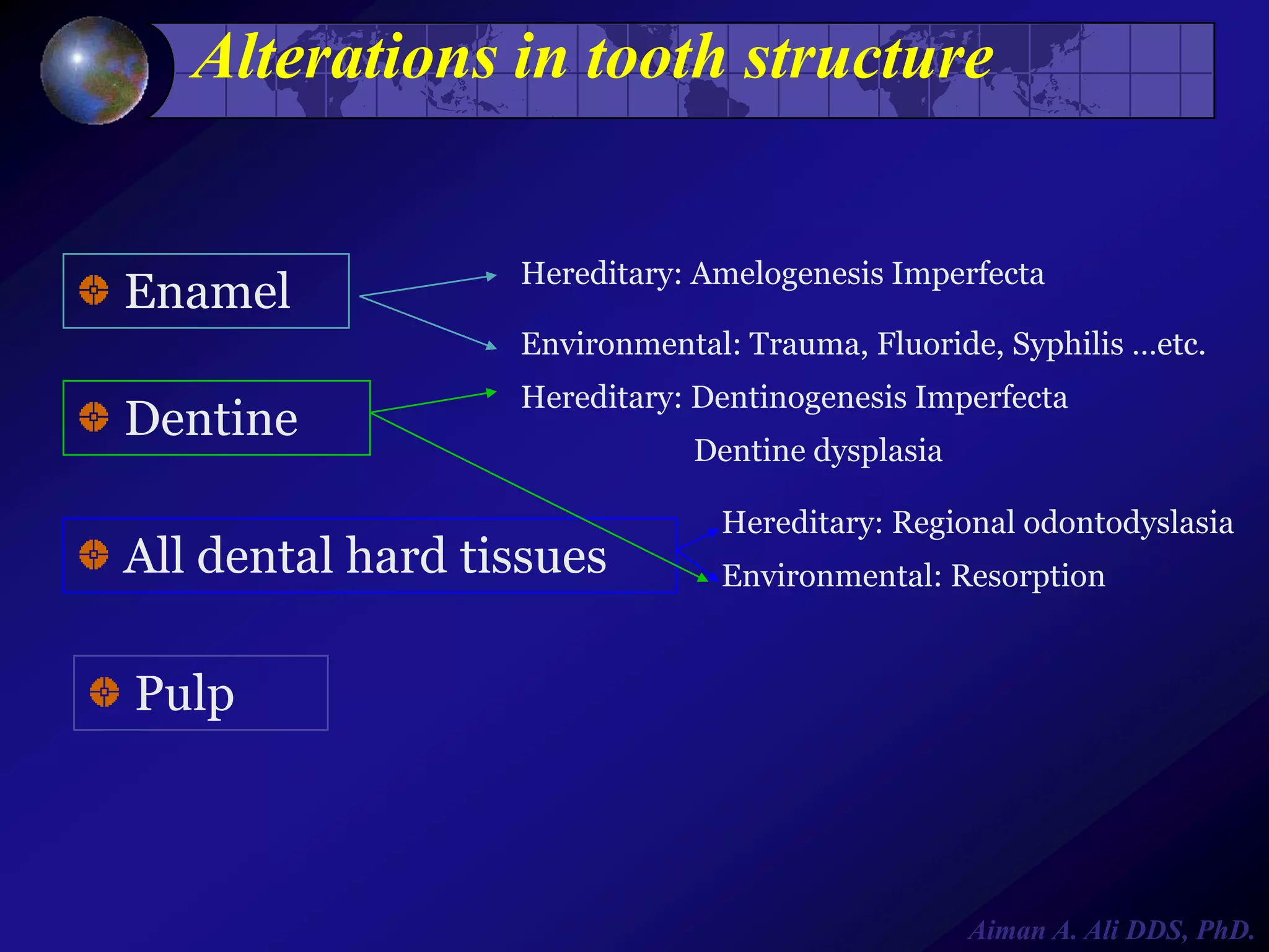
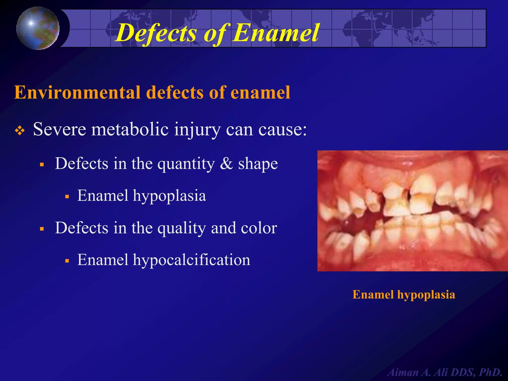
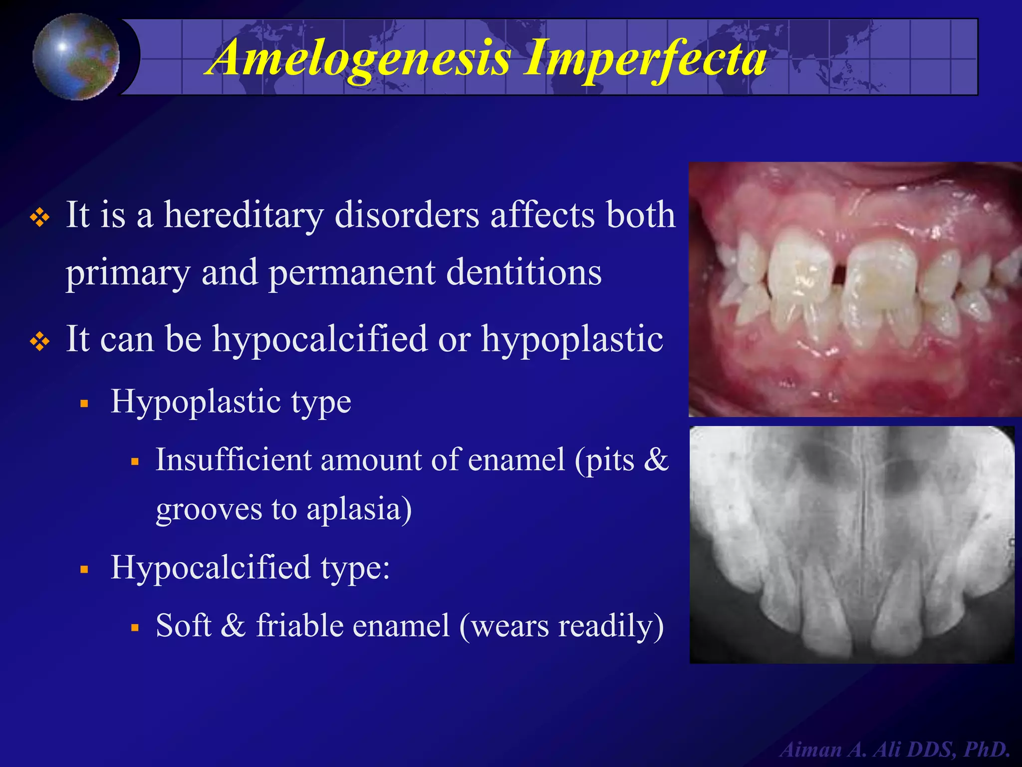
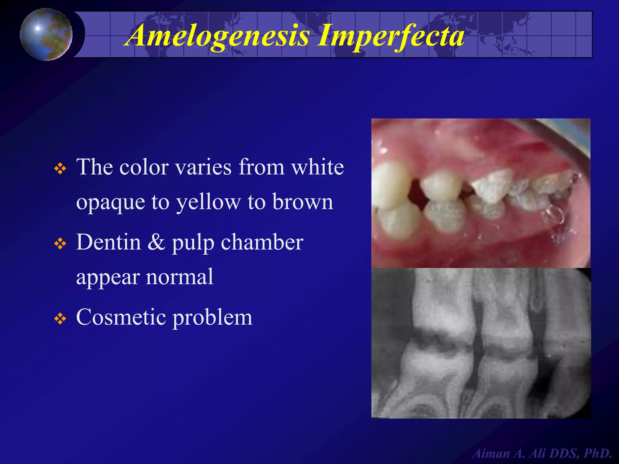
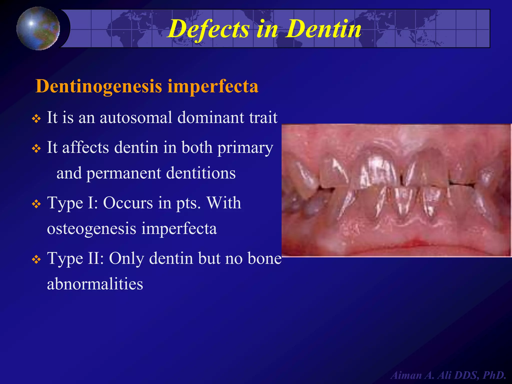
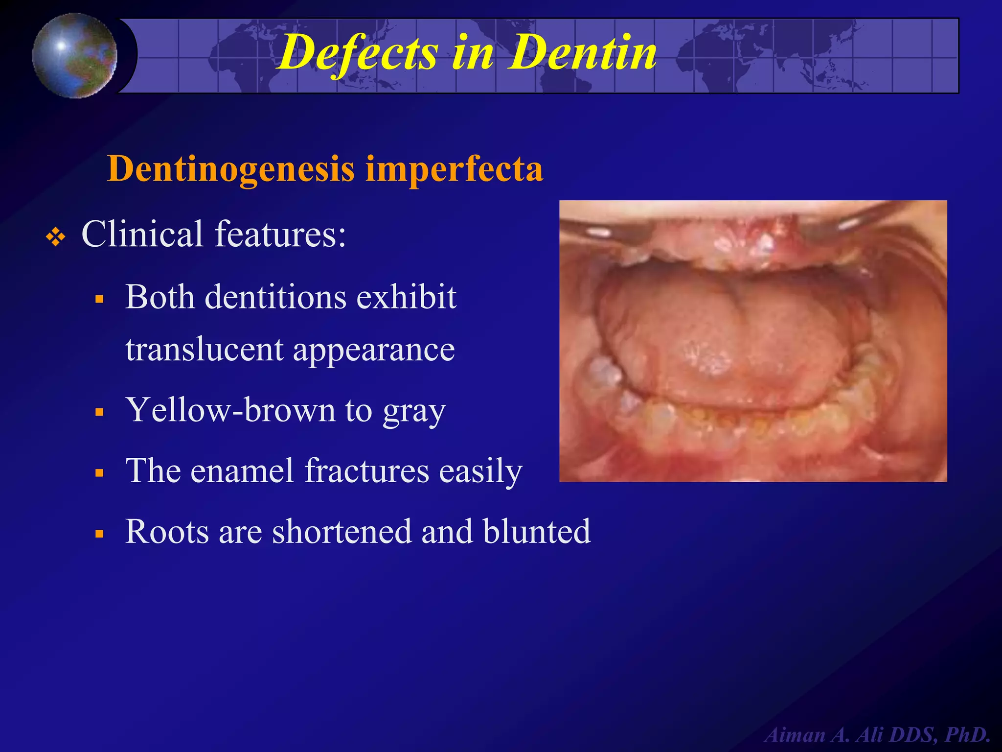
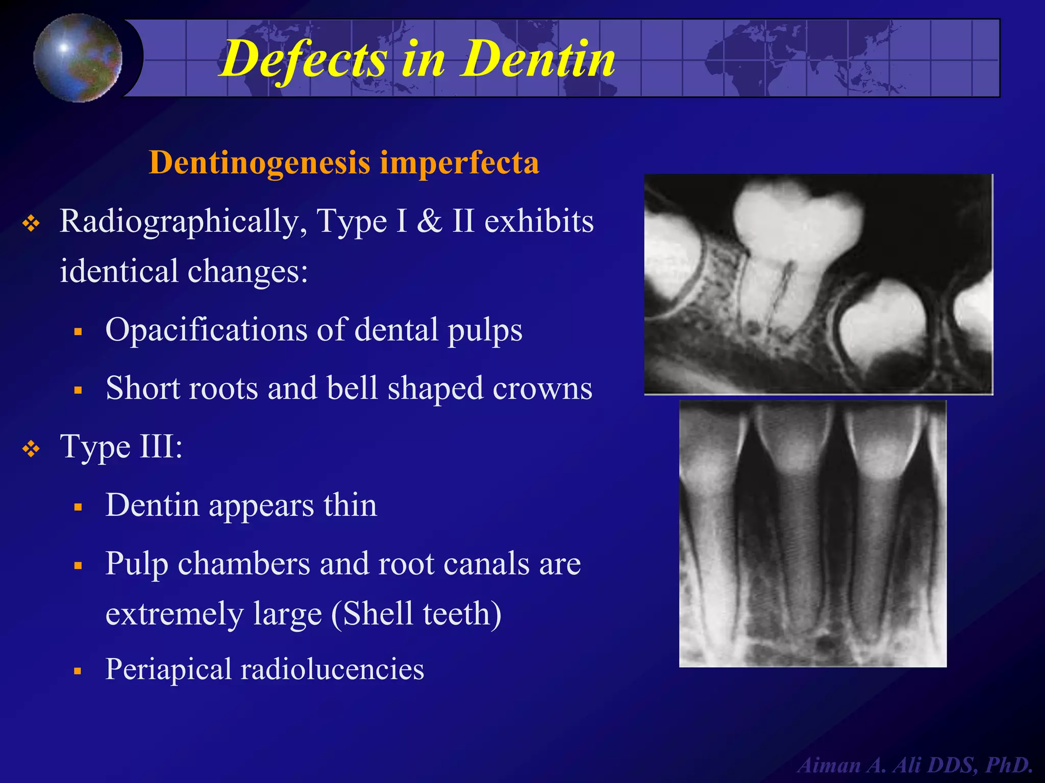
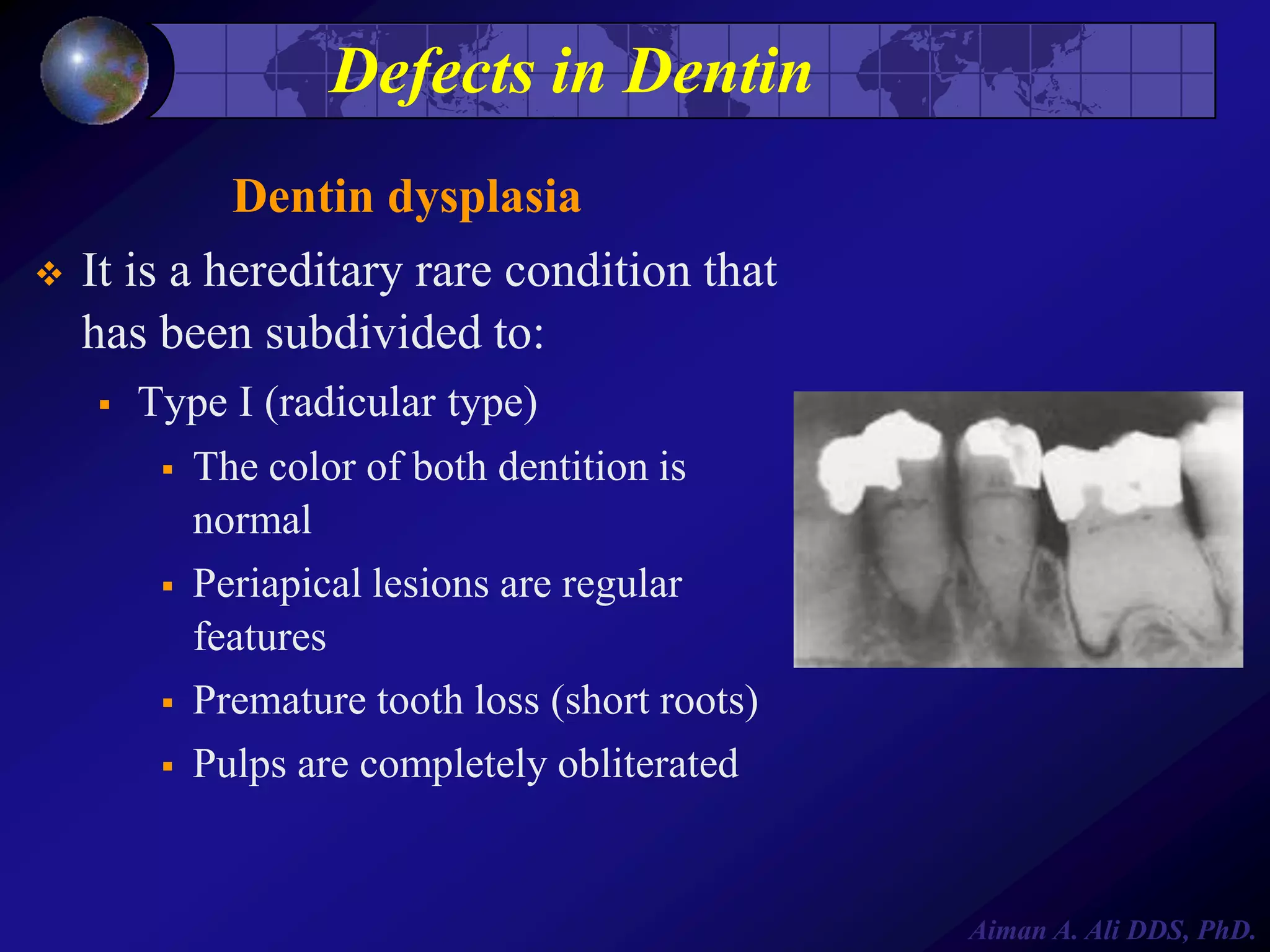
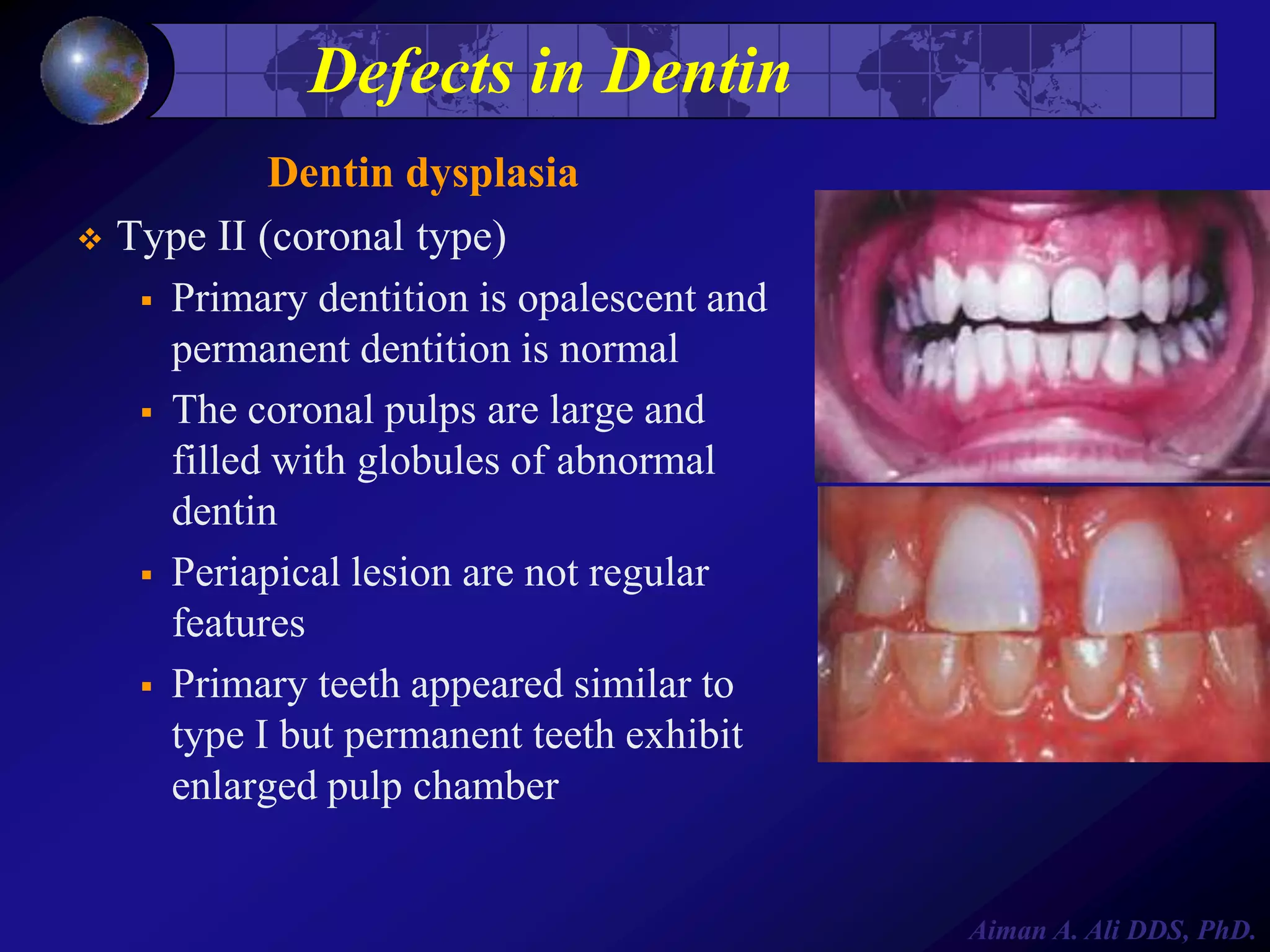
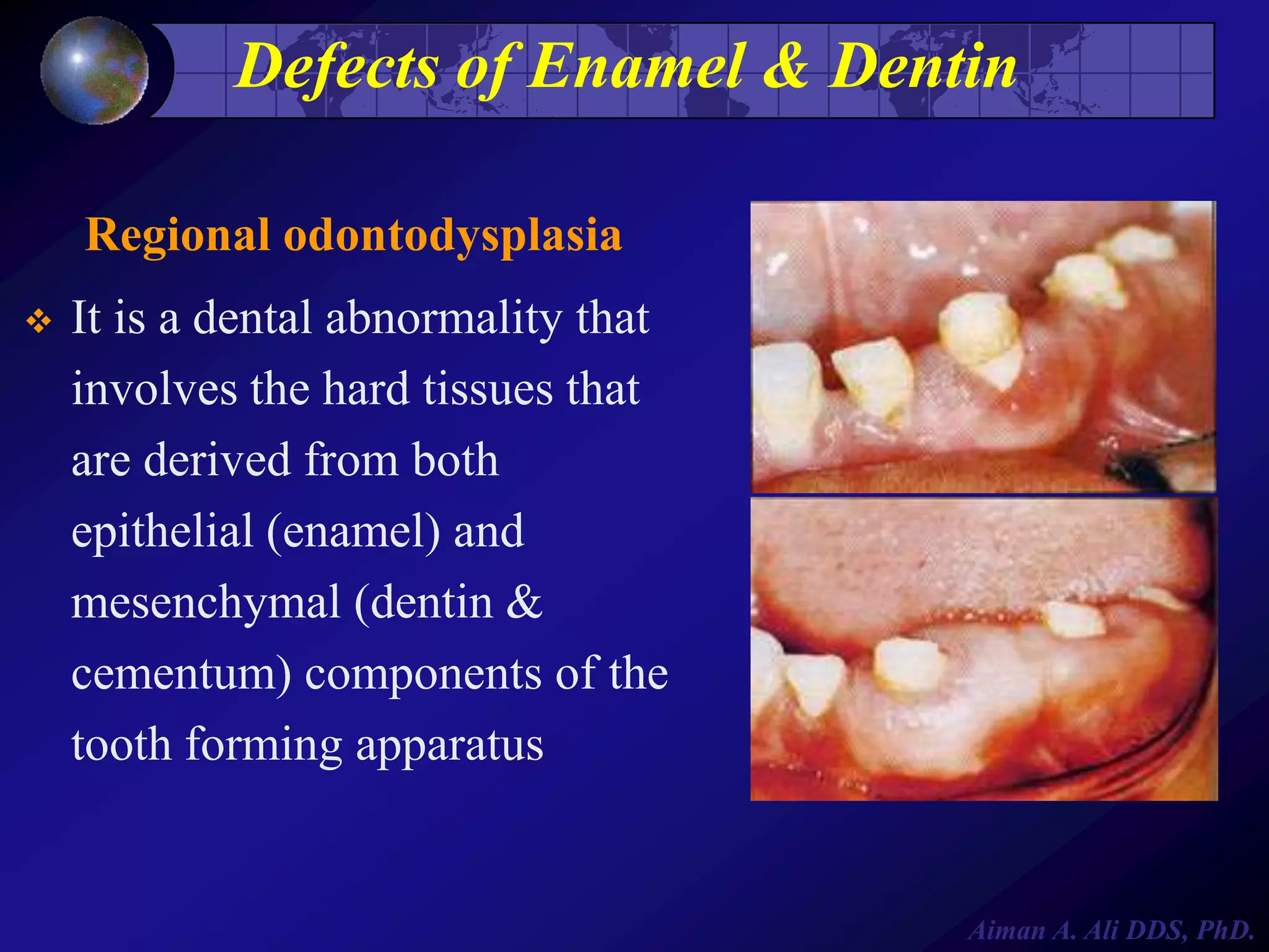
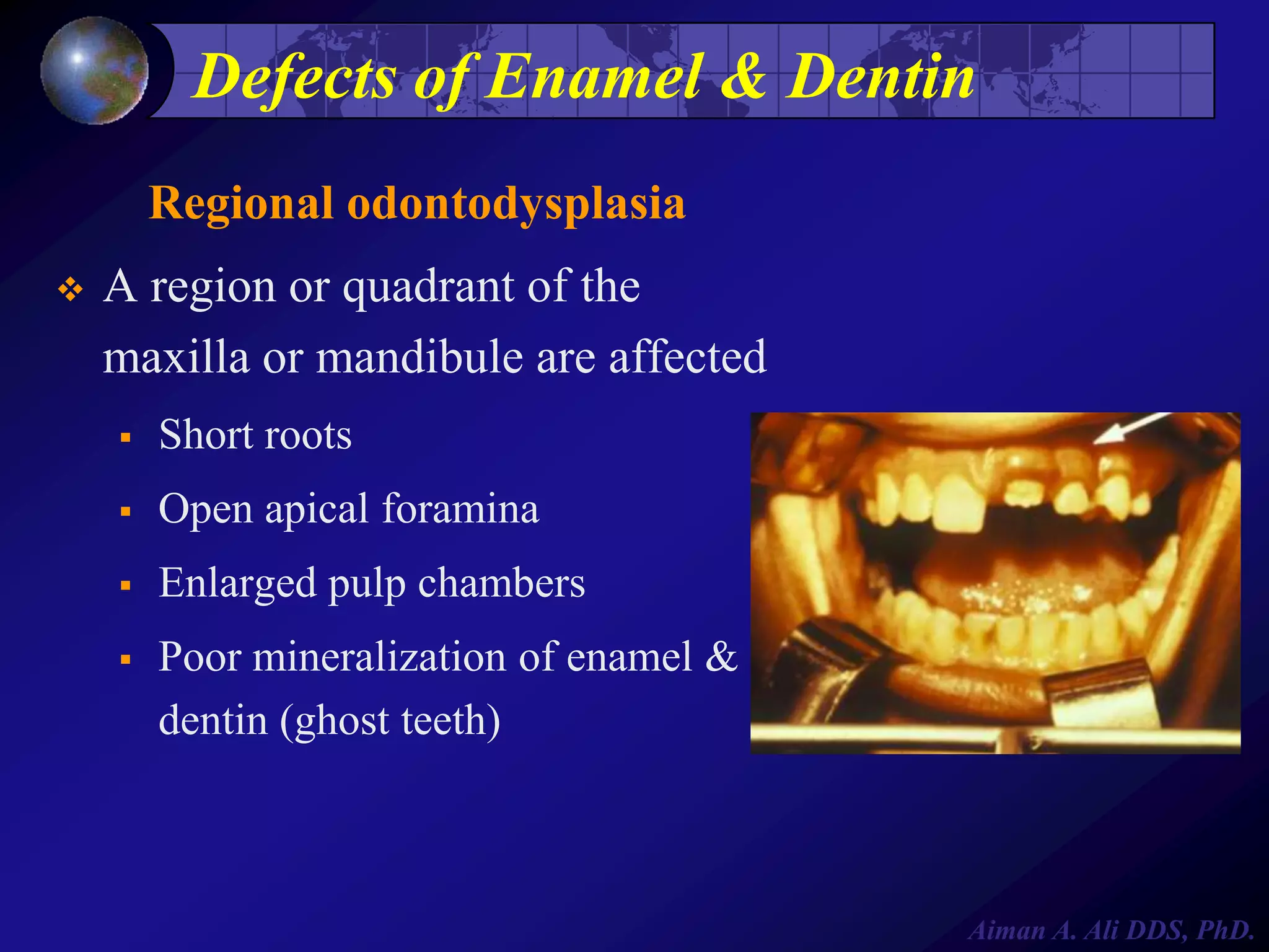
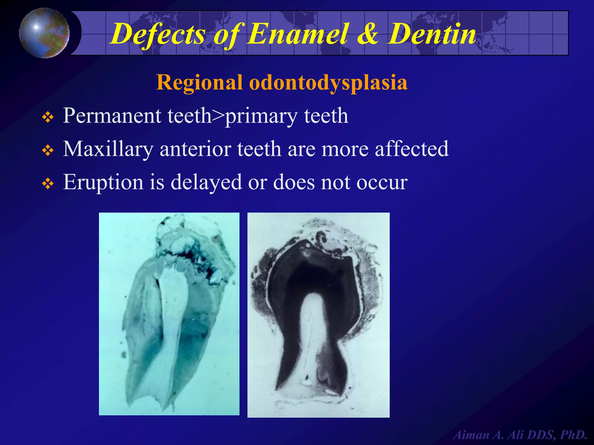
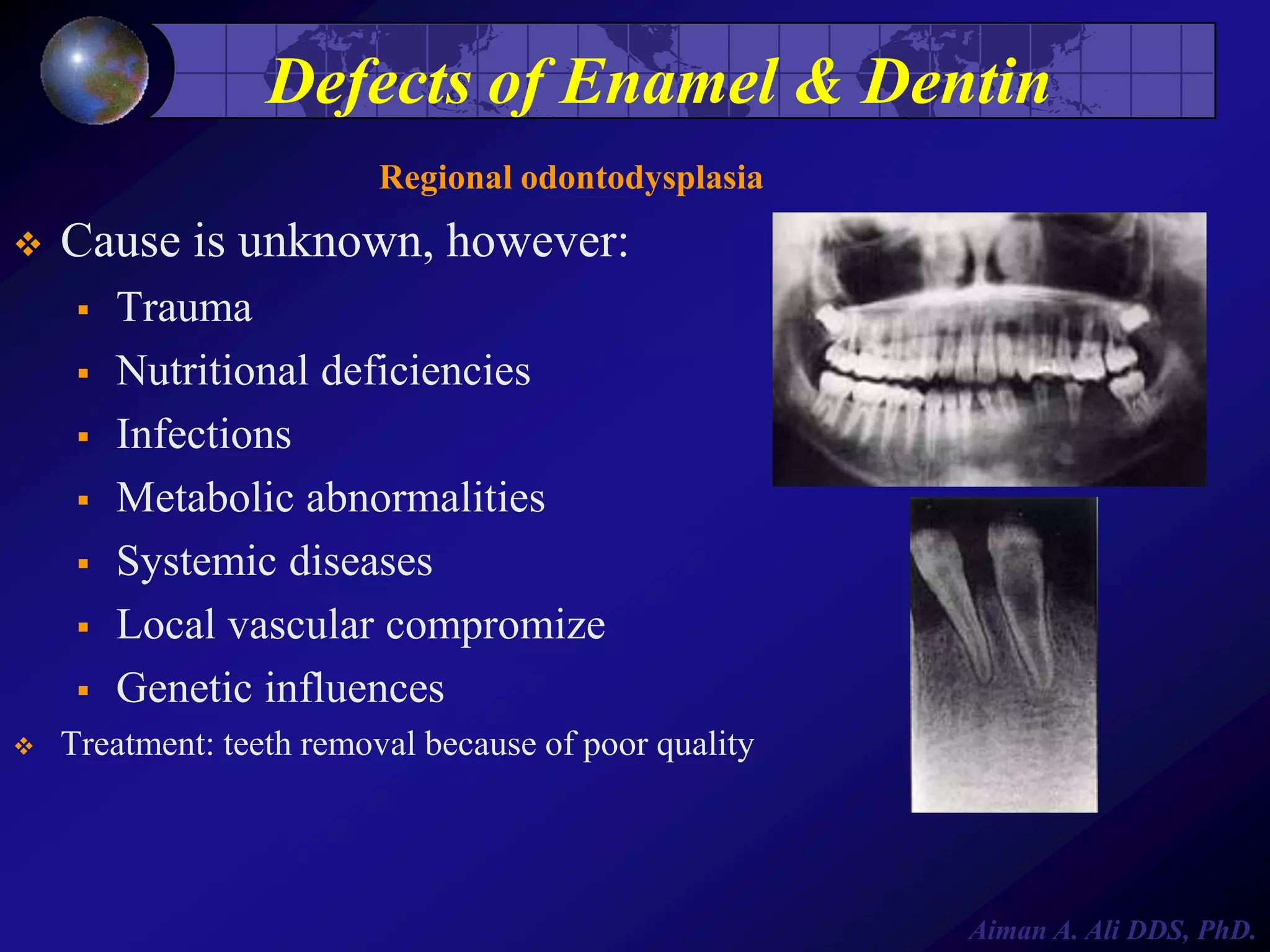
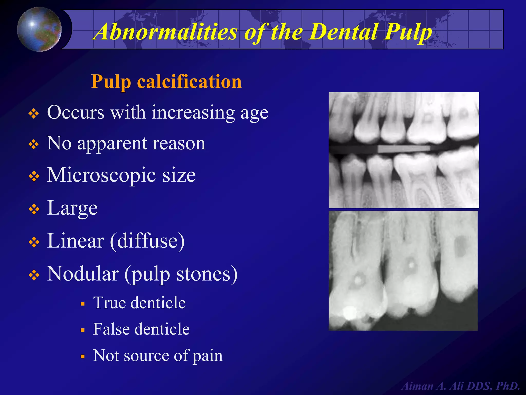
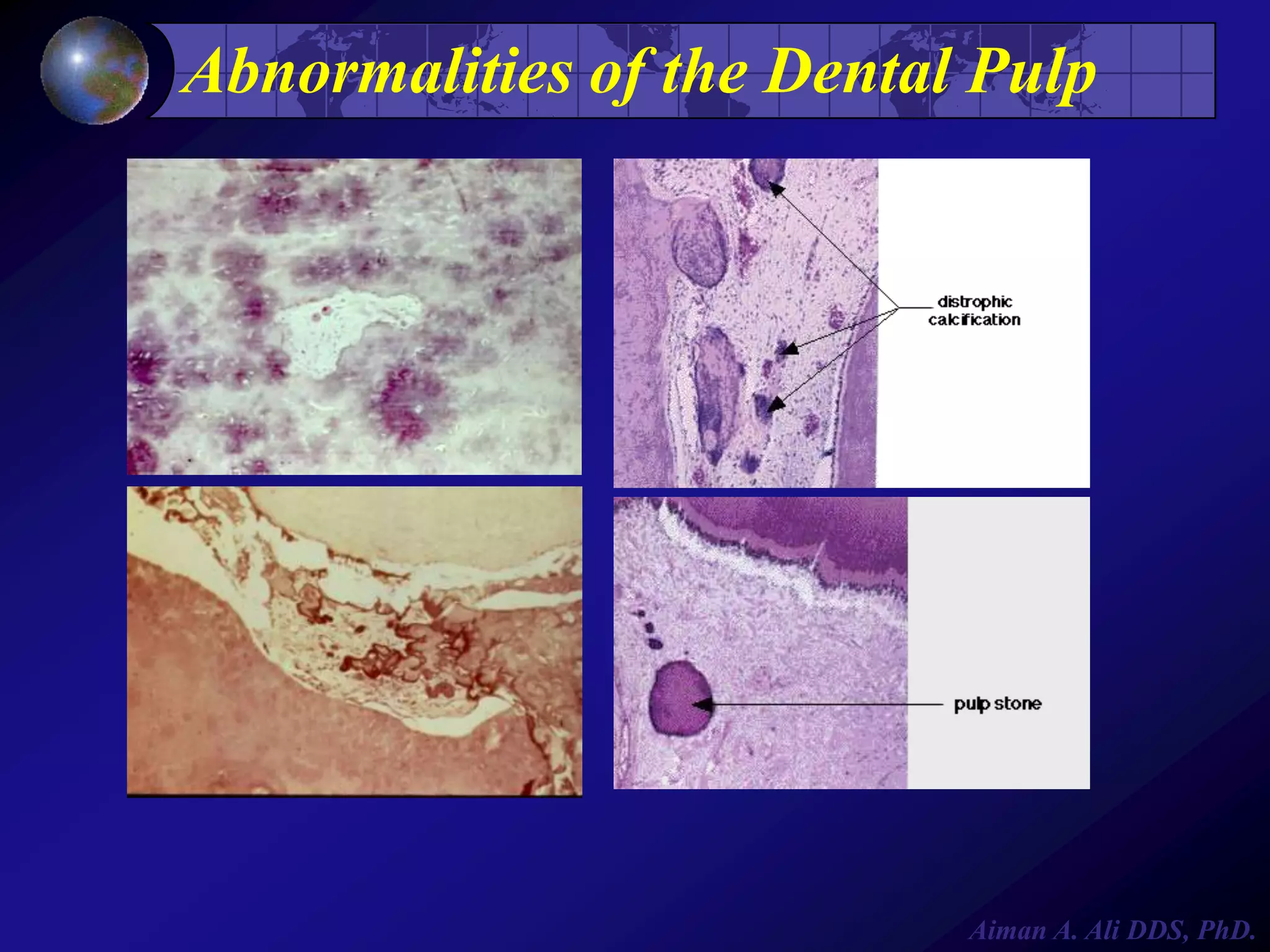
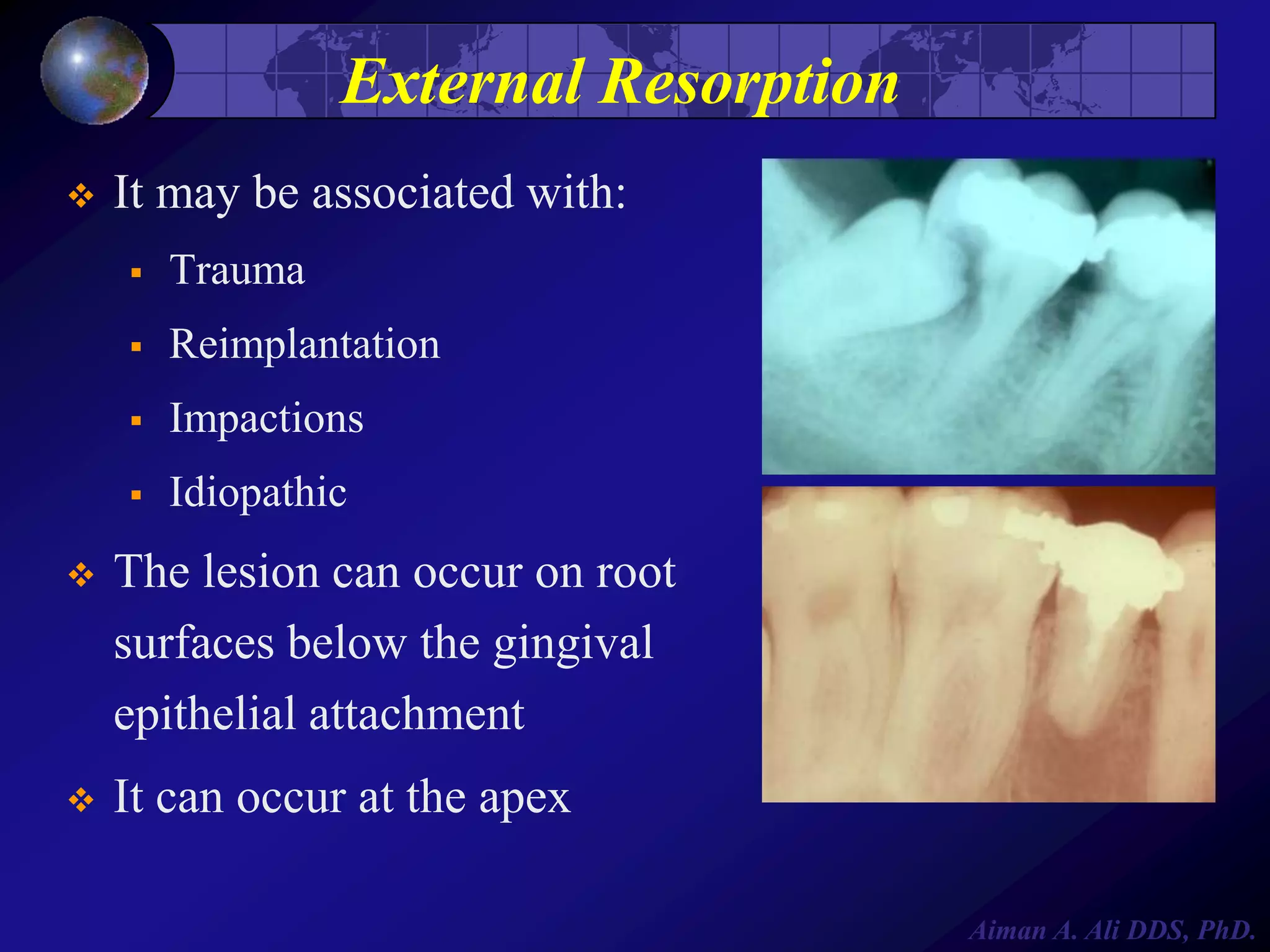
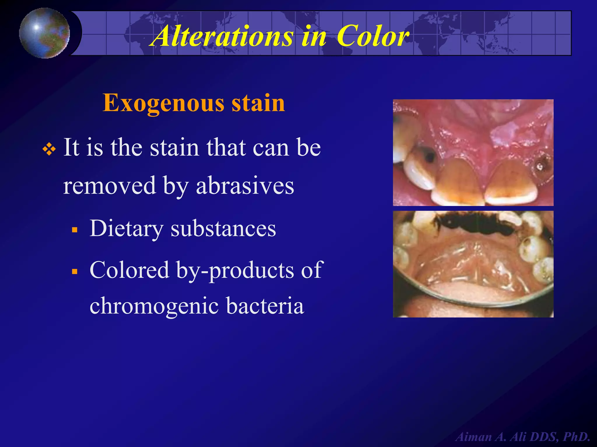
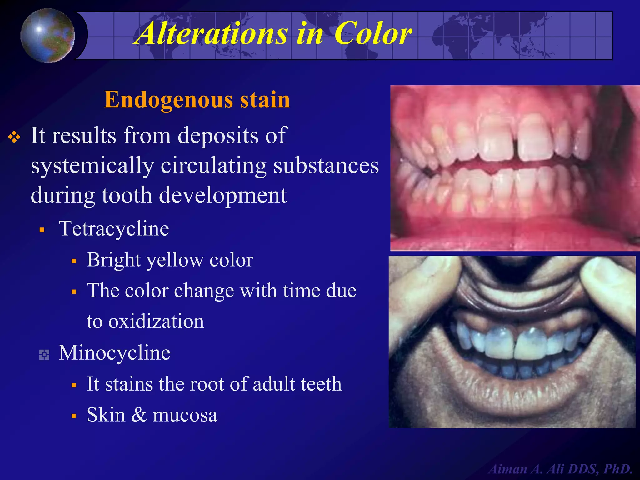
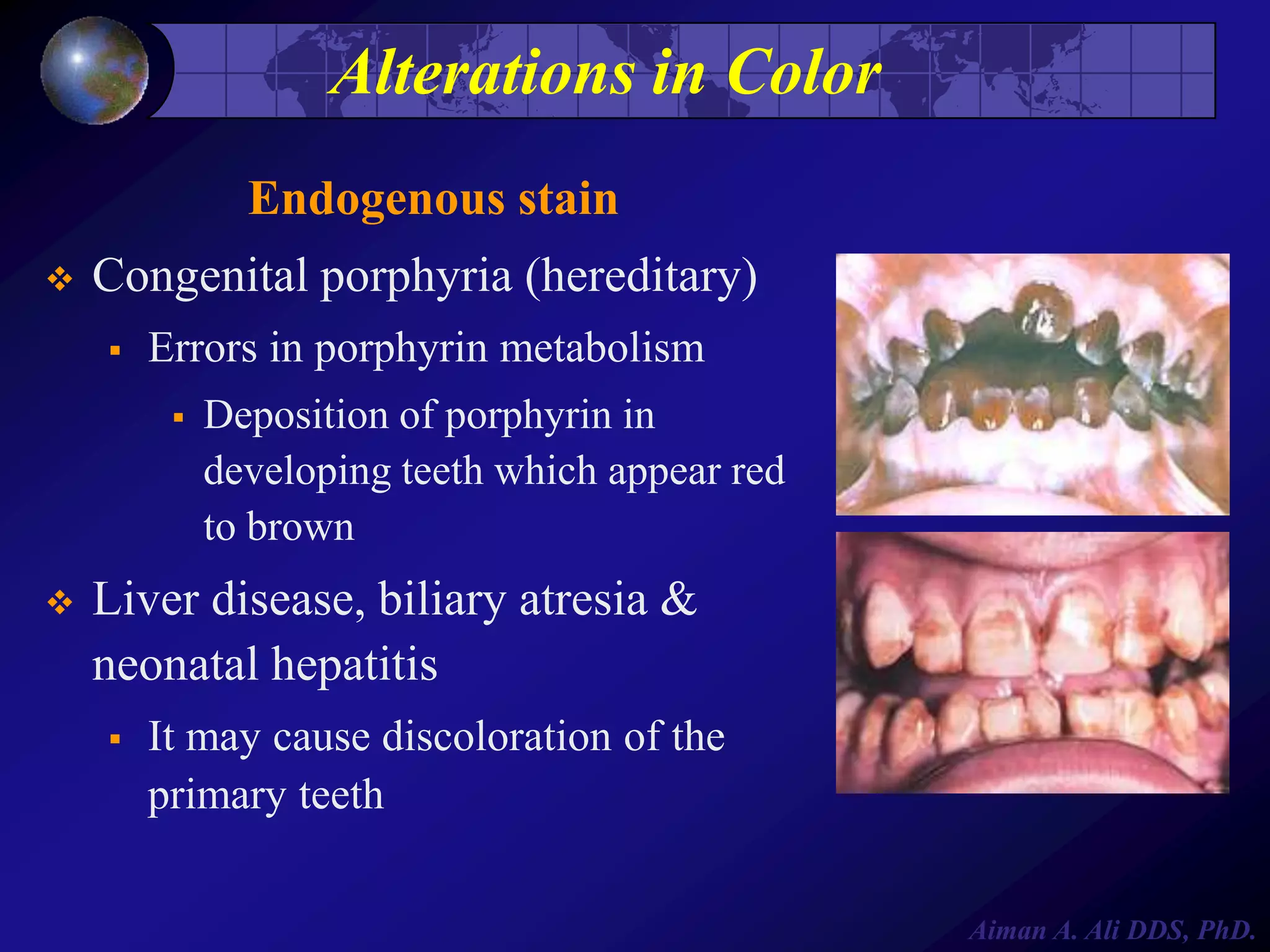
2. Oral Pathology
Oral Pathology I
Abnormalities
Dental caries
Inflammatory jaw lesions
Ulcerative lesions
Vesiculo-Bullous lesions
White lesions
Oral Pathology II
Cysts of head and neck
Odontogenic tumors
Non-Odontogenic tumors
Salivary gland diseases
---
Red blue and pigmented L.
--Aiman A. Ali DDS, PhD.
3. Oral Pathology I
Course director: Dr. Aiman A. Ali
Course contributors:
Dr Louay Jaber
Dr Wael Swelam
Theory: Saturday 1-2 pm
Laboratory: OP Lab. Saturday 2-5 pm
Aiman A. Ali DDS, PhD.
4. Course content
Week
Date
Lecturer
Topic
1
Introduction to oral pathology
2
Abnormalities of teeth
3
Dental caries
4
Inflammatory jaw lesions I
5
Inflammatory jaw lesions II
6
Pathological bases of the periodontal diseases
7
Vesiculo-Bullous disease I
8
Vesiculo-Bullous disease II
9
Ulcerative conditions I
10
Ulcerative conditions II
11
White lesions I
12
White lesions II
13
Red-Blue lesions
14
Pigmentation of oral and perioral tissues
15
Advanced diagnostic methods in oral and maxillofacial pathology
Aiman A. Ali DDS, PhD.
7. Abnormalities of teeth & oral tissues
Reference book:
• Regezi & Sciubba. Oral Pathology: clinico-pathological correlation.
• Cawson: Oral Pathology and Oral Medicine
Recommended book:
• Shafer, Hine & Levy. A text book of Oral Pathology. Chp 1.
Aiman A. Ali DDS, PhD.
8. Abnormalities of teeth
Alteration in size
Alteration in shape
Alteration in number
Alteration in structure
Alteration in color
Aiman A. Ali DDS, PhD.
9. Abnormalities of oral structures
Developmental disturbances of the jaws
Developmental disturbances of the tongue
Developmental disturbances of lip and palate
Developmental disturbances of oral mucosa
Developmental disturbances of salivary glands
Aiman A. Ali DDS, PhD.
13. Abnormalities of teeth
Alterations in shape
Gemination
Fusion
Concrescence
Dilaceration
Dens Invaginatus
Dens Evaginatus
Taurodontism
Supernumerary roots
Enamel pearls
Aiman A. Ali DDS, PhD.
20. Alteration in Shape
Taurodontism
It is a variation of tooth form.
Elongated crown
Apically displaced furcation
Increased height of pulp chamber
Associated with Down syndrome
Aiman A. Ali DDS, PhD.
22. Alteration in Shape
Taurodontism
11%
occurrence in middle east
It is of little clinical significance
No treatment is required
Aiman A. Ali DDS, PhD.
23. Alterations in Shape
Supernumerary root
Accessory roots are most
commonly seen in:
Mandibular
canines
Premolars
Molars
(thirds)
Recognition of extra-root numbers
is important for extractions or
endodontic treatment
Aiman A. Ali DDS, PhD.
25. Alterations in Shape
Enamel pearls
Droplets of ectopic enamel, they
occur most commonly on
The bi-or tri-furcation of teeth
Maxillary & mandibular molars
It may be detected on radiographic
examinations
It is of significance when
periodontal disease is present
Aiman A. Ali DDS, PhD.
27. Attrition, Abrasion & Erosion
Attrition: The loss of tooth structure from tooth
to tooth contact
Abrasion: The loss of tooth structure due to
repeated mechanical contact with objects
other than teeth
Erosion: Non-carious loss of tooth structure
due to chemical dissolution not related to
acid produced by dental plaque
Aiman A. Ali DDS, PhD.
29. Alterations in Shape
Attrition
It is a physiologic wearing of
teeth as a result of
mastication
It is an age related process &
varies from one individual to
another
Diet, dentition, musculature
& chewing habits can
influence the pattern of
attrition
Aiman A. Ali DDS, PhD.
30. Alterations in Shape
Abrasion
It is the pathologic wearing of
teeth as a result of abnormal habit
(abrasive substance)
Pipe smoking
Tobacco chewing
Aggressive toothbrushing
Abrasive dentifrices
Aiman A. Ali DDS, PhD.
31. Alterations in Shape
Erosion
It is a loss of tooth structure from
a non-bacterial chemical process
Acids
External: work environment or
in the diet
Internal: regurgitation of gastric
contents (chronic vomiting)
In many cases of tooth erosion,
no cause is found
Aiman A. Ali DDS, PhD.
32. Alterations in Number
Anodontia
It is an absence of teeth that is often
associated with hereditary ectodermal
dysplasia
It is caused by polygenic
(enviromental & genetic factors)
Complete Anodontia:
All teeth are missing
The prevalence of hypodontia is 4.6%
Partial Anodontia:
some teeth are missing
Aiman A. Ali DDS, PhD.
34. Alterations in Number
Anodontia
Partial
anodontia (hypodontia):
one or several teeth are missing
Pseudoanodontia:
when teeth are
absent clinically
Most
commonly seen in third
molars, second premolars &
maxillary lateral incisors
Aiman A. Ali DDS, PhD.
37. Alterations in Number
Impaction
It is most commonly seen in
mandibular third molars &
maxillary cuspids, it occurs
because of:
Crowding
Physical barrier
Abnormal eruption path
Ankylosis (fusion of tooth to
alveolar bone)
Aiman A. Ali DDS, PhD.
39. Alterations in Number
Supernumerary teeth
It results from continued proliferation
of permanent or primary dental lamina
The tooth can be rudimentary &
miniature
It can be an isolated event, familial or
associated with syndromes
Aiman A. Ali DDS, PhD.
40. Alterations in Number
Supernumerary teeth
Clinical significance:
Occupy space
Block eruption
Delay or mal-eruption
Natal teeth
Post-permanent dentition
Maxilla>mandible: 10:1
Anterior midline of maxilla
(mesiodens)
Fourth molar
Aiman A. Ali DDS, PhD.
41. Alterations in tooth structure
Enamel
Hereditary: Amelogenesis Imperfecta
Environmental: Trauma, Fluoride, Syphilis …etc.
Dentine
Hereditary: Dentinogenesis Imperfecta
All dental hard tissues
Dentine dysplasia
Hereditary: Regional odontodyslasia
Environmental: Resorption
Pulp
Aiman A. Ali DDS, PhD.
42. Defects of Enamel
Environmental defects of enamel
Severe metabolic injury can cause:
Defects in the quantity & shape
Enamel hypoplasia
Defects in the quality and color
Enamel hypocalcification
Enamel hypoplasia
Aiman A. Ali DDS, PhD.
43. Defects of Enamel
Environmental defects of enamel
The extent of the defect is dependent on:
The intensity of the etiologic factor
Enamel hypoplasia
The duration
The time of occurrence
Aiman A. Ali DDS, PhD.
44. Defects of Enamel
Environmental defects of enamel
Etiologic factors can be
Local: (Turner’s tooth)
Trauma
Abscess
Clinical signs
Hypocalcification or hypoplasia
Aiman A. Ali DDS, PhD.
45. Environmental Defects of Enamel
Research have shown that causes
are attributed to
infectious diseases
Nutritional defects (rickets)
Congenital syphilis
Birth trauma (neonatal line)
Fluoride (hypoplasia or
hypocalcification)
Idiopathic factors
Aiman A. Ali DDS, PhD.
51. Amelogenesis Imperfecta
It is a hereditary disorders affects both
primary and permanent dentitions
It can be hypocalcified or hypoplastic
Hypoplastic type
Insufficient amount of enamel (pits &
grooves to aplasia)
Hypocalcified type:
Soft & friable enamel (wears readily)
Aiman A. Ali DDS, PhD.
52. Amelogenesis Imperfecta
The color varies from white
opaque to yellow to brown
Dentin & pulp chamber
appear normal
Cosmetic problem
Aiman A. Ali DDS, PhD.
53. Defects in Dentin
Dentinogenesis imperfecta
It is an autosomal dominant trait
It affects dentin in both primary
and permanent dentitions
Type I: Occurs in pts. With
osteogenesis imperfecta
Type II: Only dentin but no bone
abnormalities
Aiman A. Ali DDS, PhD.
54. Defects in Dentin
Dentinogenesis imperfecta
Type III: Only dental defects occur
• Pulp exposures
• Periapical radiolucencies
• Variable radiographic appearance
Aiman A. Ali DDS, PhD.
55. Defects in Dentin
Dentinogenesis imperfecta
Clinical features:
Both dentitions exhibit
translucent appearance
Yellow-brown to gray
The enamel fractures easily
Roots are shortened and blunted
Aiman A. Ali DDS, PhD.
56. Defects in Dentin
Dentinogenesis imperfecta
Radiographically, Type I & II exhibits
identical changes:
Opacifications of dental pulps
Short roots and bell shaped crowns
Type III:
Dentin appears thin
Pulp chambers and root canals are
extremely large (Shell teeth)
Periapical radiolucencies
Aiman A. Ali DDS, PhD.
57. Defects in Dentin
Dentinogenesis imperfecta
Microscopically:
Pulp replaced by irregular dentin
The dentin contains fewer but larger and irregular
dentinal tubules
Enamel appears normal but dentinoenamel junction is
smooth
Treatment is directed toward protecting tooth from
wear and esthetic appearance
Aiman A. Ali DDS, PhD.
58. Defects in Dentin
Dentin dysplasia
It is a hereditary rare condition that
has been subdivided to:
Type I (radicular type)
The color of both dentition is
normal
Periapical lesions are regular
features
Premature tooth loss (short roots)
Pulps are completely obliterated
Aiman A. Ali DDS, PhD.
59. Defects in Dentin
Dentin dysplasia
Type II (coronal type)
Primary dentition is opalescent and
permanent dentition is normal
The coronal pulps are large and
filled with globules of abnormal
dentin
Periapical lesion are not regular
features
Primary teeth appeared similar to
type I but permanent teeth exhibit
enlarged pulp chamber
Aiman A. Ali DDS, PhD.
60. Defects in Dentin
Dentin Dysplasia
Microscopically:
Enamel and immediately subjacent dentin appear normal
Deeper layers of dentin shows atypical tubular patterns &
amorphous, atubular areas and irregular organization
Treatment is directed toward retention of teeth but
prognosis is poor
Aiman A. Ali DDS, PhD.
61. Defects of Enamel & Dentin
Regional odontodysplasia
It is a dental abnormality that
involves the hard tissues that
are derived from both
epithelial (enamel) and
mesenchymal (dentin &
cementum) components of the
tooth forming apparatus
Aiman A. Ali DDS, PhD.
62. Defects of Enamel & Dentin
Regional odontodysplasia
A region or quadrant of the
maxilla or mandibule are affected
Short roots
Open apical foramina
Enlarged pulp chambers
Poor mineralization of enamel &
dentin (ghost teeth)
Aiman A. Ali DDS, PhD.
63. Defects of Enamel & Dentin
Regional odontodysplasia
Permanent teeth>primary teeth
Maxillary anterior teeth are more affected
Eruption is delayed or does not occur
Aiman A. Ali DDS, PhD.
64. Defects of Enamel & Dentin
Regional odontodysplasia
Cause is unknown, however:
Trauma
Nutritional deficiencies
Infections
Metabolic abnormalities
Systemic diseases
Local vascular compromize
Genetic influences
Treatment: teeth removal because of poor quality
Aiman A. Ali DDS, PhD.
65. Abnormalities of the Dental Pulp
Pulp calcification
Occurs with increasing age
No apparent reason
Microscopic size
Large
Linear (diffuse)
Nodular (pulp stones)
True denticle
False denticle
Not source of pain
Aiman A. Ali DDS, PhD.
67. Internal Resorption
It may be seen as
Inflammatory response to pulpal injury
No apparent trigger
It occurs as a result of osteoclasts
activation on internal surfaces
Aiman A. Ali DDS, PhD.
68. Internal Resorption
The root or crown can be perforated
In advanced cases teeth may appear pink
Treatment is root canal therapy before
perforation
Aiman A. Ali DDS, PhD.
69. External Resorption
It may be a result of an adjacent pathologic process
Chronic inflammatory lesion
Cysts
Benign tumors
Malignant neoplasms
Aiman A. Ali DDS, PhD.
70. External Resorption
The cause has been related to:
Release of chemical mediators
Increased vascularity
Pressure
Aiman A. Ali DDS, PhD.
71. External Resorption
It may be associated with:
Trauma
Reimplantation
Impactions
Idiopathic
The lesion can occur on root
surfaces below the gingival
epithelial attachment
It can occur at the apex
Aiman A. Ali DDS, PhD.
72. Alterations in Color
Exogenous stain
It is the stain that can be
removed by abrasives
Dietary substances
Colored by-products of
chromogenic bacteria
Aiman A. Ali DDS, PhD.
73. Alterations in Color
Endogenous stain
It results from deposits of
systemically circulating substances
during tooth development
Tetracycline
Bright yellow color
The color change with time due
to oxidization
Minocycline
It stains the root of adult teeth
Skin & mucosa
Aiman A. Ali DDS, PhD.
74. Alterations in Color
Endogenous stain
Congenital porphyria (hereditary)
Errors in porphyrin metabolism
Deposition of porphyrin in
developing teeth which appear red
to brown
Liver disease, biliary atresia &
neonatal hepatitis
It may cause discoloration of the
primary teeth
Aiman A. Ali DDS, PhD.
77. Abnormalities of oral structures
Developmental disturbances of the jaws
Developmental disturbances of the tongue
Developmental disturbances of lip and palate
Developmental disturbances of oral mucosa
Developmental disturbances of salivary glands
Aiman A. Ali DDS, PhD.
78. Developmental disturbances of the
jaws
Agnathia
Micrognathia
Macrognathia
Facial Hemihypertrophy
Facial Hemiatrophy
Aiman A. Ali DDS, PhD.
79. Developmental disturbances of
the tongue
Aglossia
Microglossia
Macroglossia
Ankyloglossia
Cleft, bifid and fissured tongue
Lingual thyroid nodule
Median rhomboid glossitis
Aiman A. Ali DDS, PhD.