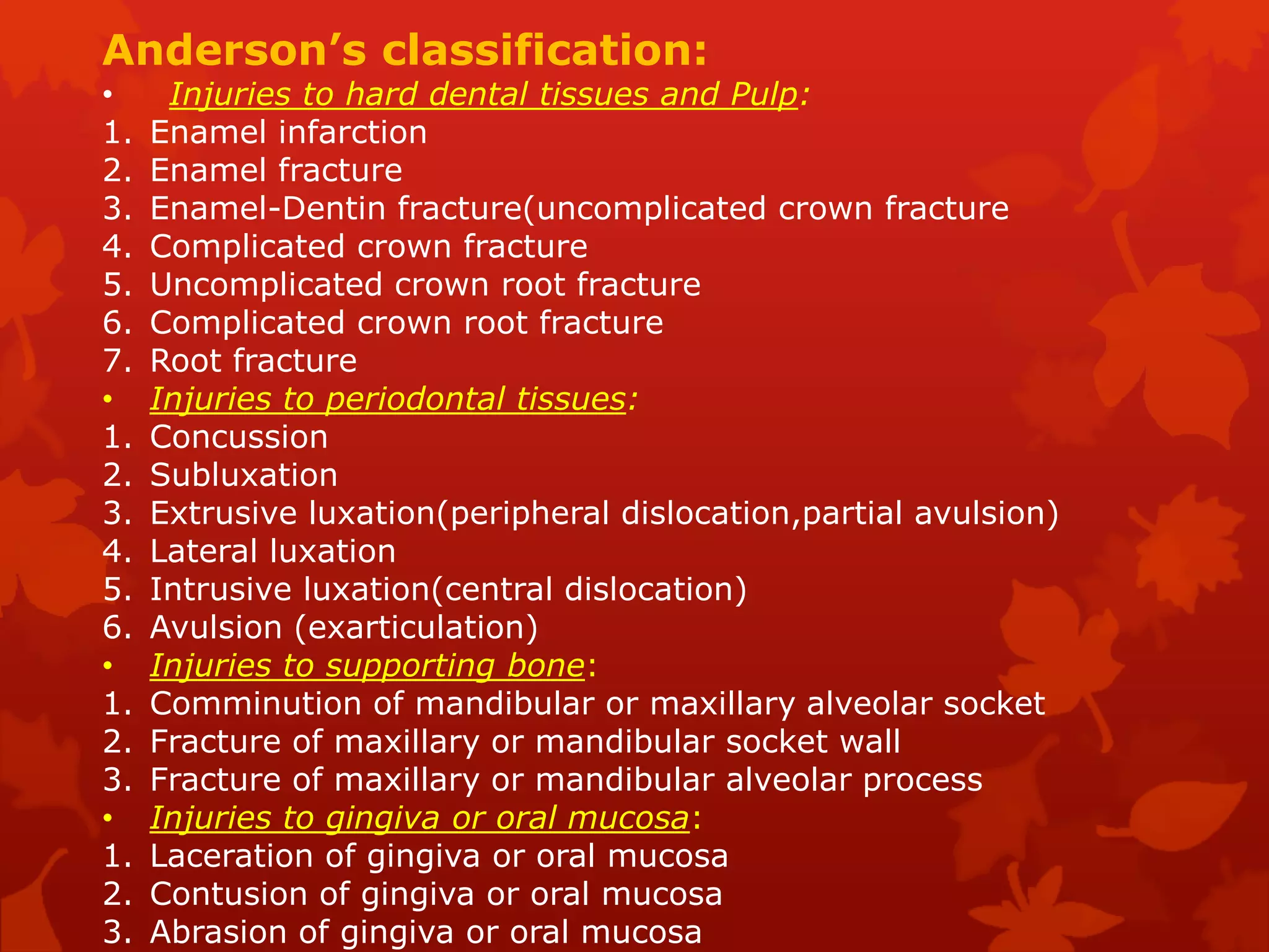
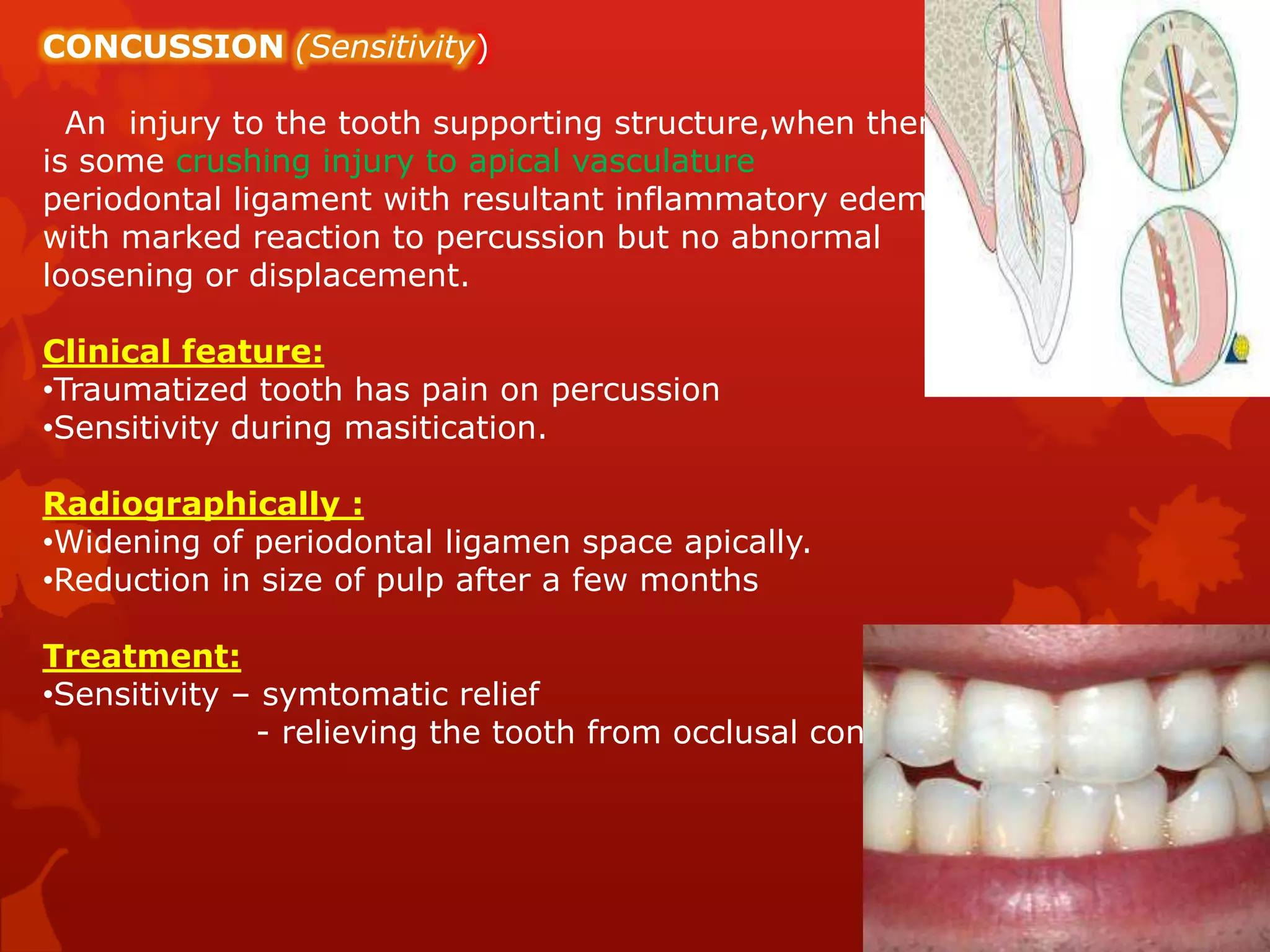
The document summarizes traumatic dental injuries and their management. It discusses the classification, clinical features, treatment, and stabilization periods for various types of dentoalveolar injuries including enamel fractures, crown fractures, root fractures, luxations, and avulsions. Splinting is described as the best method for immobilizing mobile teeth or displaced teeth, with different splinting techniques and materials discussed. Prompt treatment of dental trauma is emphasized to save injured teeth.
Apexogenesis & apexification in pediatric dentistryDr. Harsh Shah
SDDCH Parbhani
Presented by : Vipul GIratkar
Dept. of Pediatric dentitstry
Guided by . Dr. Rehan Khan
DIscussion regarding apexification and apexogenesis
One of the most painful but easy-to-treat dental emergencies is a dry socket.
• Dry socket symptoms are experienced after a tooth extraction.
• This condition requires follow-up care by the doctor who performed the surgery, an oral surgeon or a dentist who is familiar with how to treat it.
For more information, contact :-
Dr Sachdeva's Dental Aesthetic And Implant Institute,
I 101, Ashok Vihar Phase 1, Delhi- 110052
Contact us at
• Phone : +919818894041,01142464041
• Our Websites:
• www.sachdevadentalcare.com
• www.dentalclinicindelhi.com
• www.dentalimplantindia.co.in
• www.dentalcoursesdelhi.com
• www.facialaestheticsdelhi.com
#drysocket #management #thirdmolarextraction #extractioncomplications
In this lecture I explain in step-by-step fashion the basics of Endodontic Diagnosis: Pulp Vitality Tests. a photo guide is attached to the guide to aid in better understanding of the topic
DR. SWARNEET KAKPURE (DEPT OF CONSERVATIVE DENTISTRY AND ENDODONTICS)
THE TOPIC PRESENTED IN SEMINAR COVERS ALMOST ALL THE ASPECTS OF COMPLEX AMALGAM RESTORATIONS INCLUDING PIN RETAINED,SLOT RETAINED AMALGAM RESTORATIONS,CEMENTED,FRICTION LOCKED & SELF THREADING PINS, TMS SYSTEM,AMALGAM FOUNDATIONS ALONG WITH TECHNIQUES OF INSERTION AND MATRIX PLACEMENT.
The Indian Dental Academy is the Leader in continuing dental education , training dentists in all aspects of dentistry and
offering a wide range of dental certified courses in different formats.
The Indian Dental Academy is the Leader in continuing dental education , training dentists in all aspects of dentistry and
offering a wide range of dental certified courses in different formats.
Traumatic Dental Injuries to Permanent Anterior Teeth, Relation with Age and...Abu-Hussein Muhamad
Traumatic dental injury (TDI) in school children has become a serious dental public health problem in developing and developed countries. Worldwide research clearly shows that the prevalence of TDI is increasing. The purpose of this study is to clinical compare without use radiograph film in comparing between groups of boys and girls which complain of traumatic fracture of anterior permanent teeth in enamel and dentine reigns with or with out Pulp rom records of patients aged 9-12years. A descriptive cross-sectional survey in Arab Israeli schoolchildren between 2012-2015. A sample of 4262, Arab Israeli schoolchildren (2344(55%)) males and 1918(45%)) females) aged 9-12years, were interviewed and examined between 2012-2015 in different dental private clinics in Israel. Among the 4262 schoolchildren examined, 520(12,2%) had experienced traumatic dental injuries (TDIs). Males had experienced a significantly higher prevalence of trauma 340(8%) than females 180(4,2%). Overall traumatised permanent incisors were found to occur fairly frequently with males having experienced significantly more TDIs than females. The prevalence of TDIs in Arab Israeli schoolchildren was 12,2%; enamel fractures were the most frequently observed injury and falls were the leading cause of trauma.
Dentoalveolar compensations /certified fixed orthodontic courses by Indian de...Indian dental academy
The Indian Dental Academy is the Leader in continuing dental education , training dentists in all aspects of dentistry and offering a wide range of dental certified courses in different formats.
Traumatized Teeth
Copyright by Dr. Khin Swe Aye
Department of Conservative Dentistry
University of Dental Medicine, Yangon
Feel free to request to take it down this slide if you are copyright owner.
Dental trauma is one of the most common presentation in the pediatrics clinic. The fears and anxiety of these patients make management difficult. If improperly managed, it could affect the patient self-esteem and quality of life.
Knee anatomy and clinical tests 2024.pdfvimalpl1234
This includes all relevant anatomy and clinical tests compiled from standard textbooks, Campbell,netter etc..It is comprehensive and best suited for orthopaedicians and orthopaedic residents.
Ozempic: Preoperative Management of Patients on GLP-1 Receptor Agonists Saeid Safari
Preoperative Management of Patients on GLP-1 Receptor Agonists like Ozempic and Semiglutide
ASA GUIDELINE
NYSORA Guideline
2 Case Reports of Gastric Ultrasound
Tom Selleck Health: A Comprehensive Look at the Iconic Actor’s Wellness Journeygreendigital
Tom Selleck, an enduring figure in Hollywood. has captivated audiences for decades with his rugged charm, iconic moustache. and memorable roles in television and film. From his breakout role as Thomas Magnum in Magnum P.I. to his current portrayal of Frank Reagan in Blue Bloods. Selleck's career has spanned over 50 years. But beyond his professional achievements. fans have often been curious about Tom Selleck Health. especially as he has aged in the public eye.
Follow us on: Pinterest
Introduction
Many have been interested in Tom Selleck health. not only because of his enduring presence on screen but also because of the challenges. and lifestyle choices he has faced and made over the years. This article delves into the various aspects of Tom Selleck health. exploring his fitness regimen, diet, mental health. and the challenges he has encountered as he ages. We'll look at how he maintains his well-being. the health issues he has faced, and his approach to ageing .
Early Life and Career
Childhood and Athletic Beginnings
Tom Selleck was born on January 29, 1945, in Detroit, Michigan, and grew up in Sherman Oaks, California. From an early age, he was involved in sports, particularly basketball. which played a significant role in his physical development. His athletic pursuits continued into college. where he attended the University of Southern California (USC) on a basketball scholarship. This early involvement in sports laid a strong foundation for his physical health and disciplined lifestyle.
Transition to Acting
Selleck's transition from an athlete to an actor came with its physical demands. His first significant role in "Magnum P.I." required him to perform various stunts and maintain a fit appearance. This role, which he played from 1980 to 1988. necessitated a rigorous fitness routine to meet the show's demands. setting the stage for his long-term commitment to health and wellness.
Fitness Regimen
Workout Routine
Tom Selleck health and fitness regimen has evolved. adapting to his changing roles and age. During his "Magnum, P.I." days. Selleck's workouts were intense and focused on building and maintaining muscle mass. His routine included weightlifting, cardiovascular exercises. and specific training for the stunts he performed on the show.
Selleck adjusted his fitness routine as he aged to suit his body's needs. Today, his workouts focus on maintaining flexibility, strength, and cardiovascular health. He incorporates low-impact exercises such as swimming, walking, and light weightlifting. This balanced approach helps him stay fit without putting undue strain on his joints and muscles.
Importance of Flexibility and Mobility
In recent years, Selleck has emphasized the importance of flexibility and mobility in his fitness regimen. Understanding the natural decline in muscle mass and joint flexibility with age. he includes stretching and yoga in his routine. These practices help prevent injuries, improve posture, and maintain mobilit
These lecture slides, by Dr Sidra Arshad, offer a quick overview of the physiological basis of a normal electrocardiogram.
Learning objectives:
1. Define an electrocardiogram (ECG) and electrocardiography
2. Describe how dipoles generated by the heart produce the waveforms of the ECG
3. Describe the components of a normal electrocardiogram of a typical bipolar lead (limb II)
4. Differentiate between intervals and segments
5. Enlist some common indications for obtaining an ECG
6. Describe the flow of current around the heart during the cardiac cycle
7. Discuss the placement and polarity of the leads of electrocardiograph
8. Describe the normal electrocardiograms recorded from the limb leads and explain the physiological basis of the different records that are obtained
9. Define mean electrical vector (axis) of the heart and give the normal range
10. Define the mean QRS vector
11. Describe the axes of leads (hexagonal reference system)
12. Comprehend the vectorial analysis of the normal ECG
13. Determine the mean electrical axis of the ventricular QRS and appreciate the mean axis deviation
14. Explain the concepts of current of injury, J point, and their significance
Study Resources:
1. Chapter 11, Guyton and Hall Textbook of Medical Physiology, 14th edition
2. Chapter 9, Human Physiology - From Cells to Systems, Lauralee Sherwood, 9th edition
3. Chapter 29, Ganong’s Review of Medical Physiology, 26th edition
4. Electrocardiogram, StatPearls - https://www.ncbi.nlm.nih.gov/books/NBK549803/
5. ECG in Medical Practice by ABM Abdullah, 4th edition
6. Chapter 3, Cardiology Explained, https://www.ncbi.nlm.nih.gov/books/NBK2214/
7. ECG Basics, http://www.nataliescasebook.com/tag/e-c-g-basics
Basavarajeeyam is a Sreshta Sangraha grantha (Compiled book ), written by Neelkanta kotturu Basavaraja Virachita. It contains 25 Prakaranas, First 24 Chapters related to Rogas& 25th to Rasadravyas.
2. introduction-
• Dentoalveolar injuries are those injuries
involving the teeth, the alveolar portion of
the maxilla and mandible, and the adjacent
soft tissues. They are among the most
serious dental conditions.
Examples of such injuries include the avulsion
of teeth, fractures of the teeth, fractures of the
alveolar process, and lacerations of the soft
tissue.
3. HISTORY
Hippocrates of Cos , was the first to
document
treatment regiemes for dentoalveolar
traumas in
his writings . He was the one who alluded
various
splinting techniques as well as to expedit
healing
process.
4. ETIOLOGY AND INCIDENCE :
Common in
Pediatric-Falls during 1st years of life
Teenage-contact sports ,background activity ,
Adults - motor vehicle accidents, contact sports, altercations,assaults,
industrial accidents and iatrogenic medical and dental misadventures
Child abuse is one of the significant etiology causing dentoalveolar
trauma.
PREVALANCE:
Primary dentition – 11 – 30 %
Permanent dentition – 5 to 20 %
SEX RATIO:
Mem :women – 2:1
5. Other groups at increased risk :
• Seizure disorders
• Mental disorders
•Congenital abnormalities
Trauma can be
•Direct – most commonly affected teeth is Maxillary centrals
(class II division 1 is more prone for such trauma)
Primary dentition – Luxation occurs more
commonly(75%)
Permanent dentiton – Crown/crown-root fracture
(39%)
•Indirect –Forceful impact in the chin may trasmit the forces
to
the posterior teeth
6. HISTORY :
•Preinjury data – biographic
-
demographic
•Past Medical History
•Time of incident
•Occlusion
•Location of incident
•Loss of consiousness
•Nature of incident
PHYSICAL EXAMINATION:
Check for
- potential for aspiration
-Airway compromise
-Neurosensory deficit
9. CLASSIFICATIONS:
Two commonly used classifications are
-Ellis and Davey’s classification
-Andersons classification- Adopted by WHO
Ellis and Davey’s classification(1960):
Class I - Simple fracture of the crown involving only enamel with little
or no
dentin.
Class II - Extensive Fracture of crown involving considerable dentin but
not
exposing dental pulp.
ClassIII - Extensive fracture of crown involving considerable dentin and
exposing dental pulp.
Class IV - Traumatized tooth that becomes non-vital.
Class V - Total tooth loss-Avulsion.
Class VI - Fracture of the root with or with out loss of crown structure.
Class VII - Displacement of tooth with neither crown or root fracture.
ClassVIII - Fracture of crown en masse and its displacement.
Class IX - Traumatic injuries of primary teeth.
10. Anderson’s classification:
• Injuries to hard dental tissues and Pulp:
1. Enamel infarction
2. Enamel fracture
3. Enamel-Dentin fracture(uncomplicated crown fracture
4. Complicated crown fracture
5. Uncomplicated crown root fracture
6. Complicated crown root fracture
7. Root fracture
• Injuries to periodontal tissues:
1. Concussion
2. Subluxation
3. Extrusive luxation(peripheral dislocation,partial avulsion)
4. Lateral luxation
5. Intrusive luxation(central dislocation)
6. Avulsion (exarticulation)
• Injuries to supporting bone:
1. Comminution of mandibular or maxillary alveolar socket
2. Fracture of maxillary or mandibular socket wall
3. Fracture of maxillary or mandibular alveolar process
• Injuries to gingiva or oral mucosa:
1. Laceration of gingiva or oral mucosa
2. Contusion of gingiva or oral mucosa
3. Abrasion of gingiva or oral mucosa
11. MANAGEMENT OF DENTOALVEOLAR
INJURIES
ENAMEL INFARCTIONS:
• Very common
• Appear as crazing within the enamel which do not
cross the dentino-enamel junction and appear with
or without loss of tooth substance.
• Caused by direct impact
• Patterns of infarction lines depends on direction and
location of trauma
• Seen by – visualizing along the long axis of the tooth from
the incisal edge
- Fiberoptic light sources
- Transillumination
12. ENAMEL FRACTURE:
Clinical feature:
•More common in both primary and permanentdentition
then the complicated fracture
•Confined to a single tooth
•Common in maxillary region
Treatment:
•Restoration with composite resin after corrective grinding and
removal of sharp edges
13. UNCOMPLICATED CROWN FRACTURE:
Clinical feature:
•Dentin exposed after crown fracture often gives rise
to sensitivity to thermal changes and mastication
•Careful search for any minute pulp exposure to be
done during examination .
Treatment:
•Immediate provisional treatment :
Placement of calcium hydroxide paste on the exposed
dentin and restore
•Permanent treatment:
Restoration with composite resin or full coverage crown
14. COMPLICATED CROWN FRACTURE:
Clinical fracture:
•Occurs when there is a fracture of enamel ,dentin
along with exposure of pulp .
•Usuallypresents as a fractured segment of the tooth
with frank bleeding from exposed pulp.
Treatment:
Treatment depends upon the extent and time of pulp exposure
• When the exposure is small , which is not exposed for more than
4-5 minutes then it is advisable to do pulp capping .
• When the exposure is large , and is exposed for
more than 5 minutes – pulpotomy(pulp is vital)
Apexification(pulp is necrotic)
Endodontic treatment(pulpectomy)
15. CROWN –ROOT FRACTURE:
It is defined as the fracture involving enamel,dentin
and cementum .Can be either complicated or
uncomplicated fracture.
Anterior crown fracture – direct trauma
Posterior crown fracture- indirect trauma
Clinical feature:
•Fracture lines begins few millimeters incisal to marginal
gingiva or to the facial aspect of the crown (in an oblique
course below the gingival crevice )
Treatment :
Emergency treatment- acid etch split
Definitive treatment-( Before deciding the treatment the fractured fragment
to be removed to evaluate the apical extent of the
fracture)
Uncomplicated with out pulp exposure – restorable
Complicated fracture – may require RCT or extraction of root fragment
16. ROOT FRACTURE
It is the fractures involving dentin,cementum and pulp.
Mechanism of Root fracture – Frontal impact.
Clinical feature:
• Commonly seen in maxillary central incisor region
in age group of 11 to 20 years
• Coronal fragments are displaced lingually or slightly extruded
• Temporary loss of sensitivity.
Radiographically:
1. Radiolucent oblique line which is most often visible only if the
central beam is directed with in maximum range of 15-20°
CLASSIFICATION:
1.CORONAL THIRD ROOT FRACTURE
2.MIDROOT FRACTURE
3.APICAL THIRD ROOT FRACTURE
17. Coronal root fracture
Fracture in the
cervical segment
were considered to
have poor prognosis .
Treatment –
extraction of tooth
18. Mid root fracture
Prognosis and treatment plan depends on follo
wing factors
1.Position of the tooth after root fracture
2.Mobility of the coronal segment
3.Ststus of the pulp
4.Position of the fracture line.
Treatment options-1.root canal therapy of both
segments,when the segments are not separated
2.Root canal therapy of coronal segment and
removal of apical segment,when the segments are
separated.
3.Use of intra-radicular splint,eg-rigid type post to
stabilize the two root segments.
4.Root canal treatment of the coronal segment and
no treatment of apical one,when the apical segment
is vital
19. Apical third root fracture
Prognosis is favorable becouse pulp
in apical segment usually remains
vital.
If pulp of coronal segment is non
vital –rct can be done.
If tooth fails to recover,apical,
segment can be removed
surgically.
20. VERTICAL ROOT FRACTURE( Cracked tooth syndrome )
It runs lengthvise from crown towards the apex .
Etiology – mostly iatrogenic.
Clinical Features:
•Persistant dull pain of long standing origin .
•Pain is elicited by applying pressure
Radiographic Feature:
•If the central beam lies in the line of fracture it is visible
as a radiolucent line
•Widening of PDL
Treatment:
•Single rooted teeth- extraction
•Multiple rooted teeth- Hemisection and remaining tooth is
endodontically treated and restored with crown.
21. Healing patterns
1.Healing with calcified tissue-fracture line is discernible on
radiograph.
2.Healing with interproximal connective tissue-fracture
fragments appear
Separated but fracture edges appear rounded
3.Healing with interproximal connective tissue and bone-
fragments are separated by a distinct ridge.
4.Interproximal inflmmatory tissue without healing
(granulomatous tissue)
-widening of fracture line
22. CONCUSSION (Sensitivity)
An injury to the tooth supporting structure,when there
is some crushing injury to apical vasculature
periodontal ligament with resultant inflammatory edema
with marked reaction to percussion but no abnormal
loosening or displacement.
Clinical feature:
•Traumatized tooth has pain on percussion
•Sensitivity during masitication.
Radiographically :
•Widening of periodontal ligamen space apically.
•Reduction in size of pulp after a few months
Treatment:
•Sensitivity – symtomatic relief
- relieving the tooth from occlusal contact.
23. SUBLUXATION (MOBILITY, LOOSENESS)
An injury to tooth supporting structures with abnormal
loosening but with out clinically or radiographically
demonstrable displacement of the teeth.
Clinical feature:
• Tooth is tender on palpation
• Mobility
• Evidence of hemorrhage at gingival margin
Radiographically:
• Widening of PDL space
• Reduction in the size of the pulp after few months
Treatment:
• Adjustment of occlusion
• Splinting for 10 days
24. INTRUSIVE LUXATION
Displacement of the tooth into alveolar bone.
Clinical feature:
•Displacement with fracture or crushing of alveolar bone.
•Mobile tooth
•Gingival bleeding
•Metallic sound with pain on percussion
•Pain on mastication
•Clinically crown appeas shorter.
Radiographic feature:
•Obliteration of apical portion of PDL space
•Crushiong of lamina dura
Treatment:
• Mostly involves orthodontic or surgical repositioning of the
tooth
• Stabilization using splits for 2-3 weeks after
tooth has come to normal or original position
25. EXTRUSIVE LUXATION:
It is also called peripheral displacement or partial avulsion.
It is partial dispacement of tooth out of its socket.
Clinical feature:
•Crown appears longer
•Mobile tooth
•Gingival bleeding
•Pain on percussion.
Radiographically:
•Widwning of PDL
Treatment :
•Repositioning of tooth in normal position using digital
pressure.
•Splint the tooth for 2-3 weeks
26. LATERAL LUXATION
Displacement of the tooth in any direction other than axial.
Clinical features:
•Tooth is mobile and displaced
•Gingival bleeding
•Pain on percussion and mastication
Radiographically:
•Widening of the PDL space on one side and crushing of lamina dura on
other side
Treatment:
1. Repositioning of tooth followed by splinting for 2-3 weeks
27. AVULSION: (Exarticulation)
Complete displacement of tooth from its alveolus .
Clinical features:
•Bleeding socket with missing tooth
Radiographic features:
•Empty socket
•Associated bone fractures
•If the wound is recent then lamina dura is visible
Treatment:
The factors most important for determining the prognosis of the treatment ar
- the length of time the tooth has been out of the socket(sooner the better)
-Periodontal tissues
-The manner in which the tooth is preserved
28. REIMPLANTATION
The following conditions should be considered before reimplanting a
permanent tooth:
•The alveolar socket should be reasonably intact in order to provide
seat for the avulsed tooth .
•The extra alveolar period
-Short
- Long
Storage medium:
•Hank’s balanced salt solution(HBSS)
•Milk
•Saliva
•Saline
Follow up: Minimum of 1 year
Complication : Root resorption
Prognosis:
1. Tooth survival -51 to 89 %
2. PDL healing - 9 to 50 %
3. Pulp healing - 4 to 15 %
29. PROCEDURE:
The tooth is placed in saline
If contaminated ,the root surface is cleansed with stream of saline
The socket is examined for evidence of fracture.The alveolus is also cleansed
with a flow of saline to remove contaminated coagulum
Tooth to be reimplanted using slight digital pressure with light pressure. The
reimplanted tooth should fit loosely in the alveolus
Suture gingival laceration
Apply splint for 1 week only as prolonged splinting of replanted tooth
causes root resorption
Proper repositioning can now be evaluvated by the occlusion of tooth
Verify position radiographically
Tetanus prophylaxis is important
and tetracycline twice a day for
2 week
If apical foramen is closed then perform endodontic therapy after one week
prior to removal of splint
30. STABILIZATION PERIODS FOR DENTOALVEOLAR
INJURIES
DENTOALVEOLAR
INJURY
DURATION OF
IMMOBILIZATION
Mobile tooth
Tooth displacement
Root fracture
Replanted tooth (mature)
Replanted tooth(immature)
7-10 days
2-3 weeks
2-4 months
7-10days
3-4 weeks
31. METHODS OF IMMOBILISATION
SPLINTING:
It is the method of fixation is the best for treating both
dentoalveolar fracture and subluxed teeth .
Splints provide excellent immobilization and have additional
advantage that when teeth have had their crown fractured , the
splint is able to retain sedative dressing in place and provide good
protection for the traumatized tooth.
Types splinting:
•Foil /cement splint:
It is an emergency procedure , it is possible to mould a splint
using either protective lead foil from an xray pack or thin tinfoil.
It can be gently manipulated over both the subluxed tooth
and adjuscent firm tooth.
Rigidity can be gained using double thickness foil and
cemented using cold cure resin.
32. Cold-cure acrylic splint:
The material is moulded in situ with fingers to provide
temporary splinting of the subluxed tooth .
Enamel bound composite resin splint:
Hall in 1983 recommends for fixation of dentoalveolar
fracture of maxilla or mandible following repositioning
or reimplatation of the teeth.
Composite resin/acrylic resin and wire splint:
This technique is used as a rigid splint by incorporating two
adjuscent healthy teech on either side of injured teeth.
Orthodontic brackets and wires:
Used for displacement injuries and exarticulation .
They have an advantage of allowing more accurate
reduction of injury ny gentle forces .
33. Interdental Wiring:
Interdental wiring on a arch wire ligated to the teeth
with ligature wire should not be used except as
temporary measure , as it compromises gingival health.
Wiring techniques that can be followed are:
•Arch bars
•Loop wiring
•Figure of eight wiring
Thermoplastic splint:
Constructed from polyvinylacetate-poly ethylene in the
same way like a mouth caurd
34. Conclusion:
Dentoalveolar trauma being very common in dental practice
requires prompt treatment which aids in saving a tooth.
Treatment modalites in this modern world are very simple and very
effective provided the management is done on time .
After all “We can make a difference when it comes to teeth as well”
35. Reference:
.
• Contemporary Oral and maxillo facial surgery
- James.R.Hupp, Edward Ellis III, Myron R.Tucker.
• Text book of oral and Maxillo facial surgery
-Neelima Anil Malik
• Grossman’s endodontic practice(12th edition)