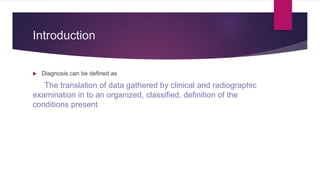
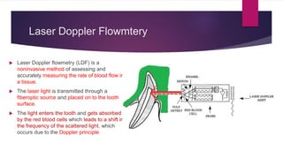
Diagnostic Aids In
Endodontics
DR. ISHAAN ADHAULIA
What is to be discussed
 Introduction
 Case history
 Clinical examination
 DIAGNOSTIC AIDS
 Bibliography
Introduction
 Diagnosis can be defined as
The translation of data gathered by clinical and radiographic
examination in to an organized, classified, definition of the
conditions present
How do we sequelize our diagnosis ?
 Demographic information with respect to name, age, sex etc
 Chief complaint and history of present illness
 Medical and dental history
 Clinical examination
 Radiographic findings
 Other laboratory findings
 Provisional diagnosis
 Treatment plan
Endodontic health questionnaire
Taken from grossman
endodontics 13th edition
Clinical Record Sheet
Taken from grossman
endodontics 13th edition
Symptoms
 Symptoms are the units of information sought in clinical diagnosis. They are
defined as phenomena or signs of a departure from the normal state and are
indicative of illness.
Symptoms can be classified as follows:
 Subjective symptoms: Those experienced and reported to the clinician by the
patient
 Objective symptoms: Those ascertained by the clinician through various tests
 Understanding of both subjective and objective symptoms is essential for the
correct identification of disease and thereby a diagnosis of the problem of the
patient.
Objective symptoms
 Objective symptoms are determined by tests and
observations performed by the clinician
 Although it may not be necessary to perform all
these tests at any one time, a combination of
supporting tests are desirable to ensure a correct
diagnosis.
 One should not rely on the results of any single
test.
Various Diagnostic Tests
 1. Visual and tactile inspection
(a) Hard tissues
(b) Soft tissues
(i) Gingiva
(ii) Periodontium
 2. Percussion
 3. Palpation
 4. Mobility and depressibility
 5. Bite test
 6. Magnification
 7. radiography
Contd.
 8. Assessment of pulp vitality
(a) Neural sensibility tests
 (i) Thermal tests
 (ii) Heat testing
 (iii) Cold testing
 (iv) Electric pulp tester (EPT)
 (v) Anesthetic test
 (vi) Test cavity
(b) Pulp vascularity tests
 (i) Pulse oximetry
 (ii) Laser Doppler flowmetry
Visual and Tactile
 The most simple clinical test is visual examination, it’s done casually
during the inspection
 The hard and soft tissues are examined for three things – Colour,
Contour & Consistency
 In hard tissues teeth should be visually examined using the “three Cs.” A
normal-appearing crown has a life-like translucency and sparkle that is
missing in pulpless teeth
Visual contd.
 less life-like in appearance should be carefully
evaluated because the pulp may already be
inflamed, degenerated, or necrotic
 Not all discolored teeth need endodontic
treatment as staining may be caused by old
amalgam restorations, root canal filling
materials and medicaments, or systemic
medication, such as tetracycline staining
Visual changes in soft tissues
 In soft tissue, such as gingiva, deviation from the healthy
pink color is readily recognized when inflammation is
present. A change in contour occurs with swelling, and the
consistency of soft, fluctuant, or spongy tissue differs from
that of normal, healthy, firm tissue
 Similar changes are observed in the periodontium, it should
be observed under good lighting and dry conditions for
fistulas which might get missed during routine inspection if
covered with saliva
 Periodontium should be examined with 3 different probes –
Williams Probe, WHO Probe & lastly UNC – 15 Probe
Tactile Diagnosis
 This test enables one to evaluate the status of the
periodontium surrounding a tooth.
 The tooth is struck with a quick, moderate blow,
initially with low intensity by the finger and then with
increasing intensity by using the handle (blunt end)
of an instrument, to determine whether the tooth is
tender
 Percussion is used in conjunction with other
periodontal tests, namely palpation, mobility, and
depressibility as these tests help to corroborate the
presence of periodontitis
Palpation – Mobility - Depressibility
This simple test is done with the fingertips, using light
pressure to examine tissue consistency and pain response.
Although simple, it is an important test. Its value lies in
locating the swelling over an involved tooth and determining
whether the -
 tissue is fluctuant and enlarged sufficiently for Incision &
Drainage
 Presence, intensity, and location of pain
 Presence and location of adenopathy or bone crepitus
Palpation – Mobility - Depressibility
 The mobility test is used to evaluate the integrity of
the attachment apparatus surrounding the tooth.
 The test consists of moving a tooth laterally in its
socket by using the fingers or, preferably, the
handles of two operative instruments
 Similarly, the test for depressibility consists of
moving a tooth vertically in its socket
Bite Test
 The bite test is useful in identifying a cracked
tooth or fractured cusp when pressure is applied
in a certain direction to one cusp or section of
the tooth.
 Also useful in diagnosing pulpal pathosis when it
has extended into the periradicular region
 The Tooth Slooth and the Frac finder are the
popular commercially available devices for the
bite test.
Magnification
 Magnification is an essential requisite in
current precision-based endodontic practice.
Initially, devices that enhanced vision were
restricted to magnification loupes
More recently, the use of dental operating
microscopes has gained momentum and its
applications in endodontic diagnosis include
 Locating hidden canals obstructed by
calcifications
 Detection of cracks and fractures
Radiography
 The radiograph is one of the most important clinical tools in
making a diagnosis. It permits visual examination of the oral
structures that would otherwise be unseen by the naked eye.
Without it, diagnosis, case selection, treatment, and
evaluation of healing would be impossible.
Intra-Oral Periapical Radiographs
 Radiographs may show the number, course,
shape, length, and width of root canal
 The presence of calcified material in the pulp
chamber or root canal
 The resorption of dentin originating within the
root canal (internal resorption) or from the root
surface (external resorption)
 Calcification or obliteration of the pulp cavity
 Thickening of the periodontal ligament
 Resorption of cementum, and nature and
extent of periapical and alveolar bone
destruction
Cone Beam Computed Tomograohy
 The introduction of cone beam computed tomography
(CBCT) or cone beam volumetric tomography (CBVT)
imaging facilitated the transition from 2D to a 3D approach in
image acquisition and interpretation
 The device utilizes a cone-shaped beam of ionizing
radiation, which passes through the center of the region of
interest
 The image is captured as three-dimensional pixels termed
voxels with a resolution ranging from 0.4 mm to 0.076 mm
 According to Scarfe et al, the following are the steps
involved in CBCT image processing are
 Acquisition configuration
 Image detection
 Image reconstruction
 Image display
Assessment Of Pulp Vitality
 The accurate diagnosis of the true
histological status of the pulp is of
significant importance for treatment
planning and diagnosis
 This is indirectly done by assessing the
neural sensitivity of the pulp although a
more accurate approach would be to
assess the vascularity of the pulp
Various tests for assessment of pulp vitality
Neural Sensibility Tests
 These are the tests that indirectly tell us about the vitality status of the pulp. They
work on the principle of stimulating the neural fibers present in the pulp.
Thermal Tests
 These tests involve the application of cold and heat to a
tooth to determine its sensitivity to thermal changes
 Heat test is performed using different techniques that
deliver different degrees of temperature on the tooth. The
area to be tested is isolated and dried and a suitable
lubricant (vaseline) is applied and heat is directed to the
exposed surface of the tooth and the patient’s response is
noted
 Whereas in cold test, cold can be applied in several
different ways, the most common method is to apply a
cotton pellet saturated with 1,1,1,2 tetra-fluoroethane to
the tooth being tested
 Dachi et al, recommended that a quarter-inch-diameter
cone of ice be placed against a tooth for 5 seconds to
quantify cold testing.
Heat test
Cold test
Thermal Tests
 The diagnostic accuracy of cold test is 86%, the electric pulp test is 81%, and
heat test is 71%
 Cold test can be used to differentiate between reversible and irreversible pulpitis
 In irreversible pulpitis, patients complain of increased pain secondary to heat test,
while in such a situation the application of cold would cause temporary relief of
pain.
Electric Pulp Test
 The electric pulp test is one of the tests used
to determine pulp vitality. The electric pulp
tester, when testing for pulp vitality, uses
nerve stimulation.
 The objective is to stimulate a pulpal response
by subjecting the tooth to an increasing
degree of electric current
 A positive response is an indication of vitality
and helps in determining the normality or
abnormality of that pulp.
 No response to the electrical stimulus can be
an indication of pulp necrosis.
Clinical Interpretations of Pulpal
Response to EPT
 Normal Response: The test tooth would be considered
normal and vital if both the teeth exhibit a positive
response at a similar numerical value of the EPT.
 Negative response: This denotes a non vital tooth, which
fails to respond even when the tester is set to the highest
electrical excitation value.
 Early response: This denotes a diseased state of pulp as
the tooth
 Delayed response: This also denotes a diseased state of
the pulp wherein the tooth responds at a significantly
higher electrical excitation level
 False Positive & False Negetive
Anaesthetic Test
 This test is restricted to patients who are in pain
at the time of the test when the usual tests have
failed to identify the tooth
 The objective is to anesthetize one tooth at a
time until the pain disappears and is localized to
a specific tooth
 The technique involves using either infiltration
or the intraligament injection
Test Cavity
 This test allows one to determine pulp vitality. It is
performed when other methods of diagnosis have
failed.
 The test cavity is made by drilling through the enamel–
dentin junction of an un-anesthetized tooth. The drilling
should be done at high speed and with a water coolant
 Sensitivity or pain felt by the patient is an indication of
pulp vitality & no endodontic treatment is indicated for
the same
 If no pain is felt, cavity preparation may be continued
until the pulp chamber is reached. If the pulp is
completely necrotic, endodontic treatment can be
continued painlessly in many cases without
anesthesia.
Pulp Vascularity Test
 The most common methods to assess pulp vitality are
based on sensitivity assessment of the neural tissues of
the pulp
 These include thermal and electric pulp tests. True vitality
status can be ascertained only when we are able to
assess the vascular or blood supply to the tooth
 Two technologies widely used are Pulse Oximetry &
Laser Doppler Flowmetry
Pulse Oximetry
 Pulse oximetry is a noninvasive method to measure the
oxygen saturation levels during the administration of
anesthesia or other medications with the help of a finger,
ear, or foot probes.
 The pulse oximeter equipment consists of a pulse
oximeter monitor (POM) which gives the digital display
of oxygen saturation values
 This pulse oximeter monitor is connected to a pulse
oximeter sensor (POS) which is designed to
anatomically conform to the area where oxygen
saturation values have to be assessed e.g, ear pulse
oximeter sensor, finger sensor, and toe sensor.
Laser Doppler Flowmtery
 Laser Doppler flowmetry (LDF) is a
noninvasive method of assessing and
accurately measuring the rate of blood flow in
a tissue.
 The laser light is transmitted through a
fiberoptic source and placed on to the tooth
surface.
 The light enters the tooth and gets absorbed
by the red blood cells which leads to a shift in
the frequency of the scattered light, which
occurs due to the Doppler principle
Bibliography
Content studied and Explainatory Images widely taken from
Grossman’s Endodontics 13th Edition
THANK YOU
ISHAAN ADHAULIA
(FINAL SECOND)
Listen to your patient ..... The patient will give you the diagnosis
- Sir William Olsen

Diagnostic Aids in Endodontics

  • 1.
  • 2.
    What is tobe discussed  Introduction  Case history  Clinical examination  DIAGNOSTIC AIDS  Bibliography
  • 3.
    Introduction  Diagnosis canbe defined as The translation of data gathered by clinical and radiographic examination in to an organized, classified, definition of the conditions present
  • 4.
    How do wesequelize our diagnosis ?  Demographic information with respect to name, age, sex etc  Chief complaint and history of present illness  Medical and dental history  Clinical examination  Radiographic findings  Other laboratory findings  Provisional diagnosis  Treatment plan
  • 5.
    Endodontic health questionnaire Takenfrom grossman endodontics 13th edition
  • 6.
    Clinical Record Sheet Takenfrom grossman endodontics 13th edition
  • 7.
    Symptoms  Symptoms arethe units of information sought in clinical diagnosis. They are defined as phenomena or signs of a departure from the normal state and are indicative of illness. Symptoms can be classified as follows:  Subjective symptoms: Those experienced and reported to the clinician by the patient  Objective symptoms: Those ascertained by the clinician through various tests  Understanding of both subjective and objective symptoms is essential for the correct identification of disease and thereby a diagnosis of the problem of the patient.
  • 8.
    Objective symptoms  Objectivesymptoms are determined by tests and observations performed by the clinician  Although it may not be necessary to perform all these tests at any one time, a combination of supporting tests are desirable to ensure a correct diagnosis.  One should not rely on the results of any single test.
  • 9.
    Various Diagnostic Tests 1. Visual and tactile inspection (a) Hard tissues (b) Soft tissues (i) Gingiva (ii) Periodontium  2. Percussion  3. Palpation  4. Mobility and depressibility  5. Bite test  6. Magnification  7. radiography
  • 10.
    Contd.  8. Assessmentof pulp vitality (a) Neural sensibility tests  (i) Thermal tests  (ii) Heat testing  (iii) Cold testing  (iv) Electric pulp tester (EPT)  (v) Anesthetic test  (vi) Test cavity (b) Pulp vascularity tests  (i) Pulse oximetry  (ii) Laser Doppler flowmetry
  • 11.
    Visual and Tactile The most simple clinical test is visual examination, it’s done casually during the inspection  The hard and soft tissues are examined for three things – Colour, Contour & Consistency  In hard tissues teeth should be visually examined using the “three Cs.” A normal-appearing crown has a life-like translucency and sparkle that is missing in pulpless teeth
  • 12.
    Visual contd.  lesslife-like in appearance should be carefully evaluated because the pulp may already be inflamed, degenerated, or necrotic  Not all discolored teeth need endodontic treatment as staining may be caused by old amalgam restorations, root canal filling materials and medicaments, or systemic medication, such as tetracycline staining
  • 13.
    Visual changes insoft tissues  In soft tissue, such as gingiva, deviation from the healthy pink color is readily recognized when inflammation is present. A change in contour occurs with swelling, and the consistency of soft, fluctuant, or spongy tissue differs from that of normal, healthy, firm tissue  Similar changes are observed in the periodontium, it should be observed under good lighting and dry conditions for fistulas which might get missed during routine inspection if covered with saliva  Periodontium should be examined with 3 different probes – Williams Probe, WHO Probe & lastly UNC – 15 Probe
  • 14.
    Tactile Diagnosis  Thistest enables one to evaluate the status of the periodontium surrounding a tooth.  The tooth is struck with a quick, moderate blow, initially with low intensity by the finger and then with increasing intensity by using the handle (blunt end) of an instrument, to determine whether the tooth is tender  Percussion is used in conjunction with other periodontal tests, namely palpation, mobility, and depressibility as these tests help to corroborate the presence of periodontitis
  • 15.
    Palpation – Mobility- Depressibility This simple test is done with the fingertips, using light pressure to examine tissue consistency and pain response. Although simple, it is an important test. Its value lies in locating the swelling over an involved tooth and determining whether the -  tissue is fluctuant and enlarged sufficiently for Incision & Drainage  Presence, intensity, and location of pain  Presence and location of adenopathy or bone crepitus
  • 16.
    Palpation – Mobility- Depressibility  The mobility test is used to evaluate the integrity of the attachment apparatus surrounding the tooth.  The test consists of moving a tooth laterally in its socket by using the fingers or, preferably, the handles of two operative instruments  Similarly, the test for depressibility consists of moving a tooth vertically in its socket
  • 17.
    Bite Test  Thebite test is useful in identifying a cracked tooth or fractured cusp when pressure is applied in a certain direction to one cusp or section of the tooth.  Also useful in diagnosing pulpal pathosis when it has extended into the periradicular region  The Tooth Slooth and the Frac finder are the popular commercially available devices for the bite test.
  • 18.
    Magnification  Magnification isan essential requisite in current precision-based endodontic practice. Initially, devices that enhanced vision were restricted to magnification loupes More recently, the use of dental operating microscopes has gained momentum and its applications in endodontic diagnosis include  Locating hidden canals obstructed by calcifications  Detection of cracks and fractures
  • 19.
    Radiography  The radiographis one of the most important clinical tools in making a diagnosis. It permits visual examination of the oral structures that would otherwise be unseen by the naked eye. Without it, diagnosis, case selection, treatment, and evaluation of healing would be impossible.
  • 20.
    Intra-Oral Periapical Radiographs Radiographs may show the number, course, shape, length, and width of root canal  The presence of calcified material in the pulp chamber or root canal  The resorption of dentin originating within the root canal (internal resorption) or from the root surface (external resorption)  Calcification or obliteration of the pulp cavity  Thickening of the periodontal ligament  Resorption of cementum, and nature and extent of periapical and alveolar bone destruction
  • 21.
    Cone Beam ComputedTomograohy  The introduction of cone beam computed tomography (CBCT) or cone beam volumetric tomography (CBVT) imaging facilitated the transition from 2D to a 3D approach in image acquisition and interpretation  The device utilizes a cone-shaped beam of ionizing radiation, which passes through the center of the region of interest  The image is captured as three-dimensional pixels termed voxels with a resolution ranging from 0.4 mm to 0.076 mm  According to Scarfe et al, the following are the steps involved in CBCT image processing are  Acquisition configuration  Image detection  Image reconstruction  Image display
  • 22.
    Assessment Of PulpVitality  The accurate diagnosis of the true histological status of the pulp is of significant importance for treatment planning and diagnosis  This is indirectly done by assessing the neural sensitivity of the pulp although a more accurate approach would be to assess the vascularity of the pulp Various tests for assessment of pulp vitality
  • 23.
    Neural Sensibility Tests These are the tests that indirectly tell us about the vitality status of the pulp. They work on the principle of stimulating the neural fibers present in the pulp.
  • 24.
    Thermal Tests  Thesetests involve the application of cold and heat to a tooth to determine its sensitivity to thermal changes  Heat test is performed using different techniques that deliver different degrees of temperature on the tooth. The area to be tested is isolated and dried and a suitable lubricant (vaseline) is applied and heat is directed to the exposed surface of the tooth and the patient’s response is noted  Whereas in cold test, cold can be applied in several different ways, the most common method is to apply a cotton pellet saturated with 1,1,1,2 tetra-fluoroethane to the tooth being tested  Dachi et al, recommended that a quarter-inch-diameter cone of ice be placed against a tooth for 5 seconds to quantify cold testing. Heat test Cold test
  • 25.
    Thermal Tests  Thediagnostic accuracy of cold test is 86%, the electric pulp test is 81%, and heat test is 71%  Cold test can be used to differentiate between reversible and irreversible pulpitis  In irreversible pulpitis, patients complain of increased pain secondary to heat test, while in such a situation the application of cold would cause temporary relief of pain.
  • 26.
    Electric Pulp Test The electric pulp test is one of the tests used to determine pulp vitality. The electric pulp tester, when testing for pulp vitality, uses nerve stimulation.  The objective is to stimulate a pulpal response by subjecting the tooth to an increasing degree of electric current  A positive response is an indication of vitality and helps in determining the normality or abnormality of that pulp.  No response to the electrical stimulus can be an indication of pulp necrosis.
  • 27.
    Clinical Interpretations ofPulpal Response to EPT  Normal Response: The test tooth would be considered normal and vital if both the teeth exhibit a positive response at a similar numerical value of the EPT.  Negative response: This denotes a non vital tooth, which fails to respond even when the tester is set to the highest electrical excitation value.  Early response: This denotes a diseased state of pulp as the tooth  Delayed response: This also denotes a diseased state of the pulp wherein the tooth responds at a significantly higher electrical excitation level  False Positive & False Negetive
  • 28.
    Anaesthetic Test  Thistest is restricted to patients who are in pain at the time of the test when the usual tests have failed to identify the tooth  The objective is to anesthetize one tooth at a time until the pain disappears and is localized to a specific tooth  The technique involves using either infiltration or the intraligament injection
  • 29.
    Test Cavity  Thistest allows one to determine pulp vitality. It is performed when other methods of diagnosis have failed.  The test cavity is made by drilling through the enamel– dentin junction of an un-anesthetized tooth. The drilling should be done at high speed and with a water coolant  Sensitivity or pain felt by the patient is an indication of pulp vitality & no endodontic treatment is indicated for the same  If no pain is felt, cavity preparation may be continued until the pulp chamber is reached. If the pulp is completely necrotic, endodontic treatment can be continued painlessly in many cases without anesthesia.
  • 30.
    Pulp Vascularity Test The most common methods to assess pulp vitality are based on sensitivity assessment of the neural tissues of the pulp  These include thermal and electric pulp tests. True vitality status can be ascertained only when we are able to assess the vascular or blood supply to the tooth  Two technologies widely used are Pulse Oximetry & Laser Doppler Flowmetry
  • 31.
    Pulse Oximetry  Pulseoximetry is a noninvasive method to measure the oxygen saturation levels during the administration of anesthesia or other medications with the help of a finger, ear, or foot probes.  The pulse oximeter equipment consists of a pulse oximeter monitor (POM) which gives the digital display of oxygen saturation values  This pulse oximeter monitor is connected to a pulse oximeter sensor (POS) which is designed to anatomically conform to the area where oxygen saturation values have to be assessed e.g, ear pulse oximeter sensor, finger sensor, and toe sensor.
  • 32.
    Laser Doppler Flowmtery Laser Doppler flowmetry (LDF) is a noninvasive method of assessing and accurately measuring the rate of blood flow in a tissue.  The laser light is transmitted through a fiberoptic source and placed on to the tooth surface.  The light enters the tooth and gets absorbed by the red blood cells which leads to a shift in the frequency of the scattered light, which occurs due to the Doppler principle
  • 33.
    Bibliography Content studied andExplainatory Images widely taken from Grossman’s Endodontics 13th Edition
  • 34.
    THANK YOU ISHAAN ADHAULIA (FINALSECOND) Listen to your patient ..... The patient will give you the diagnosis - Sir William Olsen