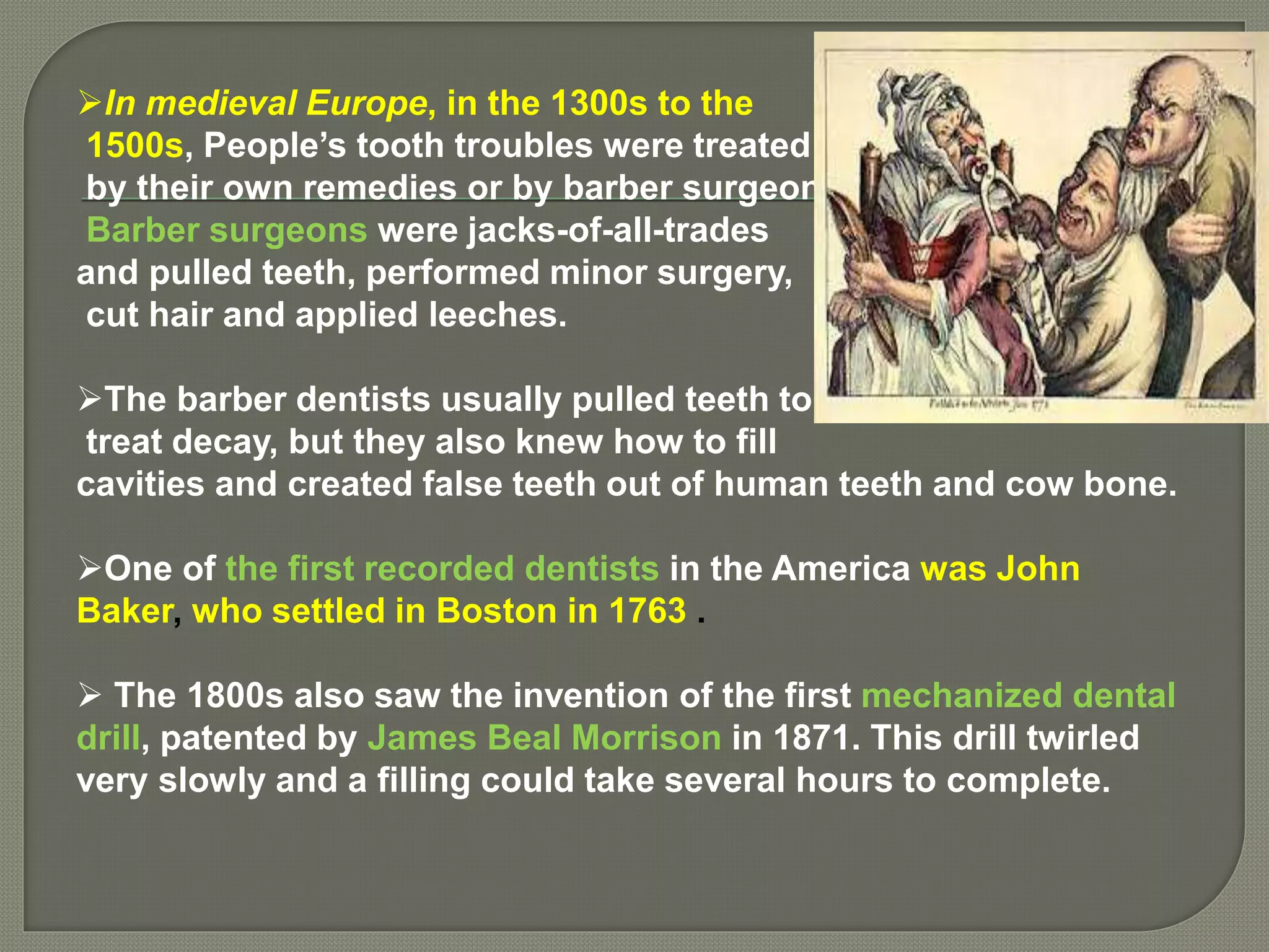
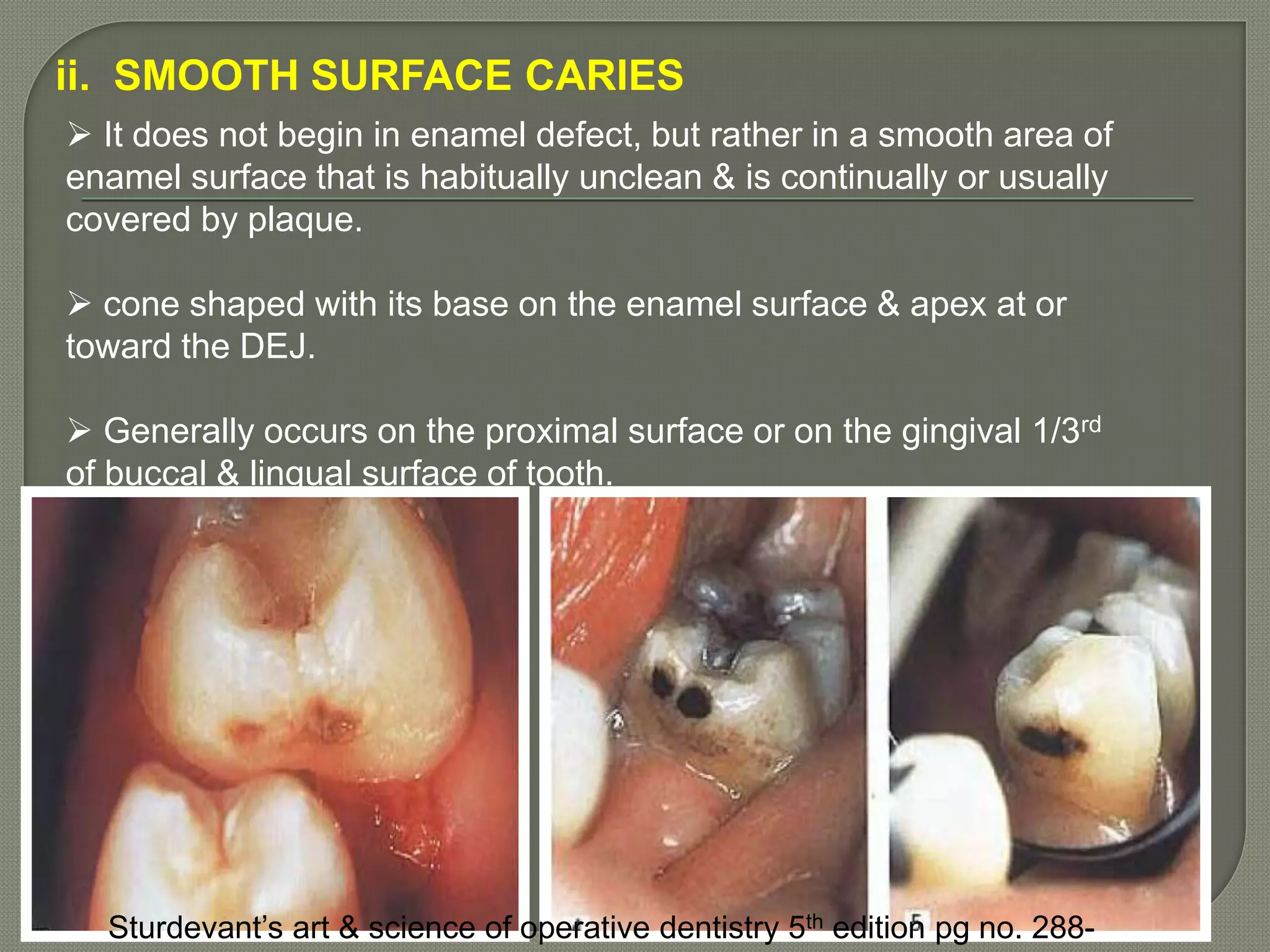
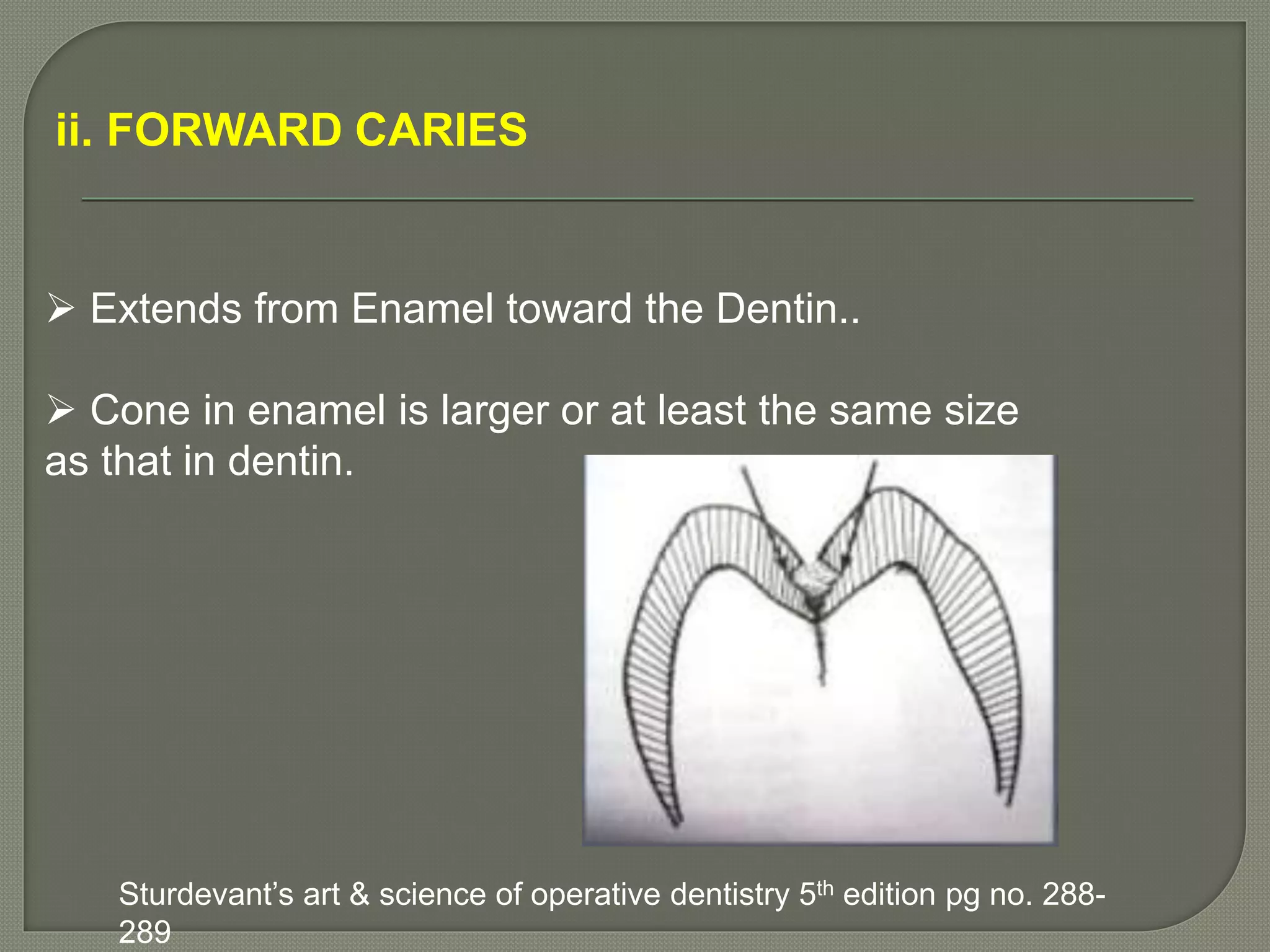
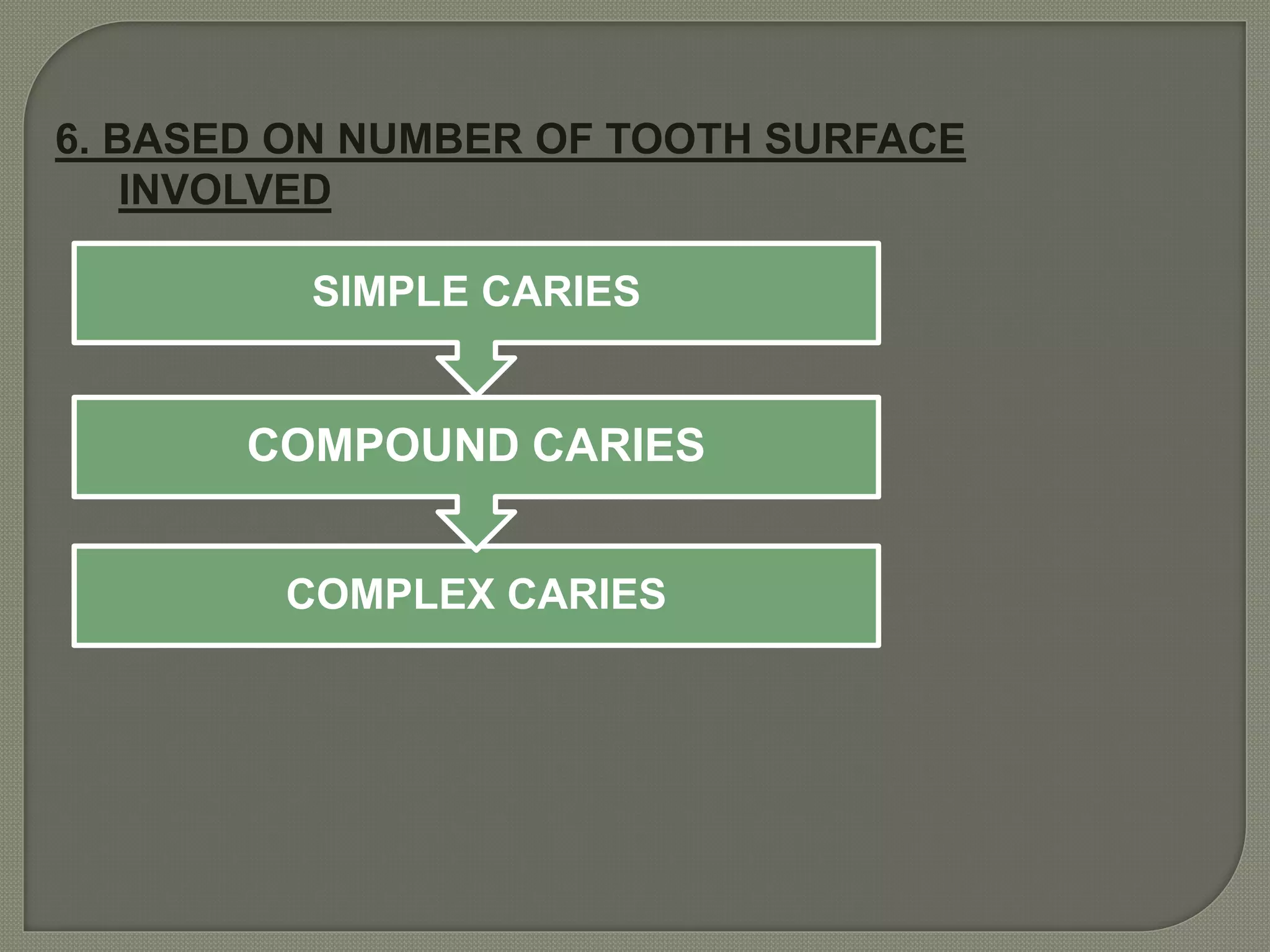
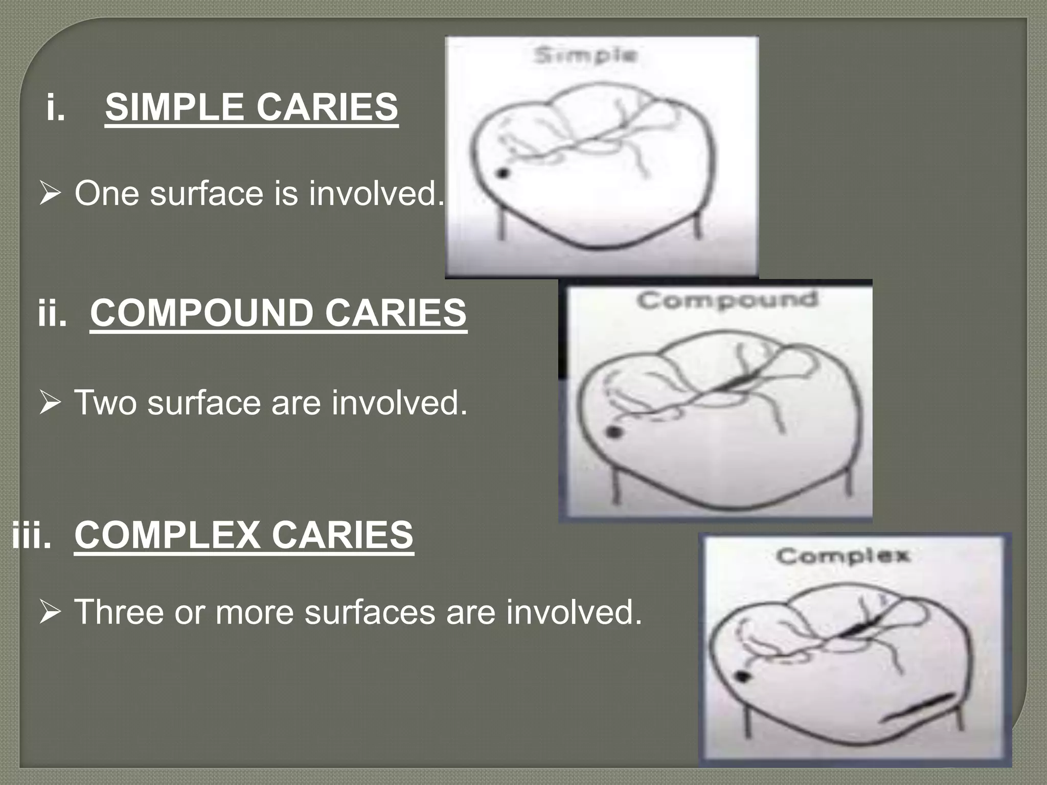
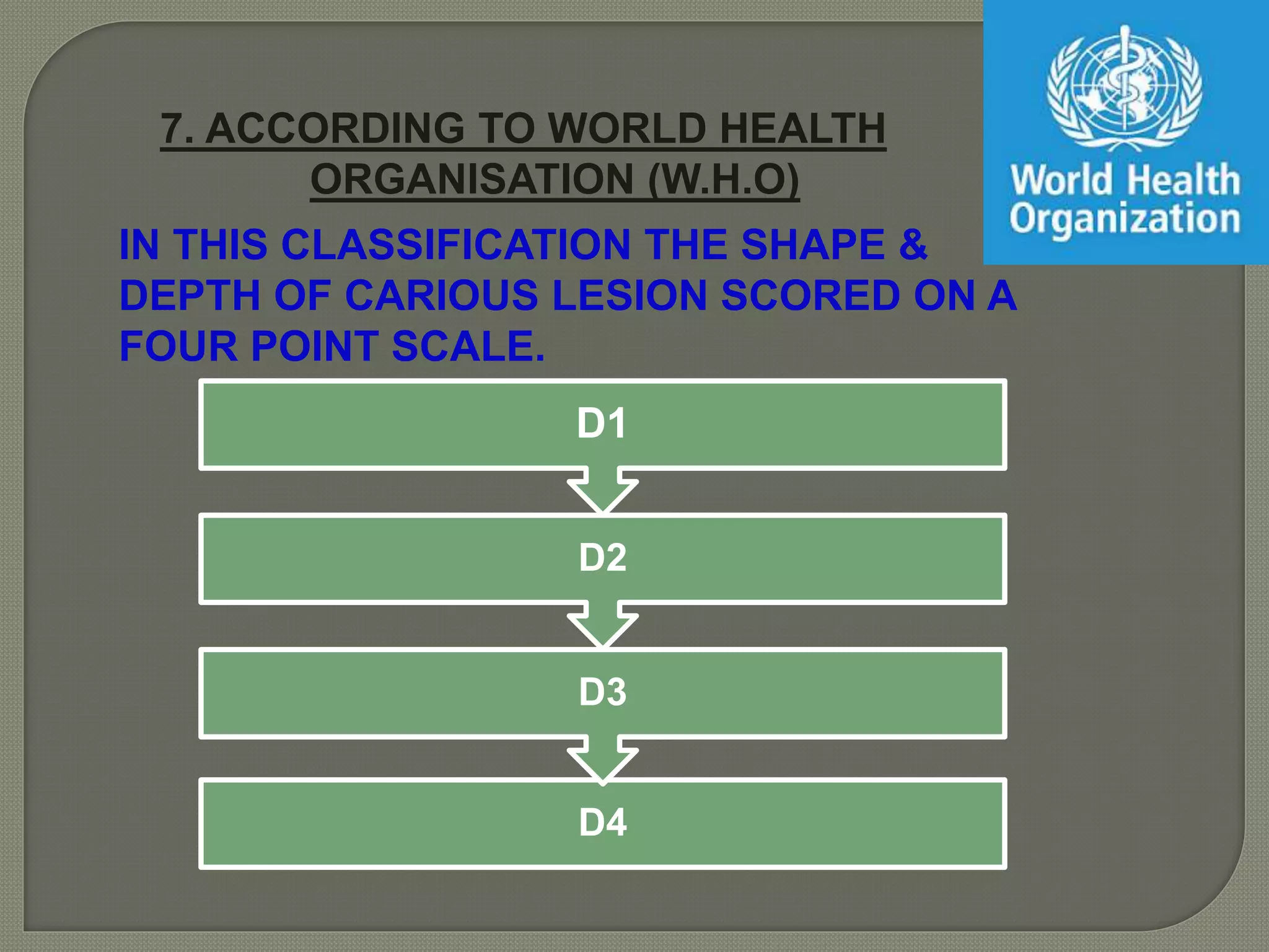
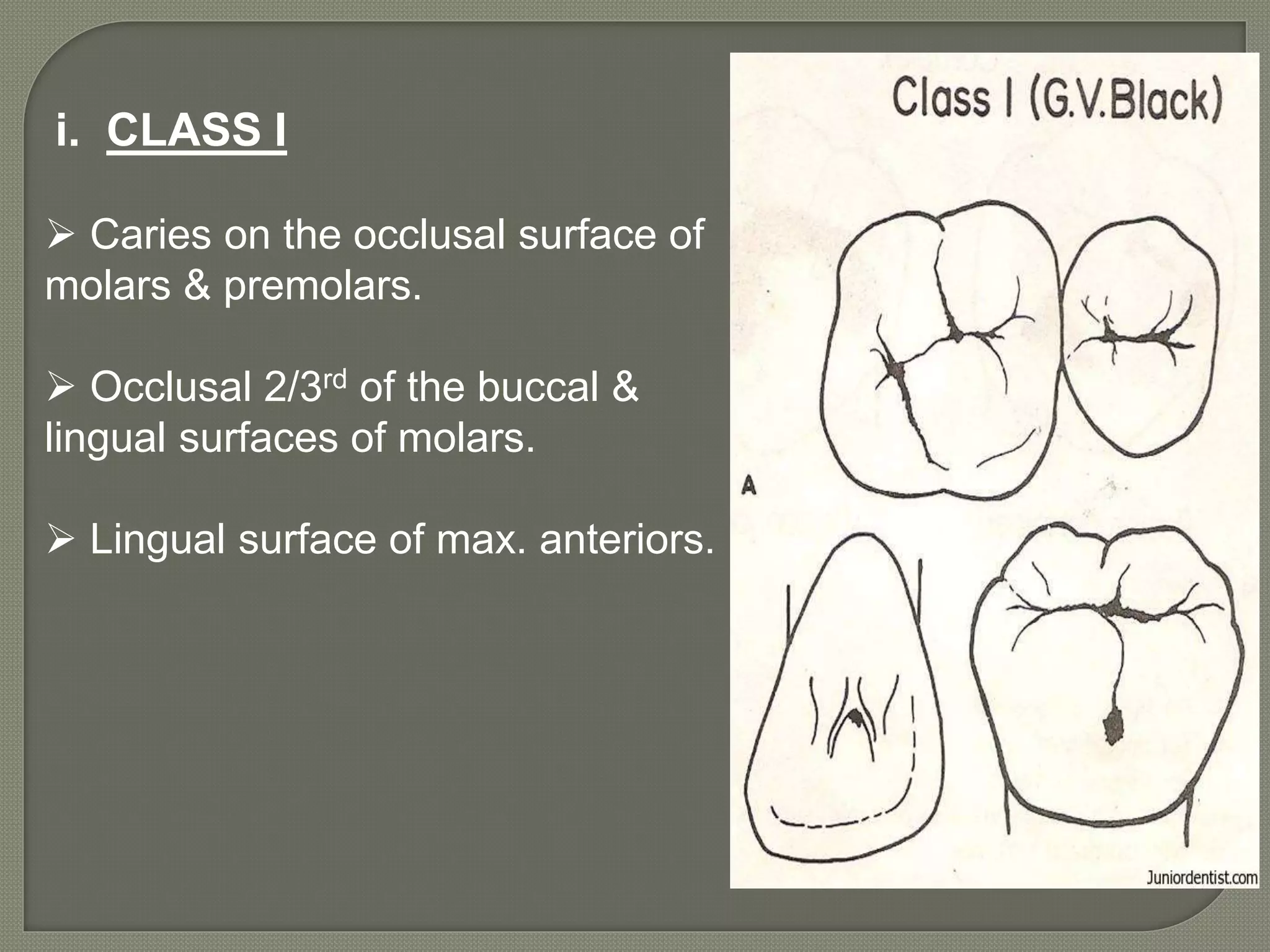
This document provides an overview of dental caries, including its history, epidemiology, classification, etiology, histogenesis, diagnosis, and treatment. It discusses the evolution of dental caries throughout history from ancient civilizations to modern times. Key points include that dental caries is caused by bacteria in the mouth, affects most people worldwide, and has been found in human remains from 25,000 years ago. The document also provides various ways of classifying dental caries based on location, progression, extent, rate, age pattern, and number of tooth surfaces involved.
Remineralization is defined as the process whereby calcium and phosphate ions are supplied from an external source to the tooth thereby, causing ion deposition into crystal voids in demineralized enamel, thus producing net mineral gain.
Remineralization Agents - Biomimetic approaches to stabilization of bioavailable calcium, phosphate, and fluoride ions and the localization of these ions to non-cavitated caries lesions for controlled remineralization.
Fermentable carbohydrates provide plaque bacteria with substrate for acid production.
This causes a rapid drop in plaque pH and when pH becomes less than 5.5, hydroxyapatite in enamel breaks down and calcium and phosphate ions diffuse out from the enamel.
5.5 is the “critical pH”, the point where equilibrium exists. There is no mineral dissolution and no mineral precipitation.
The plaque remains acidic for about 30-60 minutes after which normal pH is restored gradually.
The calcium and phosphate ions re-enter enamel when normal pH is restored and thus remineralization occurs.
Bevels and flares are very important components of resin restoration procedure. This presentation focuses on bevels and flares in restorative procedure.
This seminar includes classifcation,etiopathogenesis,Various theories of dental caries,caries patterns in primary and permanent teeth,Caries pattern in adolescets followed by caries risk assessment,CAMBRA,Differences between nursing bottle and rampant cariess,diagnosis which included the advanced digital diagnostic methods like diagnodent,QLF,etc and management with age specific management and flouride therapy age wise .
dental Cariology /certified fixed orthodontic courses by Indian dental academy Indian dental academy
Welcome to Indian Dental Academy
The Indian Dental Academy is the Leader in continuing dental education , training dentists in all aspects of dentistry and offering a wide range of dental certified courses in different formats.
Indian dental academy has a unique training program & curriculum that provides students with exceptional clinical skills and enabling them to return to their office with high level confidence and start treating patients
State of the art comprehensive training-Faculty of world wide repute &Very affordable.
CLEFT LIP &PALATE MANAGEMENT IN ORTHODONTICS /certified fixed orthodontic cou...Indian dental academy
The Indian Dental Academy is the Leader in continuing dental education , training dentists in all aspects of dentistry and offering a wide range of dental certified courses in different formats.
Indian dental academy provides dental crown & Bridge,rotary endodontics,fixed orthodontics,
Dental implants courses.for details pls visit www.indiandentalacademy.com ,or call
0091-9248678078
The Indian Dental Academy is the Leader in continuing dental education , training dentists in all aspects of dentistry and
offering a wide range of dental certified courses in different formats.for more details please visit
www.indiandentalacademy.com
Co-Existence of Synodontia & Talon Cusp in Mandible: A Rare Casepateldrona
Synodontia and Talon cusp are rare developmental anomalies present in human dentition. Talon cusp is most commonly seen on the palatal surfaces of the permanent maxillary anteriors while synodontia is seen in primary anterior dentition.
Similar to Dental caries (operative dentistry) (20)
TEST BANK for Operations Management, 14th Edition by William J. Stevenson, Ve...kevinkariuki227
TEST BANK for Operations Management, 14th Edition by William J. Stevenson, Verified Chapters 1 - 19, Complete Newest Version.pdf
- Video recording of this lecture in English language: https://youtu.be/lK81BzxMqdo
- Video recording of this lecture in Arabic language: https://youtu.be/Ve4P0COk9OI
- Link to download the book free: https://nephrotube.blogspot.com/p/nephrotube-nephrology-books.html
- Link to NephroTube website: www.NephroTube.com
- Link to NephroTube social media accounts: https://nephrotube.blogspot.com/p/join-nephrotube-on-social-media.html
Lung Cancer: Artificial Intelligence, Synergetics, Complex System Analysis, S...Oleg Kshivets
RESULTS: Overall life span (LS) was 2252.1±1742.5 days and cumulative 5-year survival (5YS) reached 73.2%, 10 years – 64.8%, 20 years – 42.5%. 513 LCP lived more than 5 years (LS=3124.6±1525.6 days), 148 LCP – more than 10 years (LS=5054.4±1504.1 days).199 LCP died because of LC (LS=562.7±374.5 days). 5YS of LCP after bi/lobectomies was significantly superior in comparison with LCP after pneumonectomies (78.1% vs.63.7%, P=0.00001 by log-rank test). AT significantly improved 5YS (66.3% vs. 34.8%) (P=0.00000 by log-rank test) only for LCP with N1-2. Cox modeling displayed that 5YS of LCP significantly depended on: phase transition (PT) early-invasive LC in terms of synergetics, PT N0—N12, cell ratio factors (ratio between cancer cells- CC and blood cells subpopulations), G1-3, histology, glucose, AT, blood cell circuit, prothrombin index, heparin tolerance, recalcification time (P=0.000-0.038). Neural networks, genetic algorithm selection and bootstrap simulation revealed relationships between 5YS and PT early-invasive LC (rank=1), PT N0—N12 (rank=2), thrombocytes/CC (3), erythrocytes/CC (4), eosinophils/CC (5), healthy cells/CC (6), lymphocytes/CC (7), segmented neutrophils/CC (8), stick neutrophils/CC (9), monocytes/CC (10); leucocytes/CC (11). Correct prediction of 5YS was 100% by neural networks computing (area under ROC curve=1.0; error=0.0).
CONCLUSIONS: 5YS of LCP after radical procedures significantly depended on: 1) PT early-invasive cancer; 2) PT N0--N12; 3) cell ratio factors; 4) blood cell circuit; 5) biochemical factors; 6) hemostasis system; 7) AT; 8) LC characteristics; 9) LC cell dynamics; 10) surgery type: lobectomy/pneumonectomy; 11) anthropometric data. Optimal diagnosis and treatment strategies for LC are: 1) screening and early detection of LC; 2) availability of experienced thoracic surgeons because of complexity of radical procedures; 3) aggressive en block surgery and adequate lymph node dissection for completeness; 4) precise prediction; 5) adjuvant chemoimmunoradiotherapy for LCP with unfavorable prognosis.
Flu Vaccine Alert in Bangalore Karnatakaaddon Scans
As flu season approaches, health officials in Bangalore, Karnataka, are urging residents to get their flu vaccinations. The seasonal flu, while common, can lead to severe health complications, particularly for vulnerable populations such as young children, the elderly, and those with underlying health conditions.
Dr. Vidisha Kumari, a leading epidemiologist in Bangalore, emphasizes the importance of getting vaccinated. "The flu vaccine is our best defense against the influenza virus. It not only protects individuals but also helps prevent the spread of the virus in our communities," he says.
This year, the flu season is expected to coincide with a potential increase in other respiratory illnesses. The Karnataka Health Department has launched an awareness campaign highlighting the significance of flu vaccinations. They have set up multiple vaccination centers across Bangalore, making it convenient for residents to receive their shots.
To encourage widespread vaccination, the government is also collaborating with local schools, workplaces, and community centers to facilitate vaccination drives. Special attention is being given to ensuring that the vaccine is accessible to all, including marginalized communities who may have limited access to healthcare.
Residents are reminded that the flu vaccine is safe and effective. Common side effects are mild and may include soreness at the injection site, mild fever, or muscle aches. These side effects are generally short-lived and far less severe than the flu itself.
Healthcare providers are also stressing the importance of continuing COVID-19 precautions. Wearing masks, practicing good hand hygiene, and maintaining social distancing are still crucial, especially in crowded places.
Protect yourself and your loved ones by getting vaccinated. Together, we can help keep Bangalore healthy and safe this flu season. For more information on vaccination centers and schedules, residents can visit the Karnataka Health Department’s official website or follow their social media pages.
Stay informed, stay safe, and get your flu shot today!
Recomendações da OMS sobre cuidados maternos e neonatais para uma experiência pós-natal positiva.
Em consonância com os ODS – Objetivos do Desenvolvimento Sustentável e a Estratégia Global para a Saúde das Mulheres, Crianças e Adolescentes, e aplicando uma abordagem baseada nos direitos humanos, os esforços de cuidados pós-natais devem expandir-se para além da cobertura e da simples sobrevivência, de modo a incluir cuidados de qualidade.
Estas diretrizes visam melhorar a qualidade dos cuidados pós-natais essenciais e de rotina prestados às mulheres e aos recém-nascidos, com o objetivo final de melhorar a saúde e o bem-estar materno e neonatal.
Uma “experiência pós-natal positiva” é um resultado importante para todas as mulheres que dão à luz e para os seus recém-nascidos, estabelecendo as bases para a melhoria da saúde e do bem-estar a curto e longo prazo. Uma experiência pós-natal positiva é definida como aquela em que as mulheres, pessoas que gestam, os recém-nascidos, os casais, os pais, os cuidadores e as famílias recebem informação consistente, garantia e apoio de profissionais de saúde motivados; e onde um sistema de saúde flexível e com recursos reconheça as necessidades das mulheres e dos bebês e respeite o seu contexto cultural.
Estas diretrizes consolidadas apresentam algumas recomendações novas e já bem fundamentadas sobre cuidados pós-natais de rotina para mulheres e neonatos que recebem cuidados no pós-parto em unidades de saúde ou na comunidade, independentemente dos recursos disponíveis.
É fornecido um conjunto abrangente de recomendações para cuidados durante o período puerperal, com ênfase nos cuidados essenciais que todas as mulheres e recém-nascidos devem receber, e com a devida atenção à qualidade dos cuidados; isto é, a entrega e a experiência do cuidado recebido. Estas diretrizes atualizam e ampliam as recomendações da OMS de 2014 sobre cuidados pós-natais da mãe e do recém-nascido e complementam as atuais diretrizes da OMS sobre a gestão de complicações pós-natais.
O estabelecimento da amamentação e o manejo das principais intercorrências é contemplada.
Recomendamos muito.
Vamos discutir essas recomendações no nosso curso de pós-graduação em Aleitamento no Instituto Ciclos.
Esta publicação só está disponível em inglês até o momento.
Prof. Marcus Renato de Carvalho
www.agostodourado.com
NVBDCP.pptx Nation vector borne disease control programSapna Thakur
NVBDCP was launched in 2003-2004 . Vector-Borne Disease: Disease that results from an infection transmitted to humans and other animals by blood-feeding arthropods, such as mosquitoes, ticks, and fleas. Examples of vector-borne diseases include Dengue fever, West Nile Virus, Lyme disease, and malaria.
263778731218 Abortion Clinic /Pills In Harare ,sisternakatoto
263778731218 Abortion Clinic /Pills In Harare ,ABORTION WOMEN’S CLINIC +27730423979 IN women clinic we believe that every woman should be able to make choices in her pregnancy. Our job is to provide compassionate care, safety,affordable and confidential services. That’s why we have won the trust from all generations of women all over the world. we use non surgical method(Abortion pills) to terminate…Dr.LISA +27730423979women Clinic is committed to providing the highest quality of obstetrical and gynecological care to women of all ages. Our dedicated staff aim to treat each patient and her health concerns with compassion and respect.Our dedicated group ABORTION WOMEN’S CLINIC +27730423979 IN women clinic we believe that every woman should be able to make choices in her pregnancy. Our job is to provide compassionate care, safety,affordable and confidential services. That’s why we have won the trust from all generations of women all over the world. we use non surgical method(Abortion pills) to terminate…Dr.LISA +27730423979women Clinic is committed to providing the highest quality of obstetrical and gynecological care to women of all ages. Our dedicated staff aim to treat each patient and her health concerns with compassion and respect.Our dedicated group of receptionists, nurses, and physicians have worked together as a teamof receptionists, nurses, and physicians have worked together as a team wwww.lisywomensclinic.co.za/
Explore natural remedies for syphilis treatment in Singapore. Discover alternative therapies, herbal remedies, and lifestyle changes that may complement conventional treatments. Learn about holistic approaches to managing syphilis symptoms and supporting overall health.
Ozempic: Preoperative Management of Patients on GLP-1 Receptor Agonists Saeid Safari
Preoperative Management of Patients on GLP-1 Receptor Agonists like Ozempic and Semiglutide
ASA GUIDELINE
NYSORA Guideline
2 Case Reports of Gastric Ultrasound
Basavarajeeyam is an important text for ayurvedic physician belonging to andhra pradehs. It is a popular compendium in various parts of our country as well as in andhra pradesh. The content of the text was presented in sanskrit and telugu language (Bilingual). One of the most famous book in ayurvedic pharmaceutics and therapeutics. This book contains 25 chapters called as prakaranas. Many rasaoushadis were explained, pioneer of dhatu druti, nadi pareeksha, mutra pareeksha etc. Belongs to the period of 15-16 century. New diseases like upadamsha, phiranga rogas are explained.
Title: Sense of Taste
Presenter: Dr. Faiza, Assistant Professor of Physiology
Qualifications:
MBBS (Best Graduate, AIMC Lahore)
FCPS Physiology
ICMT, CHPE, DHPE (STMU)
MPH (GC University, Faisalabad)
MBA (Virtual University of Pakistan)
Learning Objectives:
Describe the structure and function of taste buds.
Describe the relationship between the taste threshold and taste index of common substances.
Explain the chemical basis and signal transduction of taste perception for each type of primary taste sensation.
Recognize different abnormalities of taste perception and their causes.
Key Topics:
Significance of Taste Sensation:
Differentiation between pleasant and harmful food
Influence on behavior
Selection of food based on metabolic needs
Receptors of Taste:
Taste buds on the tongue
Influence of sense of smell, texture of food, and pain stimulation (e.g., by pepper)
Primary and Secondary Taste Sensations:
Primary taste sensations: Sweet, Sour, Salty, Bitter, Umami
Chemical basis and signal transduction mechanisms for each taste
Taste Threshold and Index:
Taste threshold values for Sweet (sucrose), Salty (NaCl), Sour (HCl), and Bitter (Quinine)
Taste index relationship: Inversely proportional to taste threshold
Taste Blindness:
Inability to taste certain substances, particularly thiourea compounds
Example: Phenylthiocarbamide
Structure and Function of Taste Buds:
Composition: Epithelial cells, Sustentacular/Supporting cells, Taste cells, Basal cells
Features: Taste pores, Taste hairs/microvilli, and Taste nerve fibers
Location of Taste Buds:
Found in papillae of the tongue (Fungiform, Circumvallate, Foliate)
Also present on the palate, tonsillar pillars, epiglottis, and proximal esophagus
Mechanism of Taste Stimulation:
Interaction of taste substances with receptors on microvilli
Signal transduction pathways for Umami, Sweet, Bitter, Sour, and Salty tastes
Taste Sensitivity and Adaptation:
Decrease in sensitivity with age
Rapid adaptation of taste sensation
Role of Saliva in Taste:
Dissolution of tastants to reach receptors
Washing away the stimulus
Taste Preferences and Aversions:
Mechanisms behind taste preference and aversion
Influence of receptors and neural pathways
Impact of Sensory Nerve Damage:
Degeneration of taste buds if the sensory nerve fiber is cut
Abnormalities of Taste Detection:
Conditions: Ageusia, Hypogeusia, Dysgeusia (parageusia)
Causes: Nerve damage, neurological disorders, infections, poor oral hygiene, adverse drug effects, deficiencies, aging, tobacco use, altered neurotransmitter levels
Neurotransmitters and Taste Threshold:
Effects of serotonin (5-HT) and norepinephrine (NE) on taste sensitivity
Supertasters:
25% of the population with heightened sensitivity to taste, especially bitterness
Increased number of fungiform papillae
3. • Dental caries is commonly known as tooth decay .
• In the minds of the lay person, and surprisingly even within dentistry,
dental caries is often thought of as holes in the teeth rather than an entire
disease process. (Dental caries: a dynamic disease process ,JDB Featherstone ,Australian
Dental Journal 2008; 53: 286–291)
• Dental caries is one of the most prevalent chronic diseases of
people worldwide; individuals are susceptible to this disease
throughout their lifetime.
(Dental caries, dr. Robert L Selwitz Volume 369, No. 9555, p51–59, 6 January 2007)
•
4. HISTORY
Caries is one of the infectious diseases more easily
observable in human remains retrieved from archaeological
excavations. (Caries Through Time: An Anthropological OverviewLuis Pezo
Lanfranco and Sabine Eggers)
Evidence of dental decay has been found in teeth from
skulls that are 25,000 years old and archaeologists have
evidence of the first dental fillings in teeth from people who
lived around 8000 BC.
The first written reference to dental
decay is found in a Sumerian text
from5000 BC that describes
"tooth worms” as a cause of dental
decay.
5. The first references to dentists are in
ancient Egyptian texts from 2700 BC,
and refer to a doctor who specializes
in treating teeth.
Greek writings from 1300 BC by
Aesculapius, a Greek physician, discuss
extracting (or pulling) diseased teeth. . (Caries
Through Time: An Anthropological OverviewLuis Pezo
Lanfranco and Sabine Eggers)
6. Around 2700 BC ancient Chinese started using acupuncture to
treat pain associated with tooth decay.(The Edwin Smith Surgical Papyrus
Sheds Light on Ancient Egyptian Dentistry)
Doctors in ancient China treated toothaches with arsenic about
A.D. 1000. They are also noted for their development of using
silver amalgam for filling teeth. The Chinese were particularly
advanced in their observation of the oral cavity.
The great Sung landscapist Li T’ang depicts a country doctor
cauterizing a patient’s arm by burning it with the powdered leaves of
an aromatic plant. The treatment is called Moxibustion , which is
widely used along with acupuncture for treatment such as relieving
toothache.
7. In medieval Europe, in the 1300s to the
1500s, People’s tooth troubles were treated
by their own remedies or by barber surgeons
Barber surgeons were jacks-of-all-trades
and pulled teeth, performed minor surgery,
cut hair and applied leeches.
The barber dentists usually pulled teeth to
treat decay, but they also knew how to fill
cavities and created false teeth out of human teeth and cow bone.
One of the first recorded dentists in the America was John
Baker, who settled in Boston in 1763 .
The 1800s also saw the invention of the first mechanized dental
drill, patented by James Beal Morrison in 1871. This drill twirled
very slowly and a filling could take several hours to complete.
8. In 1957, John Borden invented a high-speed, air-driven
hand held dental drill, which shortened the time to
prepare a tooth for a filling to a matter of minutes
With the invention of both Novocain and the high
speed drill, dentistry truly entered the modern ages.
The first dentists used chisels and hammers to knock
out decayed teeth, but today’s dentist may use advanced
technology such as Computer Aided Design to design
dental restorations that look and feel like natural teeth.
Today’s dentist might even be using lasers in their
practice to treat decay and put in fillings.
9.
10. Dental caries may be considered a disease of
modern civilization, since prehistoric man rarely
suffered from this form of tooth destruction.
Anthropologic studies of Von Lenhossek
revealed that the dolicocephalic skulls of men
from pre-Neolithic periods (12,000BC) did not
exhibit dental caries, but Brachycephalic skulls
of the Neolithic period (12,000-3000 BC)
contained carious teeth.
Essential of public health dentistry ; Soben Peter 5th edition p.g. no. 257
11. Dental caries is still a major health problem in most
industrialized countries, affecting 60-90% of school children &
vast majority of adults. . It is the most prevalent oral disease
in several Asian and Latin American countries, while it
appears to be less common and less severe in most African
countries.
The WHO records a Global DMFT of 1.61 for 12 year old in
2004, a reduction of 0.13 as compared to a DMFT of 1.74 in
the year 2001.
Essential of public health dentistry ; Soben Peter 5th edition p.g. no. 257
12. Various studies conducted in different countries at different
time periods have given evidence that a substantial decrease in
caries prevalence in the last decade has been found among
western countries whereas in case of developing and
underdeveloped countries, prevalence of caries seems to be
increasing.
Essential of public health dentistry ; Soben Peter 5th edition p.g. no. 257
13. WHO reported a DMFT score of 3.94 for India.
In India, data from the National Oral health Survey (2002-
2003) states that in children aged 12 years, the caries
prevalence was 53.8% and the mean DMFT was 1.8 whereas it
was 80.2% and 5.4 in the 35-44 year age group. In the 65-74
year age group, the prevalence was 85% and mean DMFT was
14.9.
Essential of public health dentistry ; Soben Peter 5th edition p.g. no. 257
14. Day and Tandon (1940) conducted a survey among 756
subjects aged between 5-18 years in Lahore, and the
point prevalence of caries was reported as 94.04%.
Essential of public health dentistry ; Soben Peter 5th edition p.g. no. 258
Chaudhary and Chawla (1957) conducted a survey
of 2900 school children of 5-16 years old in
Lucknow. They found the deft as 11.1 and DMFT to
1.9.
Ludwig (1960) carried out a survey among school
children and found that children in the age group of
3-5 years had an average of 0.9 deft.
15. In 2015, the caries prevalence and experience among 5, 12 and
15 years age group children from all the reviewed articles from
1999-2014 was 48.11%, 43.34% and 62.02% respectively.
Region wise distribution of dental caries in the past 15 years
(1999-2014) shows more prevalent in the Northern region
among all the index age groups with maximum prevalence
(76.06 %) among 15 year age group.
Onisi and Shinohara (1976) in their survey of 1172
children in age group of 13 years in Japan found that
mean DMFT score 7.5, and this was higher in female
than in males.
In 2007, a post-war survey conducted by Ahmed NAM,
Astrom AN and Bergen NS in12-year old school children
from Baghdad, Iraq. according to this the low prevalence
of caries among children by increasing awareness and
promoting oral health care strategies.
Essential of public health dentistry ; Soben Peter 5th edition p.g. no. 258
16. •W D Miller 1890
Dental decay is a chemo parasitic process consisting
of two stages: decalcification or softening of the hard
tissue and dissolution of soften residue.
•G V Black 1910
Disintegration of the tooth substance molecule by
molecule and was caused by the fermentation of food
inside the mouth.
•Shafer (1980)
Dental caries is an irreversible microbial disease of
the calcified tissues of the teeth, characterized by
demineralization of the inorganic tissues and
destruction of the organic substance of the tooth
which leads to cavity formation.
17. •Sturduvent (5th edition)
Dental caries is an infectious microbial disease of the
tooth that results in localized destruction and
dissolution of calcified tissues.
•WHO
Dental caries is defined as a localized post eruptive,
pathological process of external origin, involving
softening of hard tooth tissues, and proceeding into
the formation of cavity.
18. CLASSIFICATION
1. BASED ON THE LOCATION OF CARIES
Secondary Caries
Root surface caries
Smooth surface caries
Pit & Fissure caries
Sturdevant’s art & science of operative dentistry 5th edition pg no. 288-
289
19. i. PIT & FISSURE CARIES
Pit & Fissure caries can form in the regions of pit & fissures
usually resulting from the imperfect coalescence of the
developmental lobes.
Caries forms a small area of penetration in the enamel at the
bottom of a pit or fissure.
Does not spread laterally to a great extent until the DEJ is
reached.
Sturdevant’s art & science of operative dentistry 5th edition pg no. 288-
20. ii. SMOOTH SURFACE CARIES
It does not begin in enamel defect, but rather in a smooth area of
enamel surface that is habitually unclean & is continually or usually
covered by plaque.
cone shaped with its base on the enamel surface & apex at or
toward the DEJ.
Generally occurs on the proximal surface or on the gingival 1/3rd
of buccal & lingual surface of tooth.
Sturdevant’s art & science of operative dentistry 5th edition pg no. 288-
21. iii. ROOT SURFACE CARIES
Occurs on the tooth root that has been exposed to the oral
environment & habitually covered with plaque.
Usually more rapid than other forms of caries .
Should be detected & treated early.
More prevalent in older individuals.
Sturdevant’s art & science of operative dentistry 5th edition pg no. 288-
22. iv. SECONDARY (RECURRENT)
CARIES
Occurs at the junction of a restoration and the tooth .
May progress under the restoration.
This condition usually indicates that micro leakage is present,
along with other conditions conductive to caries.
Sturdevant’s art & science of operative dentistry 5th edition pg no. 288-
23. 2. BASED ON THE PATHWAY OF SPREAD OF CARIES
FORWARD CARIES
BACKWARD CARIES
Sturdevant’s art & science of operative dentistry 5th edition pg no. 288-
24. i. BACKWARD CARIES
Extends from DEJ toward the contiguous
enamel.
Triangle in shape with base at the DEJ &
apex towards the enamel.
Sturdevant’s art & science of operative dentistry 5th edition pg no. 288-
25. ii. FORWARD CARIES
Extends from Enamel toward the Dentin..
Cone in enamel is larger or at least the same size
as that in dentin.
Sturdevant’s art & science of operative dentistry 5th edition pg no. 288-
289
26. 3. BASED ON THE EXTENT OF CARIES
CAVITATED CARIES (NON
REVERSIBLE)
INCIPIENT CARIES (REVERSIBLE)
Sturdevant’s art & science of operative dentistry 5th edition pg no. 290
27. i. INCIPIENT CARIES ( REVERSIBLE)
First evidence of caries activity in the enamel.
Lesion appears opaque white when air dried & seems to
disappear by wetting.
Sturdevant’s art & science of operative dentistry 5th edition pg no. 290
28. Lesion of demineralized enamel has not extended to
the DEJ , & enamel surface is fairly hard & still intact.
Lesion can be remineralized if immediately corrective
measures alter the oral environment. (plaque removal &
control)
Sturdevant’s art & science of operative dentistry 5th edition pg no. 290
29. ii. CAVITATED CARIES (NONREVERSIBLE)
Enamel surface is broken ( not intact ).
Lesion has advanced in to the dentin.
Usually remineralization is not possible.
Tooth preparation & restoration is usually indicated.
30. 4. BASED ON THE RATE (SPEED) OF CARIES
CHRONIC (SLOW) OR ARRESTED
CARIES
ACUTE (RAMPANT) CARIES
Sturdevant’s art & science of operative dentistry 5th edition pg no. 290
31. i. ACUTE (RAMPANT) CARIES
Suddenly appearing wide spread , rapidly burrowing type
of caries resulting in early involvement of pulp.
Occurs due to radiation therapy, xerostomia.
Other causes feeding of sweetened milk through the night
etc.
Sturdevant’s art & science of operative dentistry 5th edition pg no.
32. Common sites : Labial surface of maxillary anterior
teeth.
Proximal surface of lower ant.
Teeth.
Soft consistency & light yellow in color.
Can be seen in all age groups.
33. ii. CHRONIC (SLOW) OR ARRESTED
CARIES
Caries which become stationary or static & does not
show any tendency for further progression.
Slow in progression or it may be arrested after several
active phases.
Lesion can be observed clinically intact.
34. Discolored lesions , usually brown & black spots.
Involving dentin shows a brown pigmentation &
induration of the lesion ( termed as EBURNATED
DENTIN).
35. 5. BASED ON THE AGE PATTERN
GERIATRIC CARIES
ADOLESCENT CARIES
NURSING BOTTLE CARIES
36. i. NURSING BOTTLE CARIES
Occurs in infants or toddlers.
Caries caused by a prolonged use of bottle filled with any
liquid other than the water.
Nursing bottle can effectively block the salivary access to
the tooth surfaces, thereby increasing the cariogenecity of
the oral flora.
37. Initially, a demineralized dull, white
area
Is seen along the gum line on the labial
aspect of max. incisors.
These white lesions become cavities
which Involves the neck of the teeth in a
ring like lesion.
38. ii. ADOLESCENT CARIES
This type of caries is a variant of rampant caries.
Rapidly burrowing type, with small enamel opening.
The presence of large pulp chamber causes early pulp
involvement.
Occurs at 11 to 18 yrs of
age.
39. iii. GERIATRIC CARIES
Caries which occurs in older adults.
Occurs around the age of 50 or so is referred to as
geriatric caries
Usually caries of cementum falls under this category.
40. 6. BASED ON NUMBER OF TOOTH SURFACE
INVOLVED
COMPLEX CARIES
COMPOUND CARIES
SIMPLE CARIES
41. i. SIMPLE CARIES
One surface is involved.
Two surface are involved.
Three or more surfaces are involved.
ii. COMPOUND CARIES
iii. COMPLEX CARIES
42. 7. ACCORDING TO WORLD HEALTH
ORGANISATION (W.H.O)
D4
D3
D2
D1
IN THIS CLASSIFICATION THE SHAPE &
DEPTH OF CARIOUS LESION SCORED ON A
FOUR POINT SCALE.
43. D1 : Clinically detectable enamel lesion with
intact (non cavitated) surfaces.
D2 : Clinically detectable cavities limited to
enamel.
D3 : Clinically detectable cavities limited to
dentin.
D4 : Lesions extending in to the pulp.
44. 7. G.V BLACK’S CLASSIFICATION
CLASS VI
CLASS IV
CLASS V
CLASS III
CLASS I
CLASS II
45. i. CLASS I
Caries on the occlusal surface of
molars & premolars.
Occlusal 2/3rd of the buccal &
lingual surfaces of molars.
Lingual surface of max. anteriors.
46. ii. CLASS II
Carious lesion present on the proximal surfaces of
posterior teeth.
47. iii. CLASS III
Carious lesion present on the proximal surfaces of anterior
teeth that do not involve the incisal angle.
48. iV. CLASS IV
Carious lesion present on the proximal surfaces of anterior
teeth that do involve the incisal edge.
49. V. CLASS V
Carious lesion present on the gingival third of the facial &
lingual surfaces of all the teeth (except pit & fissure lesions).
50. Vi. CLASS VI
Carious lesion on the incisal edge of anterior teeth or the
occlusal cusp heights of posterior teeth.
51.
52. Etiology of dental caries is generally agreed to be a
complex problem complicated by many indirect
factors that obscure the direct cause or causes.
There is no universally accepted opinion of the
etiology of dental caries.
Numerous references on dental caries, including
early theories attempting to explain its etiology.
However, many theories have evolved through years of
investigation & observation;
I. ACIDOGENIC THEORY (Millers chemicoparasitic
theory)
II. The PROTEOLYTIC THEORY
III. The PROTEOLYSIS CHELATION THEORY
SHAFER’S TEXTBOOK OF ORAL PATHOLOGY PG NO. 572
53. THE EARLY THEORIES
1.
• The legend of worms
2.
• Endogenous Theories
3.
• Chemical Theory
4.
• Parasitic Theory
SHAFER’S TEXTBOOK OF ORAL PATHOLOGY PG NO. 572
54. The Legend Of Worm Theory
Earliest theory probably from ancient sumerian
text known as “ LEGENDS OF WORMS”.
According to this theory the cause of dental
caries was thought to be invasion of worms into
teeth.
SHAFER’S TEXTBOOK OF ORAL PATHOLOGY PG NO. 572
55. The Endogenous Theory
According to Galen, dental caries was produced by
internal action of acids & corroding humors.
The four recognized humors of the body were blood,
phlegm, black bile & yellow bile.
The imbalance in these humors resulted in the
disease process.
Some authors favoring this concept added that
accumulated debris around the teeth helped in
corroding action.
They further stressed that stagnation of juices over
tooth surfaces caused toothache.
SHAFER’S TEXTBOOK OF ORAL PATHOLOGY PG NO.
56. The Chemical Theory
In the early 18th century, a new concept was
emerged, that teeth were destroyed by acids
formed in the oral cavity.
The exact nature of acids & the exact
mechanism of their formation were not known.
Robertson (1835) proposed that acids cause
dental decay, which was formed by fermentation
of food particles.
SHAFER’S TEXTBOOK OF ORAL PATHOLOGY PG NO. 572
57. The Parasitic Theory
Erdl 1843, described filamentous parasites in
the membrane of tooth surfaces .
Dental caries was thought to develop as a result
of infiltration & decomposition of enamel
cuticle.
SHAFER’S TEXTBOOK OF ORAL PATHOLOGY PG NO. 572
58. This theory is a blend of the previous two theories because it
states that caries is caused by acids produced by
microorganisms of the mouth.
W.D. Miller stated that “Dental decay is a chemico-parasitic
process consisting of two stages
1. Decalcification of enamel, which results in total
destruction & decalcification of dentin.
2. Followed by dissolution of softened residue.
Acid derived from the fermentation of starches & sugar
lodged in the retaining centers of the teeth.
SHAFER’S TEXTBOOK OF ORAL PATHOLOGY PG NO. 572
59. Miller found that bread, meat & sugar incubated in vitro with
saliva at body temperature, produced enough acid in 48 hrs
to decalcify the sound dentin.
The significance of Miller’s observation is that he assigned
an essential role to three factors in the caries process
1. The oral microorganism in acid production & proteolysis
2. The carbohydrate substrate.
3. The acid which causes dissolution of tooth minerals.
Miller’s chemico parasitic theory was unable to explain the
predilection of specific sites on a tooth to dental caries &
initiation of smooth surface was not accounted by this
theory.
SHAFER’S TEXTBOOK OF ORAL PATHOLOGY PG NO. 572
60. Miller’s theory doesn’t explain why some people are
caries free & the phenomenon of arrested caries.
This theory has been accepted by majority of investigators in a
form essentially unchanged since its inception.
SHAFER’S TEXTBOOK OF ORAL PATHOLOGY PG NO. 572
61. This theory proposed that the organic & protein elements
were the initial pathway of invasion by micro-organisms.
Gottlieve 1944 , stated that instead of decalcification of
inorganic part the initial action was due to the proteolytic
enzymes attacking lamellae, rod sheaths, tufts & walls of
tubules i.e. the organic component.
He also stated that yellow pigmentation observed with dental
caries was due pigment production by proteolytic enzymes.
Pincus (1949) proposed that the Nysmith's membrane
&enamel proteins are muco proteins which were acted upon by
sulphatase enzyme of the bacilli yielding sulphuric acid.
SHAFER’S TEXTBOOK OF ORAL PATHOLOGY 5th edition
PG NO. 586
62. But significant loss of enamel tissue through proteolytic activity
could not be proved experimentally .
SHAFER’S TEXTBOOK OF ORAL PATHOLOGY 5th edition
PG NO. 586
63. Schatz et al . 1955 proposed the theory of simultaneous
microbial degradation of organic components ( hence,
proteolysis) & dissolution of inorganic components of tooth by
process called chelation.
The word “chelate” refers to compound that are able to bind
metallic ions such as calcium, iron, copper, zinc etc. by valence
bond.
This theory considers dental caries to be a bacterial
destruction of organic component of enamel & the
breakdown products of these organic components to have
chelating properties & thereby dissolve the minerals in the
enamel even at the neutral/alkaline ph.
Textbook of operative dentistry 2nd edition vimal k sikri. Pg no. 44
64. A variety of complexing agents such as amino acids, amines &
peptides etc. are the breakdown products of organic components
of enamel & dentin which can act as chelates.
Since the enamel contains very little amount of organic
components (1%) so it is doubtful that chelation can disintegrate
the rest of inorganic enamel (96.6%).
Textbook of operative dentistry 2nd edition vimal k sikri. Pg no. 44
65. Dental caries is multi factorial disease , we have gone through
the etiology of dental caries & no single theory old or new can
justify all aspect of caries.
Basically caries occurs when there is interaction of four
principle factors; The Host, The Micro flora, The Substrate & The
Time.
HOST
FACTOR
MICRO
FLORA
SUBSTRAT
E
TIME
DENTAL
CARIES
KEYE’S CIRCLE
66. A. TOOTH
MORPHOLOGY & POSITION IN THE ARCH CHEMICAL NATURE
• Deep pits & fissures make tooth
susceptible to caries because of
food impaction & bacterial
stagnation.
• Irregularities in the arch form ,
CROWDING & OVERLAPPING
also favor the development of
caries.
• Partially impacted third molars
are more prone to caries.
• The surface of enamel has more
minerals & organic matter &
relatively less water.
• In addition certain elements like
fluoride , chloride , zinc , lead
accumulate more on the surface
enamel than the sub surface
enamel.
• With passage of time teeth
become more resistant to caries
because of decrease in
permeability & increase in nitrogenSHAFER’S TEXTBOOK OF ORAL PATHOLOGY 5th edition PG NO. 589
67. B. SALIVA
QUANTITY & VISCOSITY OF
FLOW
COMPOSITION, PH &
ANTIBACTERIAL ACTIVITY.
COMPOSITION
The composition of saliva varies
considerably.
Caries prone individuals have low
calcium & phosphorus levels.
The caries immune persons exhibit a
greater Amonnia content in saliva which
retards the plaque formation &
neutralizes acid formation to a certain
extent.
The quantity & viscosity of
saliva has definite influence on
caries.
Human beings suffering from
decreased flow of saliva or lack
of salivary secretions usually
sed rate of dental caries.
Certain drugs influence salivary
flow & in turn results in caries.
Drugs which lead to xerostomia
include anti depressant,
antihistamines etc.
SHAFER’S TEXTBOOK OF ORAL PATHOLOGY 5th edition PG NO. 590 - 594
68. PH OF SALIVA
The PH of saliva shows great variations as
compared to blood PH.
The PH at which any particular saliva ceases to
be saturated with calcium & phosphorus is
reffered to as critical PH (5.5)
The normal ph of saliva is 6-7 , a fall in buffer
capacity of saliva leads to increase in caries
incidence.
ANTIBACTERIAL PROPERTIES
Lysozyme an antibacterial agent present in saliva – can inhibit airborne &
water borne micro-organism in oral cavity to some extent, but its role in
caries inhibition is doubtful.
Antibodies like IgA & IgG against specific bacteria have been reported in
human saliva.
SHAFER’S TEXTBOOK OF ORAL PATHOLOGY 5th edition PG NO. 590 - 594
69. By now it is agreed that caries cannot occur without micro-
organisms.
The experimental studies on caries indicate that different
organisms display some selectivity as to which tooth surface
they would prefer.
Further studies on streptococcus mutans plays a vital role
in initiation of caries.
The main etiological micro-organisms in occlusal & pit
&fissure caries is the streptococcus mutans .
A. Ferments mannitol & sorbitol.
B. Are lactic acid formers which easily colonise on the tooth
surface.
70. C. Are more aciduric than other streptococci.
In Deep dentinal caries predominantly present micro-
organism are lactobacilli which account 1/3rd of the oral
flora.
The organism involved in root caries (cemental caries) are
different from those in smooth surface lesions .
Predominantly Actinomyces viscosus have been
isolated .
71. Diet is the third factor in initiation & progression of caries.
A variety of factors have been seen regarding role of diet in
caries production.
PHYSICAL NATURE OF DIET
The diet of primitive man consisted of raw unrefined food
containing roughage including sand & soil coatings which led to
attrition & also cleansing the teeth of adhering debris.
Modern diet includes refined foods, soft drinks & eatables
which lead to collection of debris predisposing to more caries. It
has been demonstrated that Mastication of food reduces the no.
of micro-organisms.
SHAFER’S TEXTBOOK OF ORAL PATHOLOGY 5th edition PG NO. 595
72. CHEMICAL NATURE OF DIET
The carbohydrate content of diet has been almost universally
accepted as one of the most important factors in dental caries
process.
Only refined carbohydrates are effective.
For caries production following factors are responsible :
a) Type of carbohydrate e.g. monosaccharide's, disaccharides
or polysaccharides.
b) Frequency of intake.
c) Time of stagnation.
73. The principle carbohydrates available in human diets are
starch, sucrose & some lactose with less glucose, fructose &
maltose.
Sucrose is unique in that it can serve in formation of insoluble
extracellular polysaccharides & thereby enhance plaque
formation & microbial aggregation on the tooth surface.
Another factor promoting caries is the consumption of snacks
between meals. Frequent consumption of cooked starches,
sweets, etc.
74. . VITAMIN CONTENT OF DIET: -
Of all vitamins, only Vit D and Vit K appear to have
some role in the caries process.
Vit D may have an indirect effect on caries process.
Its deficiency can cause enamel hypoplasia which
can make the tooth more susceptible to caries.
Vit K has enzyme inhibiting action in carbohydrate
degradation cycle. Can be utilized as an
anticariogenic agent.
SHAFER’S TEXTBOOK OF ORAL PATHOLOGY 5th edition PG NO. 597
75. CALCIUM & PHOSHORUS CONTENT:-
Available evidence indicates that there is no
relation between dietary calcium and phosphorus
and dental caries.
FLUORINE CONTENT: -
While topical and water fluoridation has been
known to be effective in caries control, dietary
fluorine may have no role as it is unavailable
metabolically.
SHAFER’S TEXTBOOK OF ORAL PATHOLOGY 5th edition PG NO. 597
76. Time is another significant factor in the development of dental
caries.
Earlier it was a keye’s traid consisting of host , microflora &
substrate , later time factor was added by Newbrum & then this
termed as keye’s circle.
During long intervals of undisturbed plaque stagnation , the
plaque PH is lowered favoring the production of organic acids
that demineralize tooth structure.
77. HEREDITY: -
Racial tendency for high or low caries may be
explained by heredity. However, local factors like
change in dietary habits can change this tendency.
Possible that caries tendency may be inherited
through tooth form & structure.
PREGNANCY & LACTATION: -
Commonly observed that during pregnancy, women
tend to neglect their oral health owing to all her
attention being diverted to that of care for the
newborn.
SYSTEMIC FACTORS
78. Thus increased caries incidence during pregnancy
& lactation is more a problem of neglect .
79.
80.
81. ENAMEL PELLICLE + BACTERIA
PLAQUE FORMATION
PLAQUE BACTERIA + FERMENTABLE CARBOHYDRATE
ACID PRODUCTION
ACID + ENAMEL
DEMINERALISATION AND DISSOLUTION OF INORGANIC
AND ORGANIC STRUCTURES OF TOOTH
DENTAL CARIES
Sturdevant’s art & science of operative dentistry 5th edition pg no. 92
82.
83. DENTAL BIOFILM
Dental plaque is a biofilm or mass of bacteria that
grows on surfaces within the mouth.
It is also known as DENTAL BIOFILM, MICROBIAL
PLAQUE
In order to persist, oral micro-organisms have to attach
to a surface & grow; otherwise they will be lost from
habitat.
ORAL BIOFILM is a structure of vital significance as
contributing factor to the initiation of dental caries.
DENTAL CARIES the disease & its clinical management 2nd edition ; OLE
FEJERSKOV & EDWINA KIDD pg.no. 166
84. The development of dental biofilm can be divided in to
several arbitrary stages :
1. Pellicle formation
2. Microbial colonization
3. Microbial succession
4. Mature biofilm
DEVELOPMENT OF DENTAL
BIOFILM
DENTAL CARIES the disease & its clinical management 2nd edition ; OLE
FEJERSKOV & EDWINA KIDD pg.no. 166
85. 1. PELLICLE FORMATION
Micro-organisms do not colonize directly on the
mineralized tooth surface.
The teeth are always covered by an acellular
proteinaceous film; this is the pellicle that forms on
the‘naked’ tooth surface within minutes to hours.
DENTAL CARIES the disease & its clinical management 2nd edition ; OLE
FEJERSKOV & EDWINA KIDD pg.no. 167
86. The major constituents of the pellicle are salivary
glycoproteins, phosphoproteins, lipids and, to a lesser
extent, components from the GCF.
The pellicle plays an important modifying role in caries
and erosion because of its permeable-selective nature
restricting transport of ions in and out of the dental hard
tissues. The presence of a pellicle inhibits subsurface
demineralization of enamel.
Frequent mouth rinses with milk or cream increase the
thickness and electron density of the pellicle but it is not
clear whether such modification of the pellicle provides
additional protection against demineralization of the enamel.
DENTAL CARIES the disease & its clinical management 2nd edition ; OLE
FEJERSKOV & EDWINA KIDD pg.no. 166
87. 2. MICROBIAL COLONIZATION
Microbial colonization of teeth requires that bacteria adhere
to the surface.
As the microbial cell approaches the pellicle-coated
surface, long-range but relatively weak physicochemical
forces between the two surfaces are generated & these
forces may facilitate the attachment.
Colonization occurs in 4-24 hrs after the pellicle formation.
The initial colonizers a highly selected part of the
oral microflora, mainly S. sanguinis, S. oralis and S.
mitis.
DENTAL CARIES the disease & its clinical management 2nd edition ; OLE
FEJERSKOV & EDWINA KIDD pg.no. 167
88. 3. MICROBIAL SUCCESSION
As the microbiota ages the most striking change is a shift
from a Streptococcus-dominated plaque to a plaque
dominated by Actinomyces.
Thus, the initial establishment of a streptococcal flora
appears to be a necessary precursor for the subsequent
proliferation of other organisms.
Such population shift are known as MICROBIAL
SUCCESSION.
This shift will occur within 7 -9 days after microbial
colonization.
DENTAL CARIES the disease & its clinical management 2nd edition ; OLE
FEJERSKOV & EDWINA KIDD pg.no. 168
89. 4. MATURE BIOFILM
After about a week of undisturbed growth, the microflora
develops into a climax community that harbors a broad
range of bacterial species.
The climax community is characterized by microbial
homeostasis that tends to prevent colonization by invading
species.
DENTAL CARIES the disease & its clinical management 2nd edition ; OLE
FEJERSKOV & EDWINA KIDD pg.no. 168-169
90. PLAQUE HYPOTHESIS
TO EXPLAIN THE ROLE OF DENTAL BIOFILM
IN THE ETIOLOGY OF DENTAL CARIES…!!!
DENTAL CARIES the disease & its clinical management 2nd edition ; OLE
FEJERSKOV & EDWINA KIDD pg.no. 183
91. For many years, there were two main schools of
thought on the role of plaque bacteria in the etiology of
dental caries.
First, The Specific Plaque Hypothesis , which
proposed that out of the diverse collection of organisms
comprising the resident plaque microflora, only a single or
few no. of species were actively involved in disease.
This hypothesis focused efforts on controlling disease
by targeting preventive measures & treatment against a
limited no. of organisms, such as by vaccination or
gene therapy or by antimicrobial treatment.
DENTAL CARIES the disease & its clinical management 2nd edition ; OLE
FEJERSKOV & EDWINA KIDD pg.no. 183
92. Second, The Non-specific Plaque Hypothesis,
which considered that disease is the outcome of the overall
activity of the total plaque microflora.
More recently an alternative hypothesis has been proposed
, THE ECOLOGICAL PLAQUE HYPOTHESIS which
reconciles key elements of the earlier two hypothesis.
The ecological plaque hypothesis proposes that the
organisms associated with disease may also be present at
sound site , but at too low level .
Disease is a result of a shift in the balance of the
resident microflora driven by a change in local
environmental conditions.
DENTAL CARIES the disease & its clinical management 2nd edition ; OLE
FEJERSKOV & EDWINA KIDD pg.no. 183-184
95. ZONES OF ENAMEL CARIES
ZONE 1- TRANSLUCENT ZONE
It is the deepest zone representing
the advancing front of the enamel
lesion.
In this zone, the pores or voids form along the enamel prism
(rod) boundaries & junctional sites.
The pore volume is 1%, 10 times greater than normal enamel.
No evidence of protein loss was seen in this zone.
SHAFER’S TEXTBOOK OF ORAL PATHOLOGY 5th edition PG NO. 616
96. ZONE 2-DARK ZONE
Lies adjacent & superficial to the translucent zone.
Referred to as positive zone , because it is usually present.
Dark zone has a pore volume 2 – 4%.
This zone is formed as a result of demineralization & appears
dark brown in ground section.
Therefore, when a ground section is examined in a mounting
medium (Quinoline) , the large molecules of the medium are
unable to penetrate the micro pores of dark zone .since the
micropores remain filled with air ,light is scattered on passing
through the zone causing brown discoloration of this zone.
97. ZONE 3 – THE BODY OF THE LESION
Lies between the surface layer & the dark zone.
It is the area of greatest demineralization.
Pore volume 5% in spaces near periphery to 25% in the
center of intact lesion.
The striae of retzius within the region are well marked &
therefore appear enhanced in contrast to the translucency of
the area.
There is reduction of 24% of mineral per unit volume
compared to sound enamel & corresponding increase in
unbound water & organic content due to the ingress of
bacteria & saliva.SHAFER’S TEXTBOOK OF ORAL PATHOLOGY 5th edition PG NO. 618
98. ZONE 4: SURFACE ZONE
The surface zone is relatively unaffected by the caries
attack.
It has a lower pore volume than the body of the lesion (less
than 5%).
It has radio opacity comparable to unaffected adjacent
enamel.
It has been hypothesized that hyper mineralization and
increased fluoride content of the superficial enamel are
responsible for the relative immunity of the enamel surface.
SHAFER’S TEXTBOOK OF ORAL PATHOLOGY 5th edition PG NO. 619
99. However, removal of the hyper mineralized surface by
polishing fails to prevent the reformation of a typical,
well-mineralized surface over the carious lesion.
The intact surface serves as a barrier to bacterial
invasion.
As the enamel lesion progresses, conical-shaped
defects in the surface zone can be seen by SEM.
These are probably the first sites where bacteria can
gain entry into a carious lesion.
Arresting the caries process at this stage results in a
hard surface that may at times be rough, though
cleanable.
SHAFER’S TEXTBOOK OF ORAL PATHOLOGY 5th edition PG NO. 619
100. ZONES OF DENTINAL CARIES
Caries advancement in dentin proceeds through three
changes:
Weak organic acids demineralize the dentin.
The organic material of the dentin , particularly
collagen , degenerates and dissolves.
Loss of structural integrity is followed by invasion of
bacteria.
ZONE 1 :NORMAL DENTIN
The deepest area is normal dentin , which has tubules
with odontoblastic processes that are smooth & no
crystals are in the lumens.
Sturdevant’s art & science of operative dentistry 5th edition pg no. 101
101. No bacteria are in the tubules.
The intertubular dentin has
normal cross banded collagen
& normal dense apatite crystals.
ZONE 2: SUBTRANSPARENT DENTIN
This is the zone of demineralization of the intertubular
dentin & initial formation of very fine crystals in the tubule
lumen at the advancing front.
Damage to the odontoblastic process is evident however
no bacteria are found in the zone.
Dentin is capable of remineralization.
Sturdevant’s art & science of operative dentistry 5th edition pg no. 101
102. ZONE 3 :TRANSPARENT ZONE
•This is the zone of carious dentin that is softer than normal
dentin & shows further loss of minerals from the intertubular
dentin & many large crystals in the lumen of the dentinal
tubules.
•No bacteria are present.
•Although organic acids attack both the mineral & organic
content of the dentin, the collagen cross – linking remains
intact in this zone.
•The intact collagen can serve as a template for
remineralization of the intertubular dentin, & thus this region
remains capable of self repair provided the pulp remains vital
Sturdevant’s art & science of operative dentistry 5th edition pg no. 101
103. ZONE 4: TURBID DENTIN
•It is the zone of bacterial invasion & is marked by widening &
distortion of dentinal tubules, which are filled with bacteria.
•There is very little mineral present and the collagen in this
region is irreversibly denatured.
•This zone cannot be remineralized & must be removed
before restoration.
Sturdevant’s art & science of operative dentistry 5th edition pg no. 101
104. ZONE 5: INFECTED DENTIN
•The outermost zone, infected dentin, consists of decomposed
dentin that is teeming with bacteria.
•There is no recognizable structure to the dentin & collagen &
mineral seems to be absent.
•Great number of bacteria are dispersed in this granular
material.
•Removal of infected dentin is essential to sound successful
restorative procedures as well as prevention of spreading the
infection.
105. PULPAL RESPONSE TO THE SPREAD
OF CARIES IN DENTIN
Since the dentin & pulp are intimately related, once caries
attacks the dentin the pulp-dentin complex produces a protective
response by blocking the open dentinal tubules.
This response depends on the severity of the caries attack :
1. In slowly advance caries : Formation of sclerotic dentin
by deposition of mineral ahead of the demineralized area.
Clinically it is shiny & more darkly colored & fells harder to
the explorer tip.
Radiographically it is more opaque.
Textbook of operative dentistry 2nd edition Ramya raghu p.g no. 64
106. 2. Moderate type of caries : Infected dentin has high levels of
acid, pathogenic materials, hydrolytic enzymes which causes
death of involved odontoblasts & their processes . The
tubules are thus empty .
Group of these dead, empty tubules are called dead tracts.
Beneath dead tracts the pulpal mesenchymal cells form
secondary odontoblast which produce reparative dentin in
the affected portion of the pulp.
3. Very rapidly spreading caries : Can engulf pulpal
defenses leading to pulpal infection, abscess & necrosis of
the pulp.
107. CLINICAL SIGN & SYMPTOMS
Caries initially involves only the enamel and produces no
symptoms.
A cavity that invades the dentin causes pain, first when
hot, cold, or sweet foods or beverages contact the involved
tooth, and later with chewing or percussion.
Pain can be intense and persistent when the pulp is
severely involved.
108. CLINICAL EXAMINATION :
It is the “hands-on” process
of observing normal &
abnormal conditions.
Sturdevant’s art & science of operative dentistry 5th edition pg no. 414
DIAGNOSIS:
It is a determination &
judgment of variations from
normal.
109. The primary objectives of caries examination &
diagnosis are to identify :
I. Patients with lesions that require surgical
(restorative) treatment.
II. Patients that requires nonsurgical treatment.
III. Patients who are at high risk for developing
carious lesion.
Sturdevant’s art & science of operative dentistry 5th edition pg no.
110. DIAGNOSIS OF DENTAL CARIES
Chief complaint
Visual tactile examination
Dental floss or tape
Caries activity test
Radiographs Conventional IOPA & Bitewing
Xeroradiography
Digital i. Enhancement
ii. Subtraction
iii. Tuned Aperture Computed Tomography(TACT)
iv. Cone beam computed tomography. (CBCT)
Dyes – enamel & dentin
Based on visible light FOTI & DIFOTI
QLF & Diagnodent
Based on electrical current- electrical impedance & electrical conductance
Endoscopy & Videoscopy
Ultrasound
111. CHIEF COMPLAINT
The patient ’s chief complaint is a symptom or a group of
symptoms , which the patient describes to the doctor in his
own words.
The patient’s complaint itself may provide a hint about the
presence of caries.
Symptoms like food lodgment , sensitivity to hot & cold etc
gives a indication about the disease.
112. VISUAL-TACTILE EXAMINATION
Detection of white spot, discoloration or cavitations.
Without aids, unreliable.
Magnification loupes- Head worn prism loupes (X 4.5) or
surgical microscopes(X 16) may be used
(comfort, relatively inexpensive, available in various
magnification)
Use of temporary elective tooth
Separation.
DENTAL CARIES the disease & its clinical management 2nd edition ; OLE
FEJERSKOV & EDWINA KIDD pg.no. 64-65
113. An explorer was essential; if the tip
caught in a pit or fissure, or a cavity, a restoration
was indicated.
Today, the role of explorers in caries detection has
become a controversial issue.
There is also consistent evidence that explorers do not
improve the accuracy of caries diagnosis.
Applied with slight force, an explorer could damage a
tooth surface, converting a white-spot lesion (non-
cavitated) into a cavity.
Infective microorganisms may be transferred to uninfected
areas.
114. Today’s probing is limited for the following purpose:
To assess surface texture of a lesion.
Removal of plaque- enhancing visibility
Mode of usage has changed too – blunt probe
manipulated at a 20-40˚ angle to the surface, is
recommended against a sharp probe acting
perpendicular to the tooth surface.
115. In an attempt to propose an internationally accepted caries
detection system, a new index for caries diagnosis, the
International and Caries Detection Assessment
System(ICDAS), was created in 2002 by a group of
cariologists and epidemiologists, based on visual
examination aided by a WHO probe.
The code ranges from measurement of first visual changes
in the enamel to extensive cavitation.
Before examination, teeth have to be carefully cleaned and
examinations must be performed with light illumination, an air
syringe, plane buccal mirror and, if necessary, a WHO
periodontal probe.
116.
117. DENTAL FLOSS OR TAPES
Dental floss was sawed through the contact
areas between the teeth, if it frayed or shredded
then considered presence of proximal caries.
118. Caries Activity Tests
Lactobacillus colony count test:
Introduced by Hadley in 1933
Stimulated saliva collected & diluted with distilled
water. Spread evenly on Rogasa’s SL agar plate.
Incubated at 37C for 3-4 days. No.of colonies
developed counted
No.of org/ ml Degree of caries activity
0 – 1000 Little / none
1000 – 5000 Slight
5000 – 10,000 Moderate
> 10,000 marked
119. Calorimetric Snyder test:
Measures the ability of micro organisms to form
organic acids in carbohydrate
0.2 ml of patient’s saliva is pipetted into melted
medium at 50C. Incubated for 72 hrs. medium
contains bromocresol green which changes color
from green to yellow in the range of pH5.4 – 3.8
If yellow
Marked caries
activity
If yellow
Definite caries
activity
If yellow
Limited caries
activity
If green
Observe – 48hrs
If green
Observe –72hrs
If green
Caries inactive
24 hrs 48 hrs 72 hrs
120. Swab Test:
Developed by Grainger in 1965
Based on the principle of Snyder test
Swab is taken from the teeth & incubated in
medium
pH change after 48 hrs is read on a pH meter
pH 4.1or less Marked caries activity
pH 4.2 – 4.4 Active
pH 4.5 – 4.6 Slightly active
pH 4.6 0r more Caries inactive
121. Salivary buffer capacity test
Tests the buffering capacity of bicarbonate ion in
saliva
2 ml of stimulated saliva + 4 ml of distilled water
Set up is placed under paraffin seal to prevent loss
of volatile bicarbonate ion
Micro-burette & micro glass electrode are
introduced under the seal & the amount of 0.5 N
HCl required to bring saliva to pH 5 is measured
Samples requiring less than 0.45 ml of HCl indicate
low buffering capacity & vice-versa
122. Alban test
Simplified substitute of Snyder test
Alban test medium – 60 g Snyder test agar + 1 liter
water
Patient to expectorate saliva in test tube containing
Alban test medium. Incubated at 37C upto 4 days
Tubes are observed daily for:
- change of colour from green to yellow
- depth in the medium to which change has
occurred
123. Scale for scoring:
color change is noted After 72 hrs/ 96 hrs of incubation
1. No color change
2. Beginning of color change = +
(from top to bottom)
3. One half color change = ++
4. ¾ color change = +++
5. Total color change = ++++
124. RADIOGRAPHIC EXAMINATION
CONVENTIONAL RADIOGRAPHY
Carious lesions are detectable radiographically when there
has been enough demineralization to allow it to be differentiate
from normal.
They are valuable in detecting proximal caries which may go
undetected during clinical examination.
On average they have around 50% to 70% sensitivity in
detecting carious lesions.
40% demineralization is required to produce radiographic
image
125. Radiographic examinations include;
Bitewing radiographs
Intra oral periapical radiographs
Bitewing radiographs have more diagnostic values.
It is very specific in detecting dentinal lesions but
detecting incipient enamel lesions in both occlusal &
approximal surfaces is low.
Advantages
Non- invasive method.
Discloses sites inaccessible to other diagnostic methods.
Permanent record for monitoring progress or arrest of the
carious lesion.
126. Disadvantages
Only two dimensional image of a three dimensional object.
The depth of the lesion is underestimated most often.
A wide range of intra observer variation has been
documented .Therefore , more false positive results were
encountered.
Do not reveal the earliest stages of caries development.
For accurate reproducibility, standardized geometric
angulations, exposure time, processing procedures, and
analyzing facilities are necessary.
127. Incipient occlusal lesions:
Not very effective.
Caries starts on the walls of the
pits & fissures and tends to
spread perpendicular to the
DEJ.
Only detectable change is a fine
gray shadow at the DEJ.
PIT & FISSURE CARIES
128. Moderate occlusal lesions:
First to induce specific changes
helping in a definitive diagnosis.
Broad based, thin radiolucent
zone in dentin with minimal or no
changes in enamel.
Presence of a band of increased
opacity between the lesion and
the pulp chamber due to
calcification within primary dentin.
129. Severe occlusal lesions:
Readily observed both clinically
and radiographically.
Appear as large cavities in the
crowns of the teeth.
However pulp exposure cannot
be determined.
130. Incipient lesions:
Commonly seen in the caries-
susceptible zone .
Presents as a notch on the outer
surface not involving more than half
of enamel.
Diagnosis can be missed, best
viewed under a magnifying glass.
Density along the proximal surface is high which does not
permit the detection of loss of small amounts of mineral
content.
131. Moderate proximal lesions:
Involve more than outer half of enamel but do not extend
into DEJ.
May have one of type of
appearance:
triangle with broad base
towards outer surface.
a diffuse radiolucent image.
1combination of both.
132. Advanced proximal lesions:
Radiolucent triangular cone invading into the dentin.
In addition, it spreads along the DEJ and
subsequently into dentin
This forms a 2nd cone with base
at DEJ.
Does not involve more than
half of dentin.
133. Severe proximal lesions:
Penetrating more than half of dentin.
Narrow path through enamel,
an expanded radiolucency at
DEJ, with a progress towards
pulp.
Lesions may or may not appear
to involve pulp.
Undermined enamel fractures
under masticatory load leaving
a large cavity.
134. XERORADIOGRAPHY
It is similar to photocopy machine.
Consists of Aluminum plate coated with selenium which
provides a uniform electrostatic charge.
135. X- rays selective discharge of particles Latent
image
Processing unit: Latent image positive image.
Very good Edge enhancement i.e., differentiating areas
with different densities.
Twice more sensitive than D speed film, but equivalent
to E speed film.
Disadvantages:
Expensive
Electrostatic charge may cause patient discomfort.
Processing to be completed by 15 minutes.
136. DIGITAL RADIOGRAPHY
Digital radiography has offered the potential to increase
the diagnostic yield of dental radiographs.
A digital image is an image formed & represented by a
spatially distributed set of discrete sensors & pixels.
There are two types of non-film receptors for recording
digital images.
1. The digital image receptor (DIR) which collects the x-
rays directly.
2. Video camera for forming digital images of a
radiograph.
137. DIRECT IMAGE RECEPTOR (DIR)
It works on a charged couple device (CCD).
CCD is a semiconductor made up of metal oxides
such as silicon that is coated x-ray sensitive
phosphorus & CCD is electronically connect to a
computer.
The intraoral DIR is placed in the mouth instead of x-ray
film.
Once the image is captures by CCD ,it can be stored in
the computer as a image.
Some examples are -RVG (radiovisiography)
-Sens-A-Ray etc.
138. ADVANTAGES:
Dark room is not required.
Instant image is viewed
Super resolution
Vey less radiation exposure.
DISADVANTAGES:
Expensive
Life expectancy of CCD is not fixed.
139. SUBSTRACTION RADIOGRAPHY
Digital radiographs offers a number of opportunities for
image enhancement, processing and manipulation.
One of the most promising technologies in this regard is
that of radiographic subtraction which has been extensively
evaluated for both the detection of caries and also the
assessment of bone loss in periodontal studies.
The basic premise of subtraction radiology is that two
radiographs of the same object can be compared using
their pixel values.
Two standardized radiographs produced with identical
exposure geometry, first one is the Reference Image & the
subsequent images for comparison.
140. The Reference Image is displayed on the screen then
the Subsequent Images are superimposed . The difference
between the original & subsequent images will show as dark
bright areas.
141. TUNED APERTURE COMPUTED TOMOGRAPHY
(TACT) & CONE BEAM COMPUTED TOMOGRAPGHY
(CBCT)
Recently introduced diagnostic methods based on digital
radiography are tuned aperture computed tomography
(TACT) and cone beam computed tomography (CBCT).
Both methods construct radiographic slices of different
thickness of teeth.
The slices can be viewed for the presence of
radiolucencies.
142. In addition, the slices can be brought together in a three-
dimensional computer model called a pseudohologram.
These slices and pseudoholograms perform adequately in the
detection of small primary and recurrent carious lesions.
TACT & CBCT utilizes the least amount of radiation to obtain
a diagnostic image while remaining cost effective for patients.
143. DYES FOR DETECTION OF CARIES
Dyes are a diagnostic aid for detecting caries in
questionable areas (i.e., for locating soft dentin that is
presumably infected).
Fusayama introduced a technique in 1972 that used a
basic fuchsin red stain to aid in differentiating layers of
carious dentin.
Because of potential carcinogenicity, basic fuchsin was
replaced by another dye, acid red 52, which showed equal
effectiveness.
144. Products based on acid red 52 are marketed by a number of
manufacturers e.g. Caries Detector, Kuraray, Osaka, Japan.
Many clinicians also have had good success with acid reds
50, 51, 54, and other commercially available caries detectors.
Some caries detection products contain a red and blue
disodium disclosing solution (e.g., Cari-D-Tect, Gresco
Products, Stafford, Texas). These products stain infected caries
dark blue to bluish-green.
Studies show dye stains are about 85% effective in detecting
all caries in a tooth. Clinical removal of caries without the aid of
a dye is 70% effective.
145. Technique:
1.The area to be tested is rinsed with water and then blotted dry
(excess water dilutes a stain).
2.The tooth is treated with a 1% acid red 52 solution for 10 seconds
3.The tooth is rinsed with water and suctioned and then excess
water is removed. After rinsing with water for 10 seconds, some
tooth structure shows Discoloration
4.Stained decay is removed with a spoon excavator and evaluated
by tactile sensation.
When removing stained caries, it is important to be conservative
near the pulp. Any questionable stained dentin should be left in
place; remineralization will occur in this area, and the bacterial
activity will be arrested once the tooth is restored.
146. QUESTIONS :
1. CERVICAL BURNOUT… ????
Diffuse radiolucent areas with ill defined borders may be
apparent radiographically on the mesial or distal aspects of the
teeth in the cervical regions between the edge of the enamel cap
and the crest of the alveolar ridge cervical burnout.
This is caused by the normal
configuration of the teeth
which results in decreased
x-ray absorption in the
mesial and distal aspects of
the teeth.
cervical burnout
147. 2. ACIDS RELEASED BY THE CARIES CAUSING
BACTERIA..????
a) Lactic acid
b) Acetic acid
c) Formic acid
d) Propionic acid
e) Butyric acid
f) Succinic acid
148. 3. FLUORIDE ACT ON WHICH ENZYME..???
Fluoride inhibits the enolase enzyme .
Aka phosphopyruvate hydratase
Converts 2-phosphoglycerate phosphophenol
pyruvate
Phosphophenol pyruvate is a key of energy for many
bacteria.
Presence of 10 – 100ppm of fluoride inhibits the acid
production by plaque bacteria.
149. 4. DIFFRENCE BETWEEN D SPEED & E SPEED
FILMS…????
Radiographic speed refers to the amount of radiation
required to produce an image of a standard density. Film
speed frequently is expressed as the reciprocal of the
exposure.
A fast film requires a relatively low exposure , whereas a
slower film requires a longer exposure for the processed film to
have the same density.
Film speed is controlled largely by the size of the silver halide
grains and their silver content.
150. The fastest dental film currently available has a speed rating of
F. Only films with a D or faster speed rating are appropriate for
intraoral radiography. Currently the types of film used most
often in the United States are Kodak Ultra-Speed (group D) and
Kodak InSight (group E or F, depending on
processing conditions).
InSight film is preferred because it requires only about half the
exposure of Ultra-Speed film and offers comparable contrast
and resolution.
151. 5. DIFF. B/W DENTAL FLUOROSIS & INCIPIENT
CARIES..???
CLINICAL FEATURES OF DENTAL FLUOROSIS ARE :
1. Initially white flecks areas on the tooth enamel.
2. Lusterless, opaque white patches in the enamel which
may become mottled , striated or pitted.
3. Mottled areas may become stained yellow or brown.
152. CLINICAL FEATURES OF INCIPIENT CARIES…???
1. Lesion appears opaque white when air dried & seems to
disappear if wetted.
2. Lesion does not extend to the DEJ.
3. Enamel surface is fairly hard & intact.
4. Lesion can be remineralized
if immediate corrective measure
alter the oral environment.
153. CARIES DIAGNOSIS METHOD BASED ON VISIBLE
LIGHT
I. FIBRE OPTIC TRANSILLUMINATION (FOTI)
II. DIGITAL FIBREOPTIC TRANSILLUMINATION
(DIFOTI)
III. QUANTITATIVE LASER FLUORESCENCE (QLF)
IV. DIAGNODENT
154. I) FIBREOPTIC TRANSILLUMINATION (FOTI)
Fiber-optic transillumination FOTI as a caries detection
technique is based on the fact that carious enamel has a lower
index of light transmission than sound enamel.
The light is absorbed more when the demineralization
process disrupts the crystalline structure of enamel and dentin.
In essence this gives that area a more darkened appearance.
A high intensity white light is used.
155.
156. This method is more appropriate for proximal surfaces but can
be used for all surfaces.
Advantages
-Non-invasive method.
-No hazards of radiation.
-Comfortable to patients.
-Useful in patients with posterior crowding.
Disadvantages
-Does not provide a permanent record of findings.
-Difficulty in placing probe in some areas.
157. The Midwest Caries ID uses LED and fiber optic
technologies to detect occlusal and interproximal caries. An
audible tone signals the presence of either type lesion,
while a visual signal on the tooth provides additional
feedback.
158. MIDWEST CARIES ID FEATURES:
Detects 92% of occlusal and 80% of interproximal caries .
High level of accuracy in wet environments..
Pre-diagnosis instrument - One probe for detecting
occlusal and interproximal caries .
Visual and Audible signals .
Light weight, cordless, uses two lithium batteries .
Autoclavable outer housing reduces the risk of cross -
contamination .
159.
160. II) DIGITAL FIBREOPTIC
TRANSILLUMINATION(DIFOTI)
This is relatively new technique which combines
fibreoptic transillumination & a digital CCD camera.
Images captured by the camera are sent to computer for
analysis, which produces digital images that can be
viewed.
This method overcomes the shortcomings of FOTI.
DIFOTI can also be used to detect fractures, cracks, and
secondary caries around restorations.
161. DIFOTI uses white light to
transilluminate each tooth and to
instantly create high-resolution
digital images of the tooth. It is
based on the principle that carious
tooth tissue scatters and absorbs
more light than surrounding healthy
tissue.
162. A single fiber-optics illuminator in the mouthpiece delivers
light to one of the tooth’s surfaces. As this light travels
through layers of enamel and dentin, it scatters in all
directions toward the nonilluminated surface usually the
opposite surface. The light is then directed through the
mouthpiece to a miniature electronic charge coupled
device CCD camera in the handpiece.
163. Advantages
Instantaneous image projection.
Image quality is easy to control.
This device creates high-resolution digital images of
occlusal, interproximal and smooth surfaces.
Can detect incipient & recurrent caries very early.
Non-invasive.
Records can be maintained.
Disadvantages
Does not measure the depth of the lesion.
Difficult to distinguish between deep fissure, stain & dental
caries.
164. A unique product with varied applications, G.C Tooth
Mousse is an effective way of managing incipient carious
lesions.
Its active ingredient is ACP-CPP
[amorphous calcium phosphate
- casein phosphopeptide].
ACP-CPP when applied onto
the tooth binds to the biofilms,
plaque, hydroxyapatite and the
surrounding soft tissue, thus increasing the bioavailability of
calcium and phosphate.
Question: G.C Tooth Mousse
165. It is effective in treating the subsurface defects seen in white
spots associated with dental caries, orthodontic treatment,
fluorosis.
It is available in various flavors to increase its acceptability
by the pediatric patients.
It can also be used in dentinal hypersensitivity, dental
erosions and associated lesions.
166. Another dental diagnostic tool for detection of early carious
lesions is quantitative light-induced fluorescence (QLF), which is
based on auto-fluorescence of teeth.
When the teeth are illuminated with high intensity blue light,
the resultant auto-fluorescence of enamel is detected by an
intraoral camera which produces a fluorescent image.
The emitted fluorescence has a direct relationship with the
mineral content of the enamel.
Thus, tooth image at a demineralised area is darker than the
sound area.
167.
168. QLF uses a blue light (488 nm) to illuminate the tooth,
which normally fluorescence a green colour.
Teeth should be dried before its application.
Sometimes red fluorescence appear which has an
extrinsic source & is emitted by porphyrins, byproducts
of bacteria in dental biofilm, calculus or an infected carious
lesion & usually indicates a high caries activity.
Red fluorescence is also helpful
for detecting leakage margins
around restorations & leaking
margins of sealants.
169. Advantages
Permits early detection of enamel demineralization.
Effective for detection of smooth surface enamel lesion.
Disadvantages
Expensive.
Blue light tends to scatter substantially within carious lesions and
therefore does not penetrate well through the lesion.
At high intensity, blue light induces free-radical production and
induces phototoxicity in live tissue, which could injure the pulp.
QLF cannot differentiate between an active or inactive lesion.
The instrument is sensitive to the presence of stains, deposits, or
calculus, which may be falsely registered as a change in enamel or
dentin.
170. The DIAGNODent (DD) instrument (KaVo, Germany) is another
device employing fluorescence to detect the presence of caries.
Using a small laser the system produces an excitation
wavelength of 655 nm which produces a red light.
Consisting two intra-oral tips; one designed for pits and
fissures, and the other for smooth surfaces.
The tip both emits the excitation light and collects the resultant
fluorescence.
Unlike the QLF system, the DD does not produce an image of
the tooth; instead it displays a numerical value on two LED
displays.
171. The first displays the current reading while the second
displays the peak reading for that examination.
The device captures this fluorescence & translates it on a
numerical scale from 0 to 99: the higher the number , the
deeper the carious lesions.
Normal healthy tooth structure shows no fluorescence
resulting in a low reading on the display.
This older version is not designed for proximal caries
detection, & a radiograph would be necessary to check the
proximal surfaces.
Due to this limitation, a new
version has been introduced
named DIAGNOdent pen (LF pen).
172. This new version permits the assessment of both occlusal
and proximal surfaces.
The device works on the principles of the old version, but
the design is different. The tip is rotatable around the axis of
its length, enabling the operator to assess mesial and distal
surfaces from both sides (buccal and lingual).
The tip designed for proximal
surfaces is made of sapphire
fiber with a prismatic shape,
and the light is directed
laterally to the longitudinal
axis of the tip.
173. Another cylindrical tip is recommended for occlusal
surfaces, and the direction of its light is perpendicular to
the axis of the length of the tip. After excitation, the tip
collects the fluorescence and translates it into a numerical
scale from 0 to 99.
Advantages
-Convenient and fast method.
-It has advantage of monitoring the progression/ regression of
the incipient lesion over time.
Disadvantages
-Expensive.
-It has a tendency to provide errors in the presence of stains,
biofilm, & fillings.
-Cannot differentiate between active or inactive lesions.
-Inspite of its modification, efficacy is less in detecting
proximal lesions.
174. i) Electrical conductance measurements
The theory behind the use of electrical conductance
measurements is that sound enamel is an insulator due to its
high inorganic content.
On the other hand, carious enamel has a measureable
conductivity which increases with the degree of
demineralization.
Based on the differences in electrical conductance of sound
and carious enamel, two devices were developed in the
1980s.
A. Vanguard electronic caries detector
B. Caries meter
175. The measured electrical conductivity indicates the degree of
demineralization.
Advantages
More accurate in diagnosing early occlusal caries than visual
method, radiographs or FOTI.
Can monitor the progress of caries.
Disadvantages
Hypomineralized areas, enamel cracks can cause misleading
readings.
Time consuming procedure.
Requires the use of sharp metal explorers which can cause
traumatic defects in pits & fissures.
176.
177. ii) Alternating current impedance spectroscopy
technique
This is a more sophisticated method to detect carious
lesions.
It characterizes the electrical properties of the tooth & lesion
by scanning multiple frequencies of alternating current.
The porosity of carious lesions is filled with fluids with high
concentration of ions from oral environment, & this more
porous tissue decreases electrical resistance or impedance
more than the sound dental tissue.
178. The method presents a probe that is directly applied in an
occlusal site, & the device shows a number that translates the
electrical resistance of the site.
Higher number indicates deeper carious lesions.
Disadvantages
This method is only fairly reliable due to technical problems.
This method cannot be applied to proximal surfaces.
179. This device is based on the proven technology of
alternating current impedance spectroscopy and involves
the passing of an insensitive level of electrical current
through the tooth to identify the presence and location of the
decay.
It is the first dental diagnostic tool to use ac impedance
spectroscopy to quantify dental caries early enough to
enhance preventative treatment.
The device is indicated for the detection, diagnosis, and
monitoring of primary coronal dental caries occlusal and
accessible smooth surfaces, which are not clearly visible to
the human eye.
180. For assessment of caries, while tufted
sensor brush contacts the tooth surface
being examined, a soft tissue contact, which
is a disposable metal clip that is placed over
the lip in the corner of the patient’s mouth,
connects to the CarieScan via a soft tissue
cable to complete the circuit.
During measurement, a green color
display indicates sound tooth tissue, while a
red color indicates deep caries requiring
operative, and a yellow color associated
with a range of numerical figures from 1 to
99 depicts varying severity caries, which
require only preventive care.
181. This method is based on the theory that when a tooth is
illuminated with blue light in the wavelength of 400-500nm,
sound enamel & carious enamel demonstrate different
fluorescence.
When this is viewed through a filter, white spot lesions
appear darker than sound enamel.
Additionally a camera can be used to store the image.
The integration of an intraoral camera with the endoscope is
called videoscope
182. Advantages
Early diagnosis of incipient enamel caries.
More accurate than radiographs.
Disadvantages
Expensive
Time consuming
183. This method uses waves with terahertz frequency(=1012 Hz
or a wavelength of approximately 30μm) for an image to be
obtained by terahertz irradiation, the object is placed in the path
of the beam.
It is possible to record terahertz images using CCD detector. It
has no adverse thermal effects, it is non ionising low signal to
noise ratio, but the cost of equipment is high, and careful
interpretation is required.
184. Dental Applications for this technique have been limited
but promising.
Longitudinal sections through three teeth have
demonstrated increased terahertz absorption by early
occlusal caries and an apparent ability to discriminate
dental caries from idiopathic enamel hypomineralization.
Work in progress to image intact teeth with early
carious lesion.
185. Infra red light of 850 nm has been used for multiphoton imaging
of teeth.
In conventional fluorescence imaging (QLF), a single blue
photon is used to excite a fluorescent compound in the tooth.
In the multiphoton technique
two infrared photons (with half
the energy of blue photon) are
absorbed simultaneously.
186. With this technique, sound tooth tissue fluoresces strongly,
whereas carious tooth tissue fluoresces to a much lesser
extent.
In practice, by using motors with micron accuracy, one can
move the plane of focus through the tissue and record the
sectional images from the tooth to form a 3D image.
Caries will appear as a dark form with in a brightly
fluorescing tooth.
To highlight the diseased tissue, the image may be
displayed in its negative form so that caries appear bright with
in dark tooth.
187. It is important to diagnose carious lesions early so
they can be promptly treated.
Equally important is to identify the caries risk status
of an individual. Only then will proper preventive &
restorative treatment be effective.
Several factors may help in identifying caries risk of
an individual.
188. CARIES RISK INDICATORS
Sturdevant’s art & science of operative dentistry 5th edition pg no. 407
HIGH RISK LOW RISK
SOCIAL HISTORY
Poor socioeconomic status Middle class
Low dental awareness Dentally aware
Irregular for dental checkups Regular visits to the dentist
MEDICAL HISTORY
Medically compromised,
handicapped
No medical or physical
problem
xerostomia Normal salivary flow
189. Sturdevant’s art & science of operative dentistry 5th edition pg no. 407
HIGH RISK LOW RISK
DIETARY HABITS
Frequent sugar intake Reduced sugar intake
Snacks between meals. No snacks between meals
FLUORIDE USE
Non-fluoridated area Fluoridated area
No fluoride toothpaste or
mouthwash
Uses fluoride toothpaste &
mouthwash
PLAQUE CONTROL
Poor oral maintenance Good oral hygiene
190. HIGH RISK LOW RISK
SALIVA
Reduced flow Normal flow
High S. mutans & lactobacillus
count
Low S.mutans & lactobacillus count
CLINICAL FINDINGS
More than two carious lesions or No clinical caries
More than two recent
restorations
No radiographic caries .
No recent restorations <1 y
193. I) MEASURES TO IMPROVE ORAL HYGIENE
Plaque control is vital in caries prevention because caries does
not occur in the absence of plaque.
1. Tooth brushes
Any toothbrush which allows the patient to
comfortably access all tooth surfaces is
acceptable
Brushes may be manual or powered.
Powered brushes are recommended for
physically handicaped patients.
Toothbrushes should be replaced every three months or
sooner if the bristles become permanently bent.
Clinical Operative Dentistry- Principles & Practice : RAMYA RAGHU pg no. 76
194. 2.Interdental cleaning aids
The approximal surfaces & areas where teeth are
malaligned cannot be effectively cleaned with an ordinary
toothbrush. For these areas, additional cleaning aids are
required.
DENTAL FLOSS
This is made of nylon, yarn or teflon
& may be waxed or unwaxed.
It is preferred in young & healthy
mouths where the interdental papila fill
the interdental spaces.
Useful in preventing proximal caries.
Clinical Operative Dentistry- Principles & Practice : RAMYA RAGHU pg no. 76
195. WOODEN STICKS
Soft,triangular,wooden toothpicks.
Used in patients with gingival
recession
INTER DENTAL BRUSHES
Cone shaped bristles.
Used in patients with wide
interdental spaces & those with
bridges.
Clinical Operative Dentistry- Principles & Practice : RAMYA RAGHU pg no. 77
196. 3. Dentifrices
Dentifrices are used along with the toothbrush to clean
teeth.
They are usually available in paste form .
They may contain added ingredients like fluorides,
desensitizing agents.
They clean & polish tooth surfaces.
Provide a pleasant sensation & odour to the oral cavity.
Act as a vehicle for supplying fluoride & other therapeutic
agents to the tooth structure.
Clinical Operative Dentistry- Principles & Practice : RAMYA RAGHU pg no. 76
197. 4.Professional tooth cleaning measures
Professional mechanical tooth cleaning is the selective
removal of plaque by the dentist using mechanically driven
instruments like prophylaxis contra-angle handpiece with
bristle brush & a fluoride prophylaxis paste.
This is recommended at frequent intervals for patients who
are at high risk for caries development.
Clinical Operative Dentistry- Principles & Practice : RAMYA RAGHU pg no. 78
198. II. DIET MODIFICATION
Since dietary sucrose plays a major role in caries activity,
modifying the diet can be an effective means of preventing
dental caries.
Diet modification is recommended to patients with active
caries & those who are at high risk for caries development.
These patients should be advised to increase their
consumption of foods with anticariogenic properties.
Clinical Operative Dentistry- Principles & Practice : RAMYA RAGHU pg no. 79
199. MILK
Cow’s milk contains lactose which is the
least cariogenic of all mono- and
disaccharides.
It also contains calcium phosphate & caesin which prevents
demineralization of enamel.
Antibacterial factors in milk may interfere with the oral microbial
flora.
Cheese contains casein phosphopeptides
(CPP) which makes it anticariogenic.
So a cube of cheese consumed after a sugary snack reduces
demineralization.
Also stimulates salivary flow.
CHEESE
Clinical Operative Dentistry- Principles & Practice : RAMYA RAGHU pg no. 79
200. FIBROUS FOODS
Raw vegetables & grains have natural protective factors like
organic & inorganic phosphates ,
polyphenols & other non
digestable fibres.
They also stimulate salivary
flow.
Green, oolong & black teas
contain fluorides & polyphenols
which suppress bacterial growth
& reduce the
acidogenic potential of sucrose.
201. ARTIFICIAL SWEETNERS
A sugar substitute is a food additive that provides a sweet
taste like that of sugar while containing significantly less food
energy. Some sugar substitutes are produced by nature, and
others produced synthetically. Those that are not produced by
nature are, in general, called artificial sweeteners.
Nutritive sugar alcohols like
sorbitol, xylitol & mannitol have
an anticariogenic effect. Of
these xylitol has a specific
anticariogenic effect.
Clinical Operative Dentistry- Principles & Practice : RAMYA RAGHU pg no. 79
202. Patients who are at high risk
for dental caries are recommended to
chew xylitol gums for 5 to 30
minutes after eating or snacking.
Artificial sweeteners like saccharin
& aspartame are also non - cariogenic.
ADVANTAGES OF XYLITOL
1. Increases salivary flow.
2. Enhances remineralization.
3. Prevents strep. Mutans from binding to sucrose.
4. Bacteriostatic (as they are non-fermentable)
Clinical Operative Dentistry- Principles & Practice : RAMYA RAGHU pg no. 79
203. III. SALIVARY STIMULANTS
Saliva has a very important role in caries prevention due to
its antibacterial, buffering & flushing actions.
In patients with xerostomia, there is an increased risk for dental
caries.
For these patients, salivary stimulants like gums, paraffin waxes
or salivary substitutes can be prescribed as adjuncts to other
preventive methods.
Artificial saliva with added protective agents like mucins are
being clinically tested on patients with xerostomia to check their
anticariogenic potential.
Clinical Operative Dentistry- Principles & Practice : RAMYA RAGHU pg no. 79
204. HYPERSALIVATION CONDITIONS:.. ?
•Pregnancy
•Excessive Starch Intake
•Gastroesophageal reflux disease
•Pancreatitis
•Liver disease
•Oral ulcers
•Medications like pilocarpine,ketamine,potassium chlorate
Drugs for treating hypersaalivation are anticholinergics
such as atropine & glycopyrrolate
205. IV. FLUORIDES
Small amounts of fluoride can increase the resistance of tooth
structure to demineralization.
So fluorides are an essential nutrient for humans & is required in
very small quantities.
Natural sources of fluorides in the diet include fish like salmon &
sardines, rock salt, tea leaves, potatoes, jowar & bananas.
Clinical Operative Dentistry- Principles & Practice : RAMYA RAGHU pg no. 80
206. Mechanism of action of fluorides
1.Increases enamel resistance.
2. Increases the rate of posteruptive maturation.
3. Remineralization of incipient caries
4. Interferes with microorganisms
.
5. Modification in tooth morphology
Essential Of Public Health Dentistry – Soben Peter 5th edition pg no. 528
207. Fluoride delivery methods
SYSTEMIC FLUORIDES
Fluoridated community water.
Salt Fluoridation
Milk Fluoridation
Fluoride tablets & drops.
TOPICAL FLUORIDES
PROFESSIONALLY APPLIED SELF- APPLIED
Prophylactic pastes Dentifrices
Solutions & gels Mouth rinses
Varnishes
Essential Of Public Health Dentistry – Soben Peter 5th edition pg no.
208. PROFESSIONALLY APPLIED FLUORIDES
The most commonly used professionally applied fluorides are
Sodium fluoride solution
Stannous fluoride solution
Acidulated phosphate fluoride gel and solution
Sodium fluoride varnishes and foams
The newer forms such as gels and foams are preferred because
of their improved handling properties.
Essential Of Public Health Dentistry – Soben Peter 5th edition pg no. 534-536
209. SELF APPLIED FLUORIDES
Self applied fluorides products are usually bought and
dispended by the individual patient but at the recommendation
of a dental professional. These fluoride products are of low
concentration ranging from 200-1000 ppm or 0.2-1.0 mgF/ml.
The self applied fluoride usually are:
FLUORIDE DENTIFRICES
FLUORIDE GELS
FLUORIDE MOUTH RINSES
Essential Of Public Health Dentistry – Soben Peter 5th edition pg no. 538-540
210. ANTIMICROBIAL THERAPY
Chlorhexidene gluconate is effective against a wide variety
of gram negative & gram positive organisms.
The drug has strong affinity to oral
structures & mechanism of action is two
fold; at lower concentration – CHX
interferes with the cell wall transportation
and metabolic pathways, whereas higher
concentraion cause precipitation of the
intracellular cytoplasm.
The bacterial population in plaque &
saliva can be reduced by 80% immediately after 0.2% CHX
rinse.
Clinical Operative Dentistry- Principles & Practice : RAMYA RAGHU pg no. 78
211. PIT AND FISSURE SEALANTS
Occlusal pits & fissure are the most likely sites for occurrence
of dental caries especially in children & adolescents.
Fluoride treatment is not so effective in preventing caries at
these regions.
Pit & fissure sealants have been specifically developed to
prevent caries in these critical regions.
Clinical Operative Dentistry- Principles & Practice : RAMYA RAGHU pg no. 82
212. FUNCTIONS OF PIT & FISSURE SEALANTS
Prevent cariogenic bacteria like Streptococcus mutans from
occupying their preferred habitat.
It renders enamel pits & fissures to be easily cleaned by
brushing.
Mechanically fills pit & fissures with an acid resistant resin or
cement.
Arrest incipient carious lesions.
Clinical Operative Dentistry- Principles & Practice : RAMYA RAGHU pg no. 82
213. INDICATIONS CONTRAINDICATIONS
1. Children & adolescents with high risk
for caries.
1. Children & adolescents with low risk
for caries.
2.Recently erupted molars showing
deep retentive pits & fissures.
3. Well coalesced fossae & grooves
which can be easily cleaned.
3. Teeth exhibiting signs of incipient
occlusal caries.
2. Teeth which have remained caries
free for 4 years or longer.
4. For adult patients with suspicious
fissures.
4. Occlusal pits & fissures which are
only stained.
5. Difficult to isolate the tooth.
6.Expectancy of primary tooth is short.
Clinical Operative Dentistry- Principles & Practice : RAMYA RAGHU pg no. 82
214. CURRENT METHODS OF CARIES PREVENTION
1.Lasers
Recent research has shown that Co2 lasers can be
efficiently absorbed by tooth minerals and rapidly
transformed into heat to form a ceramic like surface that is
highly resistant to acid attack.
2.Genetic modalities
Research is ongoing in the field of genetic engineering to
prevent caries. Several approaches are being studied :
Clinical Operative Dentistry- Principles & Practice : RAMYA RAGHU pg no. 82
215. i)Genetically modified organisms
Attempts are being made to create strains of
streptococcus mutans that lack lactate dehydrogenase
enzyme which is responsible for production of lactic
acid. This will prevent the development of caries.
Another attempt is to produce microorganisms capable
of destroying streptococcus mutants. One such
organism is lactobacillus zeae.
Clinical Operative Dentistry- Principles & Practice : RAMYA RAGHU pg no. 83
216. ii) Genetically modified foods
Scientists have developed genetically modified fruits to
protect against tooth decay.
These interferes with the enzymatic pathways of
streptococcus mutants & prevent their binding onto tooth
surfaces.
3) Polymeric coatings
A new technology is under investigation to develop a thin
polymeric coating over tooth surfaces which would
increase the resistance of tooth to dental caries.
217. DENTAL CARIES VACCINES
Vaccine is the immunobiological substance designed to
produce specific protection against a given disease. it
stimulates the production of protective antibody & other
immune mechanism.
S.mutans is the bacterium most intimately associated
with the initiation & development of carious lesion.
Types of caries vaccine
1.live modified organisms
2.inactive or killed micro-organisms
3.extracted cellular fractions, toxoids or combination of both.
218. Caries immunization techniques are :
1. Periglandular salivary immunization
2. Parenteral immunization
3. Oral sub mucous immunization
4. Ingestion of whole s.mutans
5. Murine monoclonal antibody
220. CRITERIA FOR CAVITY PREPARATION & RESTORATION
Fundamental Of Operative Dentistry SUMMIT 2ND Edition pg no. 87
221. PULP CAPPING
Pulp capping is defined as “endodontic treatment designed to
maintain the vitality of the endodontium”
Several favourable conditions must be present before
considering direct or indirect pulp capping
1. The tooth must be vital & have no history of spontaneous
pain.
2. Pain elicited during pulp testing with a hot or cold
stimulus should not linger after stimulus removal.
3. A periapical radiograpgh should show no evidence of a
periradicular lesion of endodontic origin.
4. Bacteria must be excluded from the site by the permanent
restoration.
Fundamental Of Operative Dentistry SUMMIT 2ND Edition pg no. 98
222. INDIRECT PULP CAPPING
Indirect pulp capping is defined as a procedure wherein the
deepest layer of the remaining affected dentin is covered
with a layer of biocompatible material in order to prevent
pulpal exposure & further trauma to pulp.
The ultimate objective is to preserve the vitality of the
pulp.
Clinical procedure
1. It is essential to use L.A & isolate with rubber dam.
2. Cavity outline is established using a high speed air
turbine handpiece.
Grossman’s endodontic practice 3rd edition pg no. 313
223. 3. A slow speed handpiece with large round burs is used to
remove the superficial debris & majority of soft infected dentin
without exposing the pulp.
4. The excavation is stopped as soon as the firm resistance of
sound dentin is felt.
5. Peripheral carious dentin can be removed with sharp spoon
excavators on the cavity floor & then the cavity is flushed with
saline & dried with cotton pellets.
6. The potential exposure site is covered with a commercial
hard set calcium hydroxide preparation & sealed with an
overlying base of reinforced zinc oxide eugenol preparation
Sealed cavity is not disturbed for 6-8 weeks.
Grossman’s endodontic practice 3rd edition pg no. 314
224. Treatment outcome
Remaining dentin thickness of 2.0-0.5mm have a good
prognosis & when RDT is between 0.5-0.25mm reduces
the prognonis.
Average rate of deposition of reparative dentin is
1.4 μm/day . The rate of reparative dentin formation
decreases markedly after 48hrs.
Grossman’s endodontic practice 3rd edition pg no. 314-315
225. DIRECT PULP CAPPING
Direct pulp capping is defined as the procedure in which the
exposed vital pulp is covered with a protective dressing or
base placed directly over the site of exposure in an attempt
to preserve pulpal vitality.
Indications
1. Iatrogenic mechanical exposure of pulp in an
asymptomatic vital tooth with sound dentin at that
periphery.
2. Small carious exposures in an asymptomatic tooth with
incomplete root formation.
Grossman’s endodontic practice 3rd edition pg no. 315
226. Contraindications
1. Direct pulp capping is not recommended in cases of
carious exposure of primary tooth.
2. Large carious exposures in symptomatic permanent tooth.
Clinical procedure
1. Anesthesia & isolation of the involved tooth.
2. The caries or undermined enamel is removed with carbide
bur & spoon excavator.
3. In cases of mild bleeding hemostatis is achieved with help
of cotton pellet soaked with 3-6% sodium hypochlorite &
placed on the exposure site for 1-10 mins.
Grossman’s endodontic practice 3rd edition pg no. 315
227. 4.After control of bleeding calcium hydroxide or MTA direct
pulp capping procedure can be employed.
5. In calcium hydroxide technique, a hard setting calcium
hydroxide paste is applied over the exposed pulp followed by
a glass ionomer lining.
In a one step pulp capping procedure, the
final bonded restoration can be placed on the top of the set
glass ionomer in the same sitting.
While in two
step procedure an temporary restoration is placed over the set
glass ionomer & pt is called back for the final restoration in the
next sitting.
6. In MTA DPC procedure the MTA is placed over the
exposure site . A minimum thickness of 1.5mm of MTA is
placed over the exposure site & a moist cotton is placed
completely covering the MTA.
Grossman’s endodontic practice 3rd edition pg no. 321-322
228. PREVENTIVE RESIN RESTORATION
The PRR is a minimally-invasive procedure that should
be the treatment of choice for small carious lesions in
posterior teeth.
Restoration of the lesion or defect with minimal removal
of tooth structure and often may be combined with the use
of composite or sealant to seal radiating noncarious
fissures or pits that are at high risk for subsequent caries
activity .Originally referred to as a preventive resin
restoration this type of ultraconservative restoration is
termed a conservative composite restoration (CCR) at the
University of North Carolina
Art & science of operative dentistry sturdevant’s 5th edition pg no. 572
229. There are three types of preventive resin restoration
based on the extent & depth of the carious lesion :
1. TYPE A
•Suspicious pits & fissures where caries removal is limited
to enamel.
•L.A is not required.
•A slow speed round bur is used to remove decalcified
enamel.
•Sealant is placed.
Essential Of Public Health Dentistry – Soben Peter 5th edition pg no. 566
230. 2. TYPE B
•Incipient lesion in dentin that is small & confined.
•No L.A is needed.
•An appropriate base is placed in areas of dentin exposure,
composite resin is placed & the remaining pit & fissure are
covered with a sealant.
3. TYPE C
•More extensive dentinal involvement & requires restorations with
posterior composite material.
•L.A is required
•Appropriate base is placed over dentin
•Pit & fissure are covered with a sealant
Essential Of Public Health Dentistry – Soben Peter 5th edition pg no. 567-568
231. Chemo mechanical caries removal is a noninvasive
technique eliminating infected dentine via a chemical agent.
This process not only removes infected tissues, it also
preserves healthy dental structure, avoiding pulp irritation
and patient discomfort.
Restoration of cavities prepared by this technique requires
materials such as composite resins or glass ionomer which
bond to the dentine surface rather than materials such as
amalgams which involve cutting a cavity designed to
mechanically retain.
CHEMOCHEMICAL CARIES REMOVAL
Chemochemical caries removal: a review of the techniques and latest
developments J. A. Beeley,1 H. K. Yip,2 and A. G. Stevenson, BRITISH
DENTAL JOURNAL, VOLUME 188, NO. 8, APRIL
232. It has the following advantages over traditional drilling:
(1) Less perception of pain and more comfortable for
patient.
(2) Less fear and anxiety to method, leads to less
discomfort to patients especially in children.
(3) Removes only infected layer and leads to more tissue
preservation.
(4) No pulpal irritation.
(5) Well suited to the treatment of deciduous teeth, dental
phobic’s and medically compromised patients.
(6) Better removal of caries in uncooperative patients.
Chemochemical caries removal: a review of the techniques and latest
developments J. A. Beeley,1 H. K. Yip,2 and A. G. Stevenson, BRITISH
DENTAL JOURNAL, VOLUME 188, NO. 8, APRIL
233. it is in the form of a pink gel which
can be applied to the carious lesion
with specially designed hand
instruments which have recently
been modified.
It is marketed in two syringes, one containing the sodium
hypochlorite solution and the other a pink viscous gel which
contains three amino acids, lysine, leucine and glutamic
acid, together with carboxymethylcellulose to make it viscous
and erythrocin to make it readily visible in use.
CARISOLV
Chemochemical caries removal: a review of the techniques and latest
developments J. A. Beeley,1 H. K. Yip,2 and A. G. Stevenson, BRITISH
DENTAL JOURNAL, VOLUME 188, NO. 8, APRIL
234. PROCEDURE
The contents of the two syringes are mixed by a simple
system which involves joining the two together
immediately before use as its effectiveness begins to
deteriorate after 20 minutes.
The gel is applied to the carious
lesion with one of the hand
instruments and after 30 seconds,
carious dentine can be gently
Removed.
Gel application. Let gel slide onto the
lesion. Wait 30 seconds.
Chemochemical caries removal: a review of the techniques and latest
developments J. A. Beeley,1 H. K. Yip,2 and A. G. Stevenson, BRITISH
DENTAL JOURNAL, VOLUME 188, NO. 8, APRIL
235. More gel is then applied and the procedure repeated
until no more carious dentine remains.
The time required for the
Procedure is about 9–12
minutes (range about 5–15
minutes) and the volume of
gel is only 0.2–1.0ml
Re-applied gel stays clear. Cavity
is hard with a probe.
Chemochemical caries removal: a review of the techniques and latest
developments J. A. Beeley,1 H. K. Yip,2 and A. G. Stevenson, BRITISH
DENTAL JOURNAL, VOLUME 188, NO. 8, APRIL
236. SMARTPREP INSTRUMENT
The smart prep instrument is a polymer that removes
decayed dentin ,leaving healthy dentin intact.
The hardness of the instrument is less than of that
healthy dentin & enamel but harder than the carious
dentin.
Smart burs has the capability
to self limit (selectively) what
it cuts, which means it will cut
only wear way.
Dental smart materials , journal of orofacial research;oct-dec 2015 5(4):125-129
237. It has been shown that the use of polymer bur for caries
removal provides a safety net to not over prepare the
dentin when removing caries.
They are single-patient-use rotary instruments.
Carious tissue is removed with circular movements
starting from the centre to the periphery
Dental smart materials , journal of orofacial research;oct-dec 2015 5(4):125-129
238. OZONE THERAPY FOR THE TREATMENT
OF DENTAL CARIES
Ozone(O3) is a gas with a characteristic, penetrating
odour that is present in small amounts in atmospheric air.
Ozone is an extremely strong oxidant that oxidizes
nearly all metals to the highest oxidation stage.
Ozone reacts with numerous inorganic and organic
compounds. It bleaches dyes and kills bacteria.
HealOzone is a apparatus for clinical ozone therapy of
caries.
National Board of Health Danish Center for Evaluation and Health Technology Assessment
HEALTH TECHNOLOGY ALERT March 2005
239. HealOzone converts oxygen to ozone. the ozone is thereafter
led to a hand piece fitted with a silicone cup.
Differently shaped silicone cups are available that correspond
to the form of various teeth and their surfaces. This ensures close
contact between the silicone cup and the carious area of the
tooth so that the ozone does not escape.
The ozone is led through the silicone cup over the tooth for a
minimum of 10 seconds.
The ozone in the silicone cup is collected again and
reconverted to oxygen by the apparatus.
Ozone treatment of the caries lesion is completed after 2-3
minutes. Thereafter a solution containing 2% sodium fluoride and
5% xylitol is applied to promote healing (remineralisation) of the
caries lesion.
National Board of Health Danish Center for Evaluation and Health Technology Assessment
240. Antibacterial Composites
Composites that offer antibacterial properties are promising
since several studies have shown that a greater amount of
bacteria and plaque accumulate on the surface of resin
composite than on the surface of other restorative material /
enamel surface.
Imazato et al 1994 incorporated a non-releasing newly
synthesized monomer MDPB with anti-bacterial properties
into resin composites.
MDPB is methacryloxy decyl pyridinium bromide. It was
found to be effective against various streptococci
241. However, its activity against other important species in
plaque formation like Actinomyces still needs to be
investigated.
Silver has also been added in composites to make it
antibacterial .