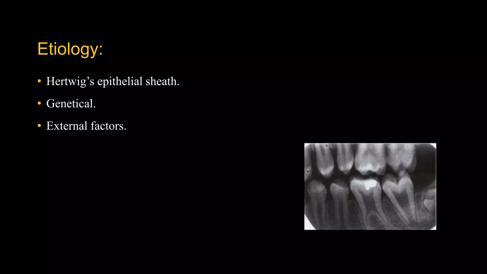
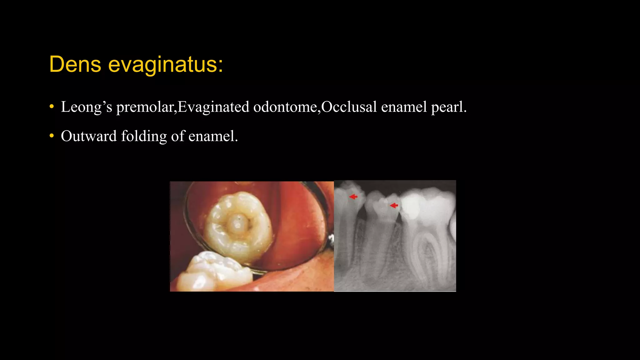
Dr.Shilpa rao
PG IIyr
CONTENTS:
• INTRODUCTION
• TERMINOLOGIES
• DEVELOPMENTAL DISTURBANCES OF JAWS
• DEVELOPMENTAL DISTURBANCES OF TEETH
• DEVELOPMENTAL CYSTS OF JAWS
• CONCLUSION
• REFERENCES
INTRODUCTION:
• Developmental Disturbances of the Jaws affect the normal growth and differentiation of
craniofacial structures. These conditions result in a variety of abnormalities, which affect the
structure, shape, organization and function of the hard and soft tissues. The causes of most of the
disturbances are of unknown etiology but some may be attributed to congenital (present at or
before birth but not necessarily inherited), hereditary (these are apparent at birth, but some may
become evident after many years) or genetic mutations caused due to altered environmental
factors.
TERMINOLOGIES:
• Allele
• Homozygous
• Heterozygous
• Autosomal dominant
• Autosomal recessive
• X linked dominant
• X linked recessive
• Developmental abnormality
• Anamoly
• Malformation
• Monostrotemies.
DEVELOPMENTAL
DISTURBANCES OF JAWS
Agnathia:
• Also called as Otocephaly, holoprosencephaly agnathia,Synotia.
• Agnathia is characterized by hypoplasia or absence of the jaws with abnormally
positioned ears having an autosomal recessive mode of inheritance.
SITE:
• Mandible – Partial or complete
Unilateral or bilateral.
• Maxilla - Maxillary process or even the premaxilla
• It is probably due to failure of migration of neural crest mesenchyme into the
maxillary prominence.
MICROGNATHIA:
• Small jaw.
• True and Apparent micrognathia.
• TRUE : Congenital
Acquired
Syndromes associated:
• Pierre Robin syndrome
• Cri du chat syndrome
• Trisomy 18
• Turner’s syndrome
• Noonan’s syndrome
• Charge syndrome
• Di George syndrome
• Treacher Collins syndrome.
Management:
• The abnormality may be corrected with surgery and orthodontic treatment.
Macrognathia:
• Abnormally large jaws.
• TRUE
• RELATIVE
CONDITIONS ASSOCIATED:
• Paget’s disease.
• Acromegaly
• Leontiasis ossea
Management:
• Surgical correction of such cases is feasible. Ostectomy, or resection of a portion
of the mandible to decrease its length, is now an established procedure, and the
results are usually excellent from both a functional and a cosmetic standpoint.
Clefts:
• The term cleft lip and palate is commonly used to represent two types of
malformation, i.e., cleft lip with or without cleft palate (CL/P) and cleft palate
(CP). Cleft lip and palate are common congenital malformations.
Etiology:
• Genetic liability.
• A defective vascular supply to the area involved.
• A mechanical disturbance in which the size of the tongue may prevent the union of
parts.
• Circulating substances, such as alcohol and certain drugs and toxins.
• Infections.
• Lack of inherent developmental force.
Cleft lip:
Cleft palate:
Classifications:
Syndromes associated with cleft palate:
1)Treacher collins syndrome
2)Pierre robbin syndrome
• Steeple palate- Downs syndrome
• High arched palate:
1)Marfan syndrome
2)Progeria
3)Apert syndrome
4)Turner syndrome
5)Crouzan syndrome
6)Oculodentodigital dysplasia
Management:
DEVELOPMENTAL
DISTURBANCES OF teeth
• Anamolies in teeth size.
• Anamolies in teeth number.
• Anamolies in teeth morphology.
• Anamolies in teeth structure.
• Anamolies in teeth position.
Anamolies in teeth size:
• Microdontia
• Macrodontia
Microdontia:
• True generalized microdontia,
• Relative generalized microdontia.
• Microdontia involving a single tooth.
Frequency of occurrence:
• Max incisors and 3rd molars then mandibular premolars.
Conditions associated:
• Dwarfism.
• Congenital heart disease.
• Down’s syndrome.
• Osteogenesis imperfecta.
• Hemiatrophy of face.
Macrodontia:
• True generalized macrodontia,
• Relative generalized macrodontia.
• Macrodontia involving a single tooth.
Frequency of occurrence:
• Primary – Max canine and mandibular second molar.
• Permanent – Max central incisors and canine and molars and mandibular 2nd PM
and 3rd M.
d/d:
• Fusion/Gemination.
ANAMOLIES IN TETH NUMBER:
• Hypodontia.
• Hyperdontia.
Hypodontia:
• Anodontia.
• Oligodontia.
• Partial anodontia.
Frequency of occurrence:
• Third molars,Premolars,Lateral incisors and mand second premolars.
Conditions associated:
• Anhidrotic ectodermal dysplasia.
• PHC syndrome / Book syndrome.
• Down syndrome.
• Otodental dysplasia.
• Rieger’s syndrome.
• Ellis van creveld ssyndrome.
• Incontinentia pigmenti.
• Chondroectodermal dysplasia.
Hyperdontia:
• Supernumerary teeth.
• Supplemental teeth.
• Accessory teeth.
Mesiodens
Distodens
Peridens
• Four different morphological types of supernumerary teeth have been described:
• Conical
• Tuberculate
• Supplemental
• Odontome.
Etiology:
• Cleavage of tooth germ.
• Extended dental lamina.
• Hyperactivity of dental lamina.
Frequency of occurrence:
• More common in permanent dentition.
• Max molars,Max paramolars,Mesiodens,Mand premolars.
Conditions associated:
• Cleido cranial dysplasia.
• Gardener’s syndrome.
• Pyknodysostosis.
Anamolies in morphology:
• Gemination.
• Fusion.
• Concresence.
• Taurodontism.
• Dilaceration.
• Dens invaginatus.
• Dens evaginatus.
• Enamel pearl.
• Talons cusp.
• Turner’s hypoplasia.
gemination:
• Schizodontia / Twinning.
Frequency of occurrence:
• More prevalent in primary dentition.
• Incisor and canine.
d/d:
• Fusion.
• Macrodontia.
Fusion:
• Syndontia.
Frequency of occurrence:
• Anterior teeth of primary and permanent dentition.
d/d:
• Gemination.
• Macrodontia.
Concresence:
• Fusion of roots.
• True and Acquired.
Taurodontism:
• Bull like teeth.
• The body of the tooth is enlarged at the expense of the roots.
• Apical extension of the pulp chamber below CEJ.
Etiology:
• Hertwig’s epithelial sheath.
• Genetical.
• External factors.
• Shaw classified into four types:
d/d:
• Teeth in conditions like : pseudo-hypoparthyroidism, hypophosphatasia, and
hypophosphatemic vitamin D-resistant and dependent rickets, early stages of
dentinogenesis imperfecta.
Conditions associated:
• Klienfelter’s syndrome.
• Amelogenesis imperfecta.
• Down’s syndrome.
• Osteoporosis.
• Mohr syndrome.
• Tricho-dento-osseous syndrome.
• Ectodermal abnormalities,
• Apert syndrome.
Dilaceration:
• Sharp bend or curve in the tooth.
• Due to mechanical trauma to the calcified portion of partially formed tooth.
BULL’S EYE APPEARANCE
Frequency of occurrence:
• Mand 3rd molars,Max lateral inciors and max premolars.
D/D:
• Fused roots.
• Condensing osteitis.
• A dense bone island.
Dens invaginatus:
Oehler’s classification:
Frequency of occurrence:
• Maxillary lateral incisors,Central incisors,Premolars,Canine and Molars
Dens evaginatus:
• Leong’s premolar,Evaginated odontome,Occlusal enamel pearl.
• Outward folding of enamel.
Talon’s cusp:
• Anamolous hyperplasia of cingulum of incisors.
• T shape.
Conditions associated:
• Rubinstein- Taybi syndrome.
• Mesiodens,Odontome,Peg laterals,Dens invaginatus,Cleft lip,Gemination and
fusion and supernumerary teeth.
d/d:
• Superneumarary teeth.
Enamel pearl:
• Enamel drop,Enamel nodule.Enameloma.
• Small globule of enamel seen on roots of molars.
• Consists of Dentin and pulp sometimes.
d/d:
• Isolated piece of calculus.
• Pulp stone.
TURNER’S HYPOPLASIA:
• Turner ’ s hypoplasia is a term used to describe a permanent tooth with a local
hypoplastic defect in its crown.
Anamolies in structure:
• AMELOGENESIS IMPERFECTA.
• DENTINOGENESIS IMPERFECTA.
• DENTIN DYSPLASIA.
• REGIONAL ODONTODYSPLASIA.
• ENAMEL HYPOPLASIA.
AMELOGENESIS IMPERFECTA:
• Hereditary enamel dysplasia, hereditary brown enamel, hereditary brown
opalescent teeth.
• A structural defect of the tooth enamel.
• Mutations in the amelogenin, enamelin, and kallikrein-4 genes have been
demonstrated to different types of AI.
HYPOPLASTIC:
HYPOCALCIFIED:
HYPOMATURATION:
D/D:
• Dentinogenesis imperfecta.
Dentinogenesis imperfecta.
• Hereditary opalescent dentin.
• Dentinogenesis imperfecta is a genetic anomaly involving primarily the dentin,
although the enamel may be thinner than normal in this condition.
• Acc. to SHIELD:
• TYPE I
• TYPE II
• TYPE III
• Modified by Mendelian to TWO types
Etiology:
• Defect in collagen type I.
• Defect in dentin sialophosphoprotein (DSPP) gene.
Type i:
Type ii:
d/d:
• Amelogenesis imperfecta.
• Dentin dysplasia.
Dentin dysplasia:
• Rootless teeth.
• Dentin dysplasia is a rare disturbance of dentin formation characterized by normal
enamel but atypical dentin formation with abnormal pulpal morphology.
• TWO TYPES according to Shield.
• RADICULAR TYPE.
• CORONAL TYPE.
TYPE 1:
TYPE II:
d/d:
• Dentinogenesis imperfecta.
REGIONAL ODONTODYSPLASIA:
• Odontogenesis imperfecta and ghost teeth.
• Both enamel and dentin are hypoplastic and hypocalcified.
• These teeth may be either primary or permanent. If the primary teeth are affected,
their successors are usually involved.
d/d:
• Dentinogenesis imperfecta.
ENAMEL HYPOPLASIA:
• Enamel hypoplasia may be defined as an incomplete or defective formation
of the organic enamel matrix of teeth.
• Heriditary type
• Environmental type
• Due to Nurtritional deficiencies.
• Congenital syphilis.
• Flourosis.
Based on position:
• IMPACTED TEETH.
• SUBMERGED TEETH.
DEVELOPMENTAL cysts
of jaws
• NASOPALATINE DUCT CYST.
• MEDIAN PALATAL CYST.
• GLOBULOMAXILLARY CYST.
• MEDIAN MANDIBULAR CYST.
• STAFNE CYST.
NASOPALATINE DUCT CYST:
D/D:
• CLINICAL: Perapical abcess RADIOLOGICAL: Enalrged foramen.
Periapical infections
MEDIAN PALATAL CYST:
D/D:
• Periapical infection.
• Palatal torus.
• Minor salivary gland tumors.
GLOBULOMAXILLARY CYST:
D/D:
• Periapical infection.
• Minor salivary gland tumors.
• OKC.
MEDIAN MANDIBULAR CYST:
D/D:
• CGCG.
• KCOT.
STAFNE CYST:
Developmental Lingual Mandibular Salivary Gland Depression (Static bone cavity or defect of the mandible,
lingual mandibular bone cavity, static bone cyst, latent bone cyst, Stafne defect)
D/D;
• Odontogenic cysts.
Conclusion:
References:
• Textbook of Oral pathology Shafer’s 7th edition.
• Text book of Oral and maxillofacial radiology White and pharaoh 4th edition.
• Text book of Oral and maxillofacial radiology Freny R Karjodkar 3rd edition.
• Text book of Diagnostic imaging of the jaws Langland and Langlais.
• Hereditary dentine disorders: dentinogenesis imperfecta and dentine dysplasia Martin J Barron Orphanet
Journal of Rare Diseases 2008, 3:31
• Taurodontism: a review of the condition and endodontic treatment challenges H. Jafarzadeh International
Endodontic Journal, 41, 375–388, 2008
• Amelogenesis Imperfecta: A Review of the Literature Dr Roma M J. Pharm. Sci. & Res. Vol. 8(9), 2016,
1042-1044
• Labial and lingual talon’s cusp A case report Journal of pediatric dentistry 27;4,2005.
• Dens invaginuts ateology,classification and treatment considerations Bader munj et al,Pakistan oral
and dental journal june 2011.
• Taurodontism a case report.karthik et al,journal of dental and orofacial research 2014 dec 2.
Thank you...

Developmental disturbances