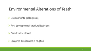
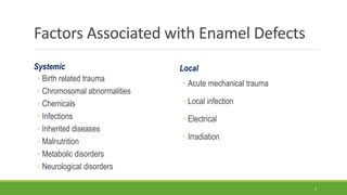
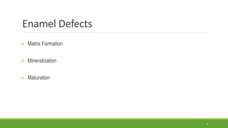
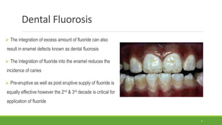
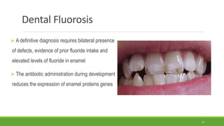
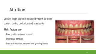
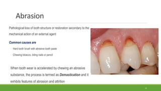
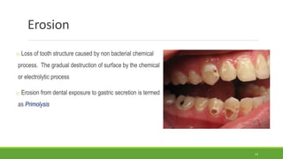
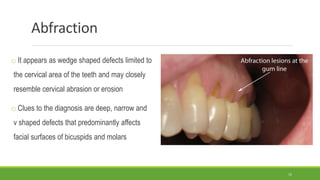
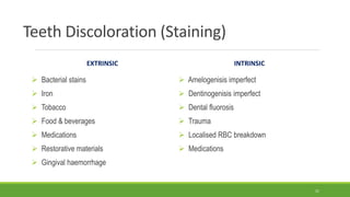
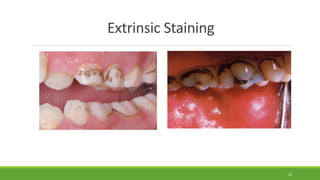
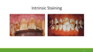
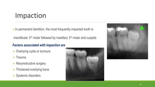
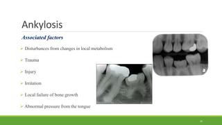
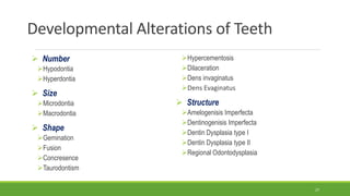
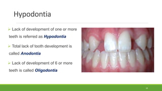
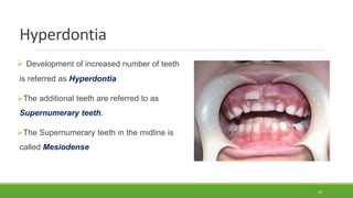
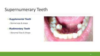
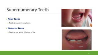
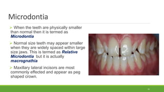
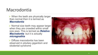
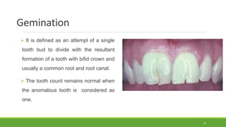
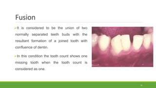
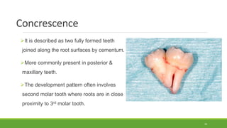
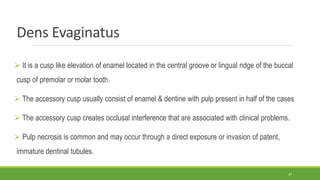
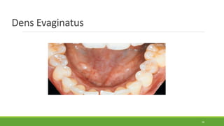
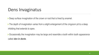
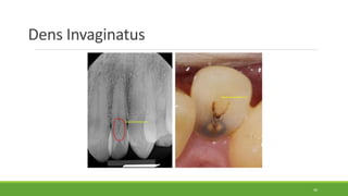
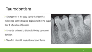
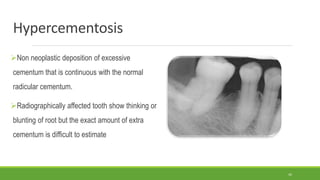
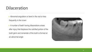
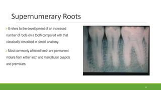
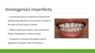
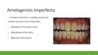
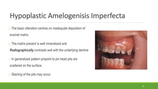
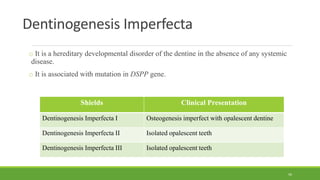
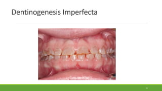
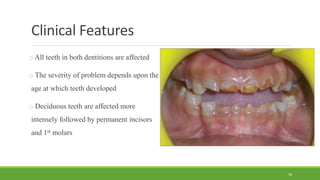
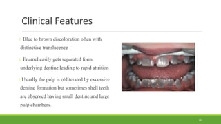
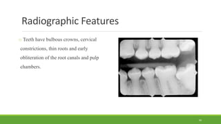
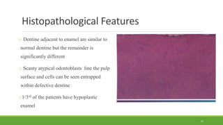
The document discusses various abnormalities and developmental alterations that can occur in teeth. It covers defects in enamel formation like enamel hypoplasia and fluorosis. It also discusses post-developmental structural loss through processes like attrition, abrasion, and erosion. Other topics include discoloration of teeth from extrinsic and intrinsic factors, localized disturbances in eruption, and developmental defects affecting tooth number, size, shape and structure. Specific conditions discussed in more depth include dentinogenesis imperfecta, amelogenesis imperfecta, and dental fluorosis. Treatment options for some of these conditions are also mentioned.
Dentin dysplasia (DD) is a rare hereditary disturbance is inherited as an autosomal dominant trait.
unknown etiology that affects approximately 1 :100,000.
In 1972, Witkop classified it into type I and type II which affect both dentitions.DD Type I
Radicular dentin dysplasia
Characterized by:-
1.Both dentitions are affected.
2.Normal appearing crowns
3.No or only rudimentary root development (rootless teeth)
4.Incomplete or total obliteration of the pulp chamber.
5.Teeth may exhibit extreme mobility and exfoliate prematurely.DD type II
coronal dentin dysplasia
Characterized by:-
1.partial pulpal obliteration.
2.Thistle-tube-or flame-shaped coronal pulp chambers
3. Thread-like root canals
4. Usually the absence of periapical radiolucencies.
5. In this type of anomaly, teeth roots are of normal shape and contour.The enamel and the immediately subjacent dentin appear normal.
Deeper layers of dentin show an atypical tubular pattern with an amorphous, atubular area, and irregular organization.
Normal dentinal tubule formation appears to have been blocked so that new dentine forms around obstacles and takes on the characteristic appearances described as “lava flowing around boulders”The radiograph revealed features of dentine dysplasia type I with normal appearance of crown but no root development Autosomal Dominant Disorder:
Manifested in heterozygous states
At least one parent of index case is usually affected
Both males and females are affected.
Clinical feature can be modified by variation in penetrance and expressivity. Some individual inherit the mutant gene but are phenotpically normal. This is reffered to as “incomplete penetrance”.
In many condition the age of onset is delayed.
Inheritance Pattern:
Typical pattern is a heterozygous affected parent with a homozygous unaffected parent.
Every child has one chance in two of having the disease
Both sexes are affected equally..Autosomal Recessive Disorder
Largest category of Mendelian disorder
Usually does not affect the parent of the affected individual, but sibling may show the disease.
Complete penetrance is common.
Onset is frequently early in life.
Usually affect enzymatic proteins.
Pattern Of Inheritance:
Typical pattern is two heterozygous unaffected (carrier) parent.
The triat does not usually affect the parent, but siblings may show the disease
Siblings have one chance in four of being affected
Both sexes affected equally.
Tooth resorption is the progressive loss of dentine and cementum by the action of osteoclasts. This is a physiological process in the exfoliation of the primary dentition, caused by osteoclast differentiation due to pressure exerted by the erupting permanent tooth
Pulmonary Thromboembolism - etilogy, types, medical- Surgical and nursing man...VarunMahajani
Disruption of blood supply to lung alveoli due to blockage of one or more pulmonary blood vessels is called as Pulmonary thromboembolism. In this presentation we will discuss its causes, types and its management in depth.
Ethanol (CH3CH2OH), or beverage alcohol, is a two-carbon alcohol
that is rapidly distributed in the body and brain. Ethanol alters many
neurochemical systems and has rewarding and addictive properties. It
is the oldest recreational drug and likely contributes to more morbidity,
mortality, and public health costs than all illicit drugs combined. The
5th edition of the Diagnostic and Statistical Manual of Mental Disorders
(DSM-5) integrates alcohol abuse and alcohol dependence into a single
disorder called alcohol use disorder (AUD), with mild, moderate,
and severe subclassifications (American Psychiatric Association, 2013).
In the DSM-5, all types of substance abuse and dependence have been
combined into a single substance use disorder (SUD) on a continuum
from mild to severe. A diagnosis of AUD requires that at least two of
the 11 DSM-5 behaviors be present within a 12-month period (mild
AUD: 2–3 criteria; moderate AUD: 4–5 criteria; severe AUD: 6–11 criteria).
The four main behavioral effects of AUD are impaired control over
drinking, negative social consequences, risky use, and altered physiological
effects (tolerance, withdrawal). This chapter presents an overview
of the prevalence and harmful consequences of AUD in the U.S.,
the systemic nature of the disease, neurocircuitry and stages of AUD,
comorbidities, fetal alcohol spectrum disorders, genetic risk factors, and
pharmacotherapies for AUD.
micro teaching on communication m.sc nursing.pdfAnurag Sharma
Microteaching is a unique model of practice teaching. It is a viable instrument for the. desired change in the teaching behavior or the behavior potential which, in specified types of real. classroom situations, tends to facilitate the achievement of specified types of objectives.
Anti ulcer drugs and their Advance pharmacology ||
Anti-ulcer drugs are medications used to prevent and treat ulcers in the stomach and upper part of the small intestine (duodenal ulcers). These ulcers are often caused by an imbalance between stomach acid and the mucosal lining, which protects the stomach lining.
||Scope: Overview of various classes of anti-ulcer drugs, their mechanisms of action, indications, side effects, and clinical considerations.
The prostate is an exocrine gland of the male mammalian reproductive system
It is a walnut-sized gland that forms part of the male reproductive system and is located in front of the rectum and just below the urinary bladder
Function is to store and secrete a clear, slightly alkaline fluid that constitutes 10-30% of the volume of the seminal fluid that along with the spermatozoa, constitutes semen
A healthy human prostate measures (4cm-vertical, by 3cm-horizontal, 2cm ant-post ).
It surrounds the urethra just below the urinary bladder. It has anterior, median, posterior and two lateral lobes
It’s work is regulated by androgens which are responsible for male sex characteristics
Generalised disease of the prostate due to hormonal derangement which leads to non malignant enlargement of the gland (increase in the number of epithelial cells and stromal tissue)to cause compression of the urethra leading to symptoms (LUTS
Explore natural remedies for syphilis treatment in Singapore. Discover alternative therapies, herbal remedies, and lifestyle changes that may complement conventional treatments. Learn about holistic approaches to managing syphilis symptoms and supporting overall health.
Title: Sense of Smell
Presenter: Dr. Faiza, Assistant Professor of Physiology
Qualifications:
MBBS (Best Graduate, AIMC Lahore)
FCPS Physiology
ICMT, CHPE, DHPE (STMU)
MPH (GC University, Faisalabad)
MBA (Virtual University of Pakistan)
Learning Objectives:
Describe the primary categories of smells and the concept of odor blindness.
Explain the structure and location of the olfactory membrane and mucosa, including the types and roles of cells involved in olfaction.
Describe the pathway and mechanisms of olfactory signal transmission from the olfactory receptors to the brain.
Illustrate the biochemical cascade triggered by odorant binding to olfactory receptors, including the role of G-proteins and second messengers in generating an action potential.
Identify different types of olfactory disorders such as anosmia, hyposmia, hyperosmia, and dysosmia, including their potential causes.
Key Topics:
Olfactory Genes:
3% of the human genome accounts for olfactory genes.
400 genes for odorant receptors.
Olfactory Membrane:
Located in the superior part of the nasal cavity.
Medially: Folds downward along the superior septum.
Laterally: Folds over the superior turbinate and upper surface of the middle turbinate.
Total surface area: 5-10 square centimeters.
Olfactory Mucosa:
Olfactory Cells: Bipolar nerve cells derived from the CNS (100 million), with 4-25 olfactory cilia per cell.
Sustentacular Cells: Produce mucus and maintain ionic and molecular environment.
Basal Cells: Replace worn-out olfactory cells with an average lifespan of 1-2 months.
Bowman’s Gland: Secretes mucus.
Stimulation of Olfactory Cells:
Odorant dissolves in mucus and attaches to receptors on olfactory cilia.
Involves a cascade effect through G-proteins and second messengers, leading to depolarization and action potential generation in the olfactory nerve.
Quality of a Good Odorant:
Small (3-20 Carbon atoms), volatile, water-soluble, and lipid-soluble.
Facilitated by odorant-binding proteins in mucus.
Membrane Potential and Action Potential:
Resting membrane potential: -55mV.
Action potential frequency in the olfactory nerve increases with odorant strength.
Adaptation Towards the Sense of Smell:
Rapid adaptation within the first second, with further slow adaptation.
Psychological adaptation greater than receptor adaptation, involving feedback inhibition from the central nervous system.
Primary Sensations of Smell:
Camphoraceous, Musky, Floral, Pepperminty, Ethereal, Pungent, Putrid.
Odor Detection Threshold:
Examples: Hydrogen sulfide (0.0005 ppm), Methyl-mercaptan (0.002 ppm).
Some toxic substances are odorless at lethal concentrations.
Characteristics of Smell:
Odor blindness for single substances due to lack of appropriate receptor protein.
Behavioral and emotional influences of smell.
Transmission of Olfactory Signals:
From olfactory cells to glomeruli in the olfactory bulb, involving lateral inhibition.
Primitive, less old, and new olfactory systems with different path
Prix Galien International 2024 Forum ProgramLevi Shapiro
June 20, 2024, Prix Galien International and Jerusalem Ethics Forum in ROME. Detailed agenda including panels:
- ADVANCES IN CARDIOLOGY: A NEW PARADIGM IS COMING
- WOMEN’S HEALTH: FERTILITY PRESERVATION
- WHAT’S NEW IN THE TREATMENT OF INFECTIOUS,
ONCOLOGICAL AND INFLAMMATORY SKIN DISEASES?
- ARTIFICIAL INTELLIGENCE AND ETHICS
- GENE THERAPY
- BEYOND BORDERS: GLOBAL INITIATIVES FOR DEMOCRATIZING LIFE SCIENCE TECHNOLOGIES AND PROMOTING ACCESS TO HEALTHCARE
- ETHICAL CHALLENGES IN LIFE SCIENCES
- Prix Galien International Awards Ceremony
These simplified slides by Dr. Sidra Arshad present an overview of the non-respiratory functions of the respiratory tract.
Learning objectives:
1. Enlist the non-respiratory functions of the respiratory tract
2. Briefly explain how these functions are carried out
3. Discuss the significance of dead space
4. Differentiate between minute ventilation and alveolar ventilation
5. Describe the cough and sneeze reflexes
Study Resources:
1. Chapter 39, Guyton and Hall Textbook of Medical Physiology, 14th edition
2. Chapter 34, Ganong’s Review of Medical Physiology, 26th edition
3. Chapter 17, Human Physiology by Lauralee Sherwood, 9th edition
4. Non-respiratory functions of the lungs https://academic.oup.com/bjaed/article/13/3/98/278874
1. Abnormalities of Teeth
Dr. Arsalan Malik
Assistant Professor & HOD (Oral Pathology)
ِيم ِحَّالر ِمنْحَّالر ِهللا ِمْسِب
1
2. Environmental Alterations of Teeth
Developmental tooth defects
Post developmental structural tooth loss
Discoloration of teeth
Localized disturbances in eruption
2
5. Enamel Defects
Enamel Hypoplasia
Occurs in form of pits, grooves or larger areas of missing enamel
Diffuse Opacities
Variation in the translucency of enamel. Normal thickness with white opacities and no boundary
Demarcated Opacities
Variation in the translucency of enamel. Normal thickness with white opacities and clear boundary
5
6. Turner’s Hypoplasia
It is caused by periapical inflammatory
disease of the overlying deciduous tooth.
Most commonly noted in permanent bicuspids
due to overlying deciduous molars
6
7. Turner’s Hypoplasia
Degree of damage depends upon stage of development, length of time of infection,
virulence of organism and the host resistance factors
If the primary tooth develops caries, the successor is twice likely to develop caries
If the primary tooth is extracted for some reason other than trauma, prevalence of
demarcated enamel is increased 5 folds.
7
8. Molar Incisor Hypomineralization
o Enamel defects of one or more first permanent molars
o The altered enamel is white, yellow or brown and often
soft and porous.
o Affected teeth are sensitive to cold and warmth and
enamel chip off easily
o Difficult to anesthetize during dental procedures
8
9. Dental Fluorosis
The integration of excess amount of fluoride can also
result in enamel defects known as dental fluorosis
The integration of fluoride into the enamel reduces the
incidence of caries
Pre-eruptive as well as post eruptive supply of fluoride is
equally effective however the 2nd & 3rd decade is critical for
application of fluoride
9
10. Dental Fluorosis
A definitive diagnosis requires bilateral presence
of defects, evidence of prior fluoride intake and
elevated levels of fluoride in enamel
The antibiotic administration during development
reduces the expression of enamel proteins genes
10
12. Attrition
Loss of tooth structure caused by tooth to tooth
contact during occlusion and mastication
Main factors are
◦ Poor quality or absent enamel
◦ Premature contacts
◦ Intra oral abrasive, erosions and grinding habits
12
13. Abrasion
Pathological loss of tooth structure or restoration secondary to the
mechanical action of an external agent
Common causes are
◦ Hard tooth brush with abrasive tooth paste
◦ Chewing tobacco, biting nails or pencil
When tooth wear is accelerated by chewing an abrasive
substance, the process is termed as Demastication and it
exhibits features of abrasion and attrition
13
14. Erosion
o Loss of tooth structure caused by non bacterial chemical
process. The gradual destruction of surface by the chemical
or electrolytic process
o Erosion from dental exposure to gastric secretion is termed
as Primolysis
14
15. Abfraction
o It appears as wedge shaped defects limited to
the cervical area of the teeth and may closely
resemble cervical abrasion or erosion
o Clues to the diagnosis are deep, narrow and
v shaped defects that predominantly affects
facial surfaces of bicuspids and molars
15
16. Resorption
Destruction of tooth can also occur through resorption, which is accomplished by cells
located in the pulp (Internal resorption) or in the periodontal ligament (External
resorption)
Internal resorption is a relatively rare occurrence and mostly it develops after trauma to
the pulpal tissues of the tooth
It continues as long as vital pulp tissue remains and may result in communication of
pulp with the PDL.
16
17. Factors Associated with External Resorption
Cysts
Dental trauma
Excessive mechanical forces
Excess occlusal forces
Hormonal imbalance
Peget’s disease of bone
Periodontal treatment
Tumours
Reimplantation of teeth
17
18. Clinical Features
Inflammatory resorption
The resorbed dentin is replaced by inflamed granulation tissues. Most commonly cervical portion is
involved and resorption continues as long
Replacement resorption
Portion of the dentinal walls are resorbed and replaced with bone or cementum like material.
Radiographically it appears as enlargement of the pulp canal and filled with a material that is less
radiodense as compared to surrounding zone.
After re-implantation of tooth, extensive resorption starts without rapid intervention
18
20. Histopathological Features
Pulp tissues are vascular and exhibit increased cellularity.
Numerous multinucleated dentinoclasts are present adjacent to
the dentinal walls.
Inflammatory infiltrate is present
In replacement resorption, the normal pulp tissues are replaced
by woven bone that is fused to adjacent dentine.
Areas of resorption are often repaired by deposition of
osteodentine
Extensive bone deposition in external resorption may lead to
ankylosis
20
24. Localized Disturbances in Eruption
Impaction
Teeth that cease to erupt before emergence are called Impacted
Ankylosis
Eruption continues after emergence of teeth to compensate for masticatory wear and the growth of
jaws. The cessation of eruption after emergence is termed Ankylosis
24
25. Impaction
In permanent dentition, the most frequently impacted tooth is
mandibular 3rd molar followed by maxillary 3rd molar and cuspids
Factors associated with impaction are
Overlying cysts or tumours
Trauma
Reconstructive surgery
Thickened overlying bone
Systemic disorders
25
26. Ankylosis
Associated factors
Disturbances from changes in local metabolism
Trauma
Injury
Irritation
Local failure of bone growth
Abnormal pressure from the tongue
26
27. Developmental Alterations of Teeth
Number
Hypodontia
Hyperdontia
Size
Microdontia
Macrodontia
Shape
Gemination
Fusion
Concresence
Taurodontism
Hypercementosis
Dilaceration
Dens invaginatus
Dens Evaginatus
Structure
Amelogenisis Imperfecta
Dentinogenisis Imperfecta
Dentin Dysplasia type I
Dentin Dysplasia type II
Regional Odontodysplasia
27
28. Hypodontia
Lack of development of one or more
teeth is referred as Hypodontia
Total lack of tooth development is
called Anodontia
Lack of development of 6 or more
teeth is called Oligodontia
28
29. Hyperdontia
Development of increased number of teeth
is referred as Hyperdontia
The additional teeth are referred to as
Supernumerary teeth.
The Supernumerary teeth in the midline is
called Mesiodense
29
32. Microdontia
When the teeth are physically smaller
than normal then it is termed as
Microdontia
Normal size teeth may appear smaller
when they are widely spaced within large
size jaws. This is termed as Relative
Microdontia but it is actually
macrognathia
Maxillary lateral incisors are most
commonly effected and appear as peg
shaped crown.
32
33. Macrodontia
When the teeth are physically larger
than normal then it is termed as
Macrodontia
Normal size teeth may appear larger
when they are crowded within small
size jaws. This is termed as Relative
Macrodontia but it is actually
micrognathia
Diffuse macrodontia has been
observed in pituitary gigantism and
otodental syndrome
33
34. Gemination
It is defined as an attempt of a single
tooth bud to divide with the resultant
formation of a tooth with bifid crown and
usually a common root and root canal.
The tooth count remains normal when
the anomalous tooth is considered as
one.
34
35. Fusion
It is considered to be the union of two
normally separated teeth buds with the
resultant formation of a joined tooth with
confluence of dentin.
In this condition the tooth count shows one
missing tooth when the tooth count is
considered as one.
35
36. Concrescence
It is described as two fully formed teeth
joined along the root surfaces by cementum.
More commonly present in posterior &
maxillary teeth.
The development pattern often involves
second molar tooth where roots are in close
proximity to 3rd molar tooth.
36
37. Dens Evaginatus
It is a cusp like elevation of enamel located in the central groove or lingual ridge of the buccal
cusp of premolar or molar tooth.
The accessory cusp usually consist of enamel & dentine with pulp present in half of the cases
The accessory cusp creates occlusal interference that are associated with clinical problems.
Pulp necrosis is common and may occur through a direct exposure or invasion of patent,
immature dentinal tubules.
37
39. Dens Invaginatus
Deep surface invagination of the crown or root that is lined by enamel.
The depth of invagination varies from a slight enlargement of the cingulum pit to a deep
infolding that extends to apex.
Occasionally the invagination may be large and resemble a tooth within tooth appearance
called den in dente.
39
41. Taurodontism
Enlargement of the body & pulp chamber of a
multirooted tooth with apical displacement of the pulpal
floor & bifurcation of the root.
It may be unilateral or bilateral affecting permanent
dentition.
Classified into mild, moderate and sever forms
41
42. Hypercementosis
Non neoplastic deposition of excessive
cementum that is continuous with the normal
radicular cementum.
Radiographically affected tooth show thinking or
blunting of root but the exact amount of extra
cementum is difficult to estimate
42
44. Dilaceration
Abnormal angulation or bend in the root or less
frequently in the crown
A number of teeth having dilacerations arises
after injury that displaces the calcified portion of the
tooth germ and remainder of the tooth is formed at
an abnormal angle
44
45. Supernumerary Roots
It refers to the development of an increased
number of roots on a tooth compared with that
classically described in dental anatomy.
Most commonly affected teeth are permanent
molars from either arch and mandibular cuspids
and premolars
45
46. Amelogenisis Imperfecta
A complicated group of conditions that demonstrate
developmental alterations in the structure of enamel in
the absence of any systemic disease.
Different classification systems were proposed but
Witkop’s Classification is widely accepted.
It is based on phenotype and pedigree (clinical
appearance & apparent mode of inheritance)
46
47. Amelogenisis Imperfecta
Formation of enamel is a multistep process and
problem may arise in any of these them.
1) Elaboration of the organic matrix
2) Mineralization of the matrix
3) Maturation of the enamel.
47
48. Hypoplastic Amelogenisis Imperfecta
o The basic alteration centres on inadequate deposition of
enamel matrix.
o The matrix present is well mineralized and
Radiographically contrasts well with the underlying dentine
o In generalized pattern pinpoint to pin head pits are
scattered on the surface.
o Staining of the pits may occur.
48
49. Hypoplastic Amelogenisis Imperfecta
o In localized pattern, the affected teeth demonstrate horizontal rows of pits, linear
depression or one large area of hypocalcification.
o In other patterns of this disorder, enamel may appear smooth, thin, hard and glossy.
o Radiographically demonstrate a thin radio-opaque line of enamel.
49
50. Hypomaturation Amelogenisis Imperfecta
• Enamel matrix is laid down properly and begin to mineralize but there is defect in
maturation of enamel crystal structure
• Affected teeth are normal in shape but show mottled appearance and tend to chip easily
• Radiographically its density is similar to dentine
50
52. Hypocalcified Amelogenisis Imperfecta
o Enamel matrix is laid down properly but failed to mineralize properly
o Affected teeth are proper in shape but very soft & easily lost
o It has more tendency of calculus deposition on it
o Radiographically the density of enamel & dentine is similar
o After some duration time of eruption, cuspal enamel is lost leaving occlusal surfaces
irregular
52
54. Histopathological Features
These are not evident in routine preparations because decalcification of the teeth is
necessary before processing and all the enamel is lost in it
Ground sections of Non decalcified teeth are prepared for examination
The alterations are highly diverse and depend upon the type of variation in enamel structure
54
55. Treatment
o Main problems are aesthetics, dental sensitivity and loss of vertical dimension
o In some types, increased prevalence of caries, anterior open bite, delayed eruption and associated
gingival enlargement are observed
o Crown placement
o Full dentures
o Veneers
oGlass Ionomer Cements restorations
55
56. Dentinogenesis Imperfecta
o It is a hereditary developmental disorder of the dentine in the absence of any systemic
disease.
o It is associated with mutation in DSPP gene.
Shields Clinical Presentation
Dentinogenesis Imperfecta I Osteogenesis imperfect with opalescent dentine
Dentinogenesis Imperfecta II Isolated opalescent teeth
Dentinogenesis Imperfecta III Isolated opalescent teeth
56
58. Clinical Features
o All teeth in both dentitions are affected
o The severity of problem depends upon the
age at which teeth developed
o Deciduous teeth are affected more
intensely followed by permanent incisors
and 1st molars
58
59. Clinical Features
o Blue to brown discoloration often with
distinctive translucence
o Enamel easily gets separated form
underlying dentine leading to rapid attrition
oUsually the pulp is obliterated by excessive
dentine formation but sometimes shell teeth
are observed having small dentine and large
pulp chambers.
59
60. Radiographic Features
o Teeth have bulbous crowns, cervical
constrictions, thin roots and early
obliteration of the root canals and pulp
chambers.
60
61. Histopathological Features
o Dentine adjacent to enamel are similar to
normal dentine but the remainder is
significantly different
o Scanty atypical odontoblasts line the pulp
surface and cells can be seen entrapped
within defective dentine
o1/3rd of the patients have hypoplastic
enamel
61
62. Treatment
o Teeth are not good candidates for crown placement because of cervical fractures
o Overly dentures placed on teeth with fluoride releasing material is being practiced in
some cases
o In cases with lose of vertical dimension, metal crowns are used with adhesive bonding
agents
o On occlusal surfaces composites are being used
62