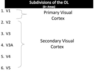
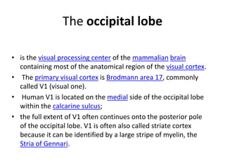
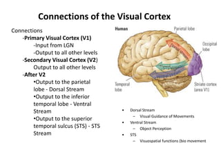
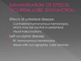
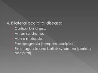
The occipital lobe is the visual processing center of the brain containing most of the visual cortex. It contains the primary visual cortex (V1) and several extrastriate areas involved in more complex visual tasks. Lesions can cause visual field defects, cortical blindness, visual agnosias or hallucinations depending on the location and extent of damage. Balint's syndrome and simultanagnosia involve bilateral lesions disrupting global visual perception while preserving local details.
the recently evolved lobe which is responsible for the higher mental process which makes human unique. the damage can result in many impairments. this presentation gives insight into a few syndromes that could occur due to the frontal lobe damage
Visual System - the visual pathway basicRituYadav112
It consists of a brief description of the visual system. The different types of cells present in the retina and their connections among each other. Signal transmission in the retina. Some basic description about the visual field , receptive field and some basic terminologies. The brief description about the visual pathway. The visual cortex and the primary and extrastriate Visual Cortex.
USMLE NEUROANATOMY 020 Orbit and globe anatomical structures of the eye soc...AHMED ASHOUR
he orbit and globe refer to the anatomical structures of the eye socket (orbit) and the eyeball (globe). Understanding the surgical anatomy of these structures is crucial for procedures related to ophthalmology and orbital surgery.
Understanding the surgical anatomy of the orbit and globe is vital for ophthalmic surgeons and other professionals involved in eye-related procedures. Surgical interventions aim to address various eye conditions, improve vision, and restore or enhance the aesthetic appearance of the eye and surrounding structures.
CONCEPT OF NODOPATHIES AND PARANODOPATHIES.pptxNeurologyKota
emergence of autoimmune neuropathies and role of nodal and paranodal regions in their pathophysiology.
Peripheral neuropathies are traditionally categorized into demyelinating or axonal.
dysfunction at nodal/paranodal region key for better understanding of patients with immune mediated neuropathies.
antibodies targeting node and paranode of myelinated nerves have been increasingly detected in patients with immune mediated neuropathies.
have clinical phenotype similar common inflammatory neuropathies like Guillain Barre syndrome and chronic inflammatory demyelinating polyradiculoneuropathy
they respond poorly to conventional first line immunotherapies like IVIG
This presentation briefs out the approach of dementia assessment in line with consideration of recent advances. Now the pattern of assessment has evolved towards examining each individual domain rather than lobar assessment.
This presentation contains information about Dementia in Young onset. Also it describes the etiologies, clinical feature of common YOD & their management.
Entrapment Syndromes of Lower Limb.pptxNeurologyKota
This presentation contains information about the various Entrapment syndromes of Lower limb in descending order of topography. It also contains information about etiology, clinical features and management of each of these entrapment syndromes with special emphasis on electrodiagnostic confirmation.
TEST BANK for Operations Management, 14th Edition by William J. Stevenson, Ve...kevinkariuki227
TEST BANK for Operations Management, 14th Edition by William J. Stevenson, Verified Chapters 1 - 19, Complete Newest Version.pdf
Prix Galien International 2024 Forum ProgramLevi Shapiro
June 20, 2024, Prix Galien International and Jerusalem Ethics Forum in ROME. Detailed agenda including panels:
- ADVANCES IN CARDIOLOGY: A NEW PARADIGM IS COMING
- WOMEN’S HEALTH: FERTILITY PRESERVATION
- WHAT’S NEW IN THE TREATMENT OF INFECTIOUS,
ONCOLOGICAL AND INFLAMMATORY SKIN DISEASES?
- ARTIFICIAL INTELLIGENCE AND ETHICS
- GENE THERAPY
- BEYOND BORDERS: GLOBAL INITIATIVES FOR DEMOCRATIZING LIFE SCIENCE TECHNOLOGIES AND PROMOTING ACCESS TO HEALTHCARE
- ETHICAL CHALLENGES IN LIFE SCIENCES
- Prix Galien International Awards Ceremony
Pulmonary Thromboembolism - etilogy, types, medical- Surgical and nursing man...VarunMahajani
Disruption of blood supply to lung alveoli due to blockage of one or more pulmonary blood vessels is called as Pulmonary thromboembolism. In this presentation we will discuss its causes, types and its management in depth.
Couples presenting to the infertility clinic- Do they really have infertility...Sujoy Dasgupta
Dr Sujoy Dasgupta presented the study on "Couples presenting to the infertility clinic- Do they really have infertility? – The unexplored stories of non-consummation" in the 13th Congress of the Asia Pacific Initiative on Reproduction (ASPIRE 2024) at Manila on 24 May, 2024.
These simplified slides by Dr. Sidra Arshad present an overview of the non-respiratory functions of the respiratory tract.
Learning objectives:
1. Enlist the non-respiratory functions of the respiratory tract
2. Briefly explain how these functions are carried out
3. Discuss the significance of dead space
4. Differentiate between minute ventilation and alveolar ventilation
5. Describe the cough and sneeze reflexes
Study Resources:
1. Chapter 39, Guyton and Hall Textbook of Medical Physiology, 14th edition
2. Chapter 34, Ganong’s Review of Medical Physiology, 26th edition
3. Chapter 17, Human Physiology by Lauralee Sherwood, 9th edition
4. Non-respiratory functions of the lungs https://academic.oup.com/bjaed/article/13/3/98/278874
Report Back from SGO 2024: What’s the Latest in Cervical Cancer?bkling
Are you curious about what’s new in cervical cancer research or unsure what the findings mean? Join Dr. Emily Ko, a gynecologic oncologist at Penn Medicine, to learn about the latest updates from the Society of Gynecologic Oncology (SGO) 2024 Annual Meeting on Women’s Cancer. Dr. Ko will discuss what the research presented at the conference means for you and answer your questions about the new developments.
Explore natural remedies for syphilis treatment in Singapore. Discover alternative therapies, herbal remedies, and lifestyle changes that may complement conventional treatments. Learn about holistic approaches to managing syphilis symptoms and supporting overall health.
- Video recording of this lecture in English language: https://youtu.be/lK81BzxMqdo
- Video recording of this lecture in Arabic language: https://youtu.be/Ve4P0COk9OI
- Link to download the book free: https://nephrotube.blogspot.com/p/nephrotube-nephrology-books.html
- Link to NephroTube website: www.NephroTube.com
- Link to NephroTube social media accounts: https://nephrotube.blogspot.com/p/join-nephrotube-on-social-media.html
These lecture slides, by Dr Sidra Arshad, offer a quick overview of physiological basis of a normal electrocardiogram.
Learning objectives:
1. Define an electrocardiogram (ECG) and electrocardiography
2. Describe how dipoles generated by the heart produce the waveforms of the ECG
3. Describe the components of a normal electrocardiogram of a typical bipolar leads (limb II)
4. Differentiate between intervals and segments
5. Enlist some common indications for obtaining an ECG
Study Resources:
1. Chapter 11, Guyton and Hall Textbook of Medical Physiology, 14th edition
2. Chapter 9, Human Physiology - From Cells to Systems, Lauralee Sherwood, 9th edition
3. Chapter 29, Ganong’s Review of Medical Physiology, 26th edition
4. Electrocardiogram, StatPearls - https://www.ncbi.nlm.nih.gov/books/NBK549803/
5. ECG in Medical Practice by ABM Abdullah, 4th edition
6. ECG Basics, http://www.nataliescasebook.com/tag/e-c-g-basics
Flu Vaccine Alert in Bangalore Karnatakaaddon Scans
As flu season approaches, health officials in Bangalore, Karnataka, are urging residents to get their flu vaccinations. The seasonal flu, while common, can lead to severe health complications, particularly for vulnerable populations such as young children, the elderly, and those with underlying health conditions.
Dr. Vidisha Kumari, a leading epidemiologist in Bangalore, emphasizes the importance of getting vaccinated. "The flu vaccine is our best defense against the influenza virus. It not only protects individuals but also helps prevent the spread of the virus in our communities," he says.
This year, the flu season is expected to coincide with a potential increase in other respiratory illnesses. The Karnataka Health Department has launched an awareness campaign highlighting the significance of flu vaccinations. They have set up multiple vaccination centers across Bangalore, making it convenient for residents to receive their shots.
To encourage widespread vaccination, the government is also collaborating with local schools, workplaces, and community centers to facilitate vaccination drives. Special attention is being given to ensuring that the vaccine is accessible to all, including marginalized communities who may have limited access to healthcare.
Residents are reminded that the flu vaccine is safe and effective. Common side effects are mild and may include soreness at the injection site, mild fever, or muscle aches. These side effects are generally short-lived and far less severe than the flu itself.
Healthcare providers are also stressing the importance of continuing COVID-19 precautions. Wearing masks, practicing good hand hygiene, and maintaining social distancing are still crucial, especially in crowded places.
Protect yourself and your loved ones by getting vaccinated. Together, we can help keep Bangalore healthy and safe this flu season. For more information on vaccination centers and schedules, residents can visit the Karnataka Health Department’s official website or follow their social media pages.
Stay informed, stay safe, and get your flu shot today!
2. The two occipital lobes
• Are the smallest of four paired lobes in the
human cerebral cortex.
• Separated from parietal lobe by: Parieto-occipital
sulcus.
• The OL 3 landmarks:
• Calcrine fissure: Div. The upper and lower halves
of the visual world.
• Lingual Gyrus:
• Fusiform Gyrus:
3.
4.
5. Four Lobes on the Lateral Surface
OCCIPITAL
LOBE
The occipital lobe is
separated from the
parietal
and temporal lobes by
the
line between the
parieto-
occipital sulcus and the
preoccipital notch
FRONTAL
LOBE
PARIETAL
LOBE
TEMPORAL
LOBE
Preocci
pital
Notch
Parieto-occipital
Sulcus
11. The occipital lobe
• is the visual processing center of the mammalian brain
containing most of the anatomical region of the visual cortex.
• The primary visual cortex is Brodmann area 17, commonly
called V1 (visual one).
• Human V1 is located on the medial side of the occipital lobe
within the calcarine sulcus;
• the full extent of V1 often continues onto the posterior pole
of the occipital lobe. V1 is often also called striate cortex
because it can be identified by a large stripe of myelin, the
Stria of Gennari.
12. Extrastriate regions
• Visually driven regions outside V1 are called
extrastriate cortex.
• There are many extrastriate regions, and these
are specialized for different visual tasks, such
as visuospatial processing, color
discrimination, and motion perception.
13. Connections of the Visual Cortex
Connections
-Primary Visual Cortex (V1)
-Input from LGN
-Output to all other levels
-Secondary Visual Cortex (V2)
Output to all other levels
-After V2
•Output to the parietal
lobe - Dorsal Stream
•Output to the inferior
temporal lobe - Ventral
Stream
•Output to the superior
temporal sulcus (STS) - STS
Stream
• Dorsal Stream
– Visual Guidance of Movements
• Ventral Stream
– Object Perception
• STS
– Visuospatial functions (bio movement
14. Connections
V1- Largest area, called striate cortex.
Receives the largest input from the
LGN and projects to ALL other occipital
regions. 1st processing level.
V2 - Also projects to all other occipital areas.
Segregates info from V1.
15. Pathways into the visual brain
Geniculo-striate system
Tecto-pulvinar system
LGN V1V2
Ventral (TempL)
“What”
Obj. perception & recog.
Tectum
(Sup Colli)
Pulvinar
(Thal)
2
Dorsal (ParL)
“How” or Where”
Visual Guidence of mov.
1
Eye
16. Functions
• A significant functional aspect of the occipital
lobe is that it contains the primary visual cortex.
• Retinal sensors convey stimuli through the optic
tracts to the lateral geniculate bodies, where
optic radiations continue to the visual cortex.
• Each visual cortex receives raw sensory
information from the outside half of the retina on
the same side of the head and from the inside
half of the retina on the other side of the head.
17. O.L Function
V1 & V2 - function like mailboxes: segregating info
to other areas., receives primary visual
impressions Color/Form/Motion/Size and
illumination.
V3, V3A, V4, V5- Visual association areas-
Recognition and identification of objects, storage
of visual memories, it functions in more complex
visual recognition and perception, revisualization,
visual association and spatial orientation.
18. The primary visual cortex (Brodmann area 17 or,
according to more recent nomenclature, V1)
• is located almost entirely on the medial surface of the
occipital lobe; just a small portion (perhaps 1 cm long)
extends around the posterior pole onto the lateral surface.
• The visual cortex also is called the striate cortex because a
white myelinated fiber layer, the white stria of Gennari,is
characteristic of this area.
• The calcarine fissure extends from the parieto-occipital
sulcus to the posterior pole, dividing the visual cortex into an
upper portion (the cuneus gyrus) and a lower part (the
lingual gyrus) most of the primary visual cortex is buried in
the tissue within the calcarine fissure.
19. are two functions of the visual cortex, and
such processing is a function of both its
horizontal and its vertical organization.
• The cells within the striate cortex are activated
only by input from the LGN, although other
cortical areas have input into the striate
cortex.
• The striate cortex communicates with the
superior colliculus and the frontal eye fields.
•Contour analysis and binocular vision
20. • Combines and analyzes the visual information
relayed from the LGN and transmits this
information to the higher visual association areas
(the extrastriate cortex), which provide further
interpretation.
• These areas surround the striate cortex and are
located on the lateral aspects of the occipital
cortex.
• Historically called Brodmann areas these areas
now are known to contain several distinct cortical
areas (designated V2, V3, V4, and V5)in which
visual processing occurs.
22. Visual field defects
• The most familiar clinical abnormality resulting from
a lesion of one occipital lobe, is a contralateral
homonymous hemianopia,.
• Extensive destruction abolishes all vision in the
corresponding half of each visual field.
• With a neoplastic lesion that eventually involves the
entire striate region, the field defect may extend
from the periphery toward the center, and loss of
color vision (hemiachromatopsia) often precedes loss
of black and white.
• Destruction of only part of the striate cortex on one
side yields characteristic field defects that accurately
indicate the loci of the lesion.
23.
24. Cortical Blindness
• With bilateral lesions of the occipital lobes
(destruction of area 17 of both hemispheres),
there is a loss of sight that can be conceptualized
as bilateral hemianopia.
• The degree of blindness may be equivalent to
that which follows severing of the optic nerves.
• The pupillary light reflexes are preserved
because they depend upon visual fibers that
terminate in the midbrain, but reflex closure of
the eyelids to threat or bright light may be
preserved
25. • No changes are detectable in the retinas.
• The eyes are still able to move through a full
range and, if there is macular sparing as there
usually is with vascular lesions, optokinetic
nystagmus can be elicited
• Visual imagination and visual imagery in dreams
are preserved.
• With rare exceptions, no cortical potentials can
be evoked in the occipital lobes by light flashes or
pattern changes (visual evoked response), and
the alpha rhythm is lost in the
electroencephalogram
26. Less-complete bilateral lesions
• leave the patient with varying degrees of visual
perception.
• There may also be visual hallucinations of either
elementary or complex types.
• The mode of recovery from cortical blindness:There
will be a regular progression from cortical blindness
through visual agnosia and partially impaired
perceptual function to recovery.
• Even with recovery, the patient may complain of visual
fatigue (asthenopia) and difficulties in fixation and
fusion.
27. The usual cause of cortical blindness
• is occlusion of the posterior cerebral arteries (most
often embolic) or the equivalent, occlusion of the distal
basilar artery.
• Macular sparing may leave the patient with an island of
barely serviceable central vision.
• The infarct may also involve the mediotemporal
regions or thalami, which share the posterior cerebral
artery supply, with a resulting Korsakoff amnesic defect
and a variety of other neurologic deficits referable to
the high midbrain and diencephalon (drowsiness,
akinetic mutism etc… )
28. Patients with bilateral occipital lobe
damage
• may have complete“cortical” blindness.
• Some patients with cortical blindness are
unaware that they cannot see, and some even
confabulate visual descriptions or blame their
poor vision on dim lighting or not having their
glasses (Anton syndrome, originally described in
1899).
• Patients with Anton syndrome may describe
objects they “see” in the room around them but
walk immediately into the wall.
29. Visual Anosognosia (Anton Syndrome )
• The main characteristic of this disorder is the
denial of blindness by a patient who obviously
cannot see.
• The patient acts as though he could see, and in
attempting to walk, collides with objects, even to
the point of injury.
• The lesions in cases of negation of blindness
extend beyond the striate cortex to involve the
visual association areas.
30. Visual Illusions (Metamorphopsias
• These may present as distortions of form, size,
movement, or color like deformation of the image,
change in size, illusion of movement, or a combination
of all three.
• Illusions of these types have been reported with
lesions confined to the occipital lobes but are more
frequently caused by shared occipitoparietal or
occipitotemporal lesions;
• The right hemisphere appears to be involved more
often than the left.
31. Illusions of movement
• occur more frequently with posterior temporal
lesions or seizures, polyopia (one object
appearing as two or more objects) more
frequently with occipital lesions (it also occurs in
hysteria), and
• palinopsia (perseveration of visual images, as in
the frames of a celluloid film) with both posterior
parietal and occipital lesions.
• Visual field defects are present in many of the
cases.
• In all these conditions the anatomic correlates
are imprecise.
32. Visual Hallucinations
• These phenomena may be elementary or complex, and both
types have sensory as well as cognitive aspects.
• Elementary (or unformed) hallucinations include flashes of
light, colors, luminous points, stars, multiple lights (like
candles), and geometric forms (circles, squares, and
hexagons).
• They may be stationary or moving (zigzag, oscillations,
vibrations, or pulsations).
• Complex hallucinations include objects, persons, or animals
and infrequently, more complete scenes that are indicative of
lesions in the visual association areas or their connections with
the temporal lobes.
33. Color vision defects
• Two types of color vision deficit are associated with
occipital lesions.
• First, a complete loss of color vision, or
achromatopsia, may occur either ipsilaterally or in one
visual hemifield with lesions that involve portions of
the visual association cortex (Brodmann areas 18 and
19).
• Second, patients with pure alexia and lesions of the
left occipital lobe fail to name colors, although their
color matching and other aspects of color perception
are normal.
34. • Patients often confabulate an incorrect color
name when asked what color an object is.
• This deficit can be called color agnosia, in the
sense that a normally perceived color cannot
be properly recognized.
• Although this deficit has been termed color
anomia, these patients can usually name the
colors of familiar objects such as a school bus
or the inside of a watermelon.
35. Balint Syndrome and Simultanagnosia
• In 1909, Balint described a syndrome in which
patients act blind, yet can describe small
details of objects in central vision.
• The disorder is usually associated with
bilateral hemisphere lesions, often involving
the parietal and frontal lobes.
36. Balint syndrome
• involves a triad of deficits:
(1) psychic paralysis of gaze, also called ocular
motor apraxia,
or difficulty directing the eyes away from central
fixation;
(2) optic ataxia, or incoordination of extremity
movement
under visual control (with normal coordination
under proprioceptive control; and
(3) impaired visual attention.
37. These deficits
• result in the perception of only small details of
a visual scene, with loss of the ability to scan
and perceive the “big picture.”
• Patients with Balint syndrome literally cannot
see the forest for the trees.
• Some but not all patients have bilateral visual
field deficits.
38. Simultanagnosia
• To establish the presence of simultanagnosic
symptoms, patients are asked to describe
complex visual displays, such as the commonly
used “Boston Cookie Theft” picture, which is a
component of the Boston Diagnostic Aphasia
Examination.
• In the picture, the sink in the kitchen is
overflowing as a boy and his sister attempt to
steal cookies from the cookie jar without their
mother noticing.
40. Patients take
• a clearly piecemeal approach to interpreting the scene by
reporting isolated items from the image. For instance, a
patient may report seeing a “boy,” “stool,” and a “woman.”
• However, when asked to interpret the overall meaning of
the picture, the patient fails to comprehend the global
whole.
• Another picture used to assess visual impairments of
patients with simultanagnosia is the “Telegraph Boy”
picture.
• Upon examination of higher nervous system functions,
patients display no general intellectual impairments.
42. In bedside neurological examination
• helpful tests include asking the patient to
interpret a complex drawing or photograph,
such as the “Cookie Theft” picture from the
Boston Diagnostic Aphasia Examination and
the National Institutes of Health Stroke Scale.
43. Partial deficits
• related to Balint syndrome have also been described,
including isolated optic ataxia, or impaired visually
guided reaching toward an object.
• Optic ataxia likely results from disruption of the
transmission of visual information for visual direction
of motor acts from the occipital cortex to the
premotor areas.
• This function involves portions of the dorsal occipital
and parietal areas as part of the “dorsal visual
Stream”
44. A second partial Balint
syndrome
• deficit is simultanagnosia, or loss of ability to perceive
more than one item at a time, first described by Wolpert
in 1924.
• The patient sees details of pictures, but not the whole.
• Many such patients have left occipital lesions and
associated pure alexia without agraphia; these patients
can often read “letter-by-letter,” or one letter at a time,
but they cannot recognize a word at a glance
45.
46.
47. Visual Object Agnosia
• Visual object agnosia is the quintessential visual
agnosia: the patient fails to recognize objects by sight,
with preserved ability to recognize them through touch
or hearing in the absence of impaired primary visual
perception or dementia
• In 1890, Lissauer distinguished two subtypes of visual
object agnosia:
apperceptive visual object agnosia,
referring to the synthesis of elementary perceptual
elements into a unified image, and
associative visual object agnosia,
in which the meaning of a perceived stimulus is
appreciated by recall of previous visual experiences.
48. Apperceptive Visual Agnosia
• The first type, apperceptive visual agnosia, is difficult to
separate from impaired perception or partial cortical
blindness.
• Any failure of object recognition in which relatively basic
visual functions (acuity, color, motion) are preserved is
apperceptive.
• Patients with apperceptive visual agnosia can pick out
features of an object correctly (e.g., lines, angles, colors,
movement), but they fail to appreciate the whole object
• Warrington and Rudge (1995) pointed to the right
parietoccipital cortex for its importance in visual processing of
objects, and they found this area critical to apperceptive
visual agnosia.
49. • Apperceptive visual agnosia related to bilateral occipital
lesions a “pseudoagnosic syndrome” associated with visual
processing defects, as compared to true visual agnosias, in
which the right parietal cortex is deficient in identifying and
recognizing visual objects.
• Recent evidence of the functions of specific cortical areas has
included the specialization of the medial occipital cortex for
appreciation of color and texture,whereas the lateral occipital
cortex is more involved with shape perception.
• Deficits in these specific visual functions can be seen in
patients with visual object agnosia
50. • apperceptive visual agnosia usually occurs in
patients with bilateral occipital lesions.
• It may represent a stage in recovery from
complete cortical blindness.
• Deficits in recognition of visual objects may be
especially apparent with recognition of degraded
images, such as drawings rather than actual objects.
• Apperceptive visual agnosia can also be part of
dementing syndromes
51. Associative visual agnosia
• It is defect in the association of the object with past
experience and memory . The inability to recognize an
object despite an apparent perception of the object
is associative agnosia.
• Some patients can copy or match drawings of objects
they cannot name, thus excluding a primary defect of
visual perception.
• Aphasia is excluded because the patient can identify
the same object presented in the tactile or auditory
modality
• occurs with bilateral occopitotemoral junction
lessions.
52. prosopagnosia
• Patients with facial agnosia cannot recognize any
previously known faces, including their own as
seen in a mirror or photograph.
• First, patients who cannot match pictures of faces
must have defective face processing,or
apperceptive prosopagnosia, whereas those who
can match faces but simply fail to recognize
familiar examples(either friends and relatives or
famous personages) have associative
prosopagnosia
53. Benton Face Recognition
• History: “facial agnosia”/ prosopagnosia
• Purpose: Measures visualoperceptual discrimination of unfamiliar faces
(not recognition/memory)
• Associated with right hemisphere: parietal, occipitoparietal and
occipitotemporal
• 3 parts:
– Match identical front view
– Match front view with ¾ view
– Match front view with various lighting conditions
54. In clinical studies
• prosopagnosia may occur either as an isolated
deficit or as part of a more general visual
agnosia for objects and colors. Faces are likely
the most complex and individualized visual
displays to recognize, but some patients with
visual object agnosia can recognize faces,
suggesting that there may be a specific brain
area devoted to facial recognition.
55. The anatomical localization of
prosopagnosia
• parallels that of the other visual agnosias.
• Most studies have reported bilateral temporo-
occipital lesions, often involving the fusiform or
occipitotemporal gyri, but cases with unilateral
posterior right hemisphere lesions have also been
described.
• Facial perception seems localized to the fusiform
gyri, but recognition of familiar faces may require
anterior temporal memory stores
56. The disconnection hypothesis
• has been invoked in prosopagnosia, reflecting
interruption of fibers passing from the
occipital cortices to the centers where
memories of faces are stored.
• Prosopagnosia also occurs in dementing
illnesses such as frontotemporal dementia and
posterior cortical atrophy
57. Optic Aphasia
• The syndrome of optic aphasia, or optic anomia, is
intermediate
• between agnosias and aphasias.
• The patient with optic aphasia cannot name objects
presented visually but can demonstrate recognition of
the objects by pantomiming or describing their use.
• The preserved recognition of the objects distinguishes
optic aphasia from associative visual agnosia.
• Like visual agnosics, patients with optic aphasia can
name objects presented in the auditory or tactile
modalities, distinguishingthem from anomic aphasics.
58. In optic aphasia
• information about the object must reach parts of
the cortex involved in recognition, perhaps in the
right hemisphere, but the information is not
available to the language cortex for naming.
• Patients with optic aphasia may confabulate
incorrect names when asked to name an object
they clearly recognize,just as the patient with
color agnosia confabulates incorrect color names.
59. • Visuospatial Agnosia
• Among this variety of disorders of spatial perception
and orientation, one disruptive form is topographical
disorientation—the inability to find one’s way around
familiar environments such as one’s neighborhood.
• People with this deficit seem unable to recognize
landmarks that would indicate the appropriate
direction in which to travel
• Most people with topographical disorientation have
other visual deficits, especially defects in facial
recognition.
• Critical area for this disorder lies in the right medial
occipitotemporal region, including the fusiform and
lingual gyri.
60. Epilepsy and occipital lobes
• Occipital lobe seizures are triggered by a flash, or
a visual image that contains multiple colors.
These are called flicker stimulation (usually
through TV) these seizures are referred to as
photo-sensitivity seizures. Patients having
experienced occipital seizures described their
seizure as seeing bright colors, and having severe
blurred vision (vomiting was also apparent in
some patients).
61. Occipital seizures
• are triggered mainly during the day, through television,
video games or any flicker stimulatory system.
• Occipital seizures originate from an epileptic focus
confined within the occipital lobes. They may be
spontaneous or triggered by external visual stimuli.
Occipital lobe epilepsies are etiologically idiopathic,
symptomatic, or cryptogenic.
• Symptomatic occipital seizures can start at any age, as well
as any stage after or during the course of the underlying
causative disorder.
• Idiopathic occipital epilepsy usually starts in childhood.
• Occipital epilepsies account for approximately 5% to 10%
of all epilepsies.