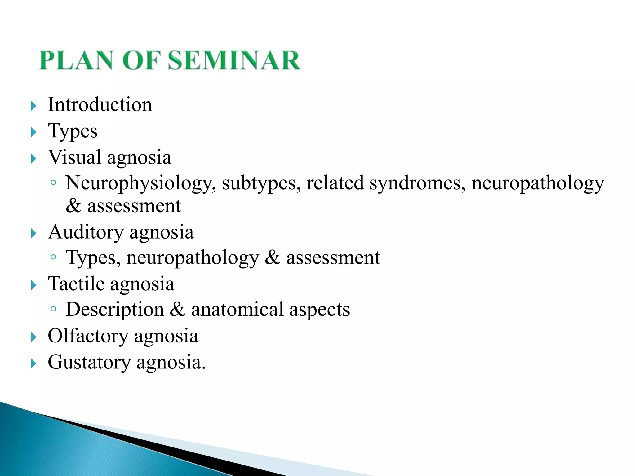
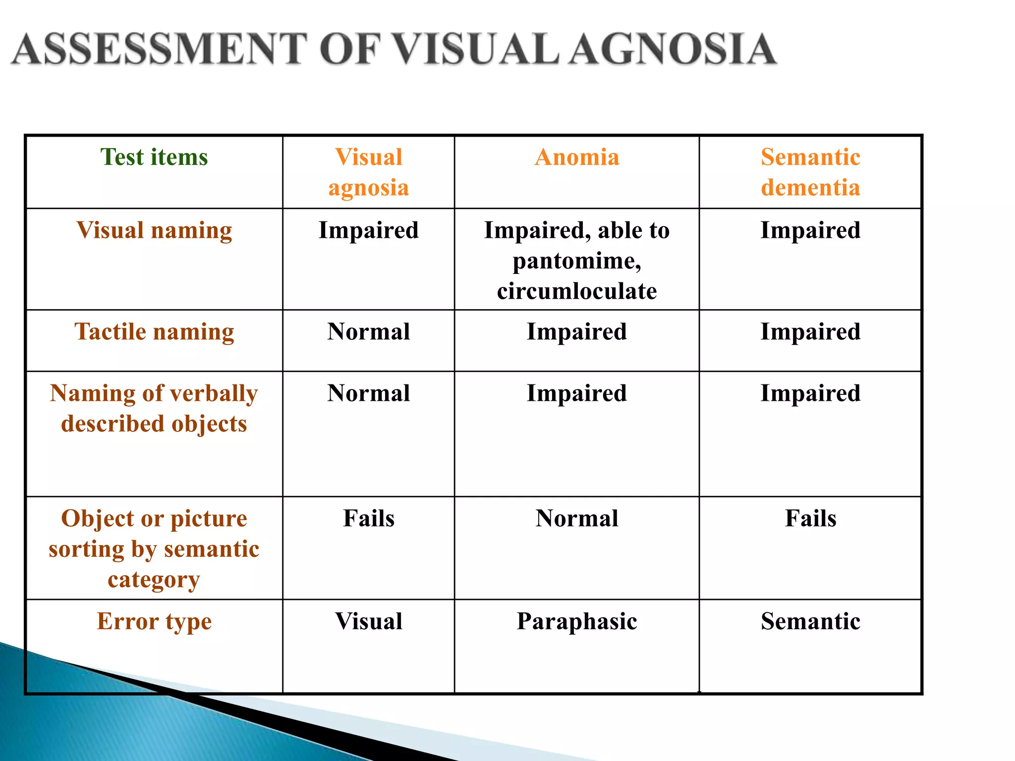
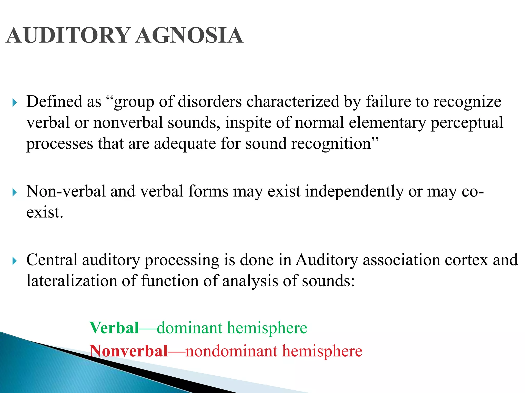
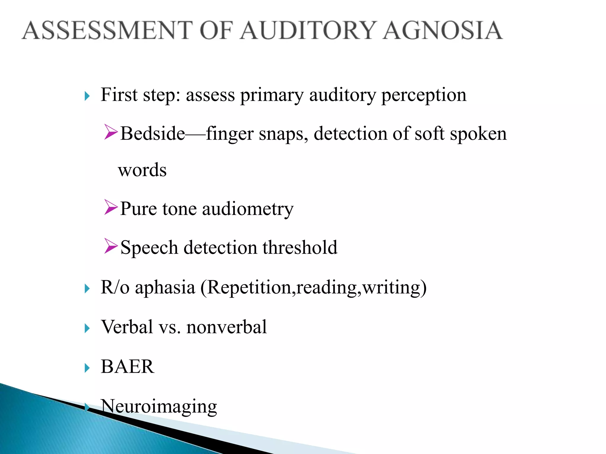
DR. MUNISH KUMAR
MD (MEDICINE), DM Neurology
G b pant, new delhi
 Introduction
 Types
 Visual agnosia
◦ Neurophysiology, subtypes, related syndromes, neuropathology
& assessment
 Auditory agnosia
◦ Types, neuropathology & assessment
 Tactile agnosia
◦ Description & anatomical aspects
 Olfactory agnosia
 Gustatory agnosia.
 Word agnosia derived from Greek meaning “not-knowing”
 “Class of neuropsychological disorders in which patients fail
to recognise familiar objects despite seemingly adequate
perception, memory, language, and general intellectual ability”
 Freud coined the term "agnosia"
 Inability to recognise everyday objects that would otherwise be
familiar with. This can’t be attributed to
◦ elementary sensory deficits
◦ mental deterioration
◦ attentional disturbances
◦ aphasic misnaming, or
◦ unfamiliarity with sensorily presented stimuli
 Can’t be better explained by any other physiological dysfunction
 Deficit is in one specific sense rather than a general deficit
◦ i.e. could recognise object through other sensory means
◦ Therefore, many patients are able to maintain daily
routines/activities to some degree.
 It is a perceptual disorder in which sensations remain
preserved but the ability to recognize a stimulus or know its
meaning is lost.
 This results from lesions that disconnect and isolate visual,
auditory and somatosensory input from higher level
processing.
 The nature and severity of perceptual impairment depends upon
modality affected and the level at which sensory processing has been
interrupted
 Examination involves assessing what the patient sees, hears or feels
when presented with objects, pictures or sounds using a combination
of clinical procedures and neuropsychological tests.
 VISUAL (most common form of agnosia)
 AUDITORY
 TACTILE
 GUSTATORY (not well described)
 OLFACTORY (not well described)
Non-striate vision via the Superior Colliculus.
Primitive vision
Spatial processing
Movement, location (e.g., Blind sight)
Striate vision via the geniculo-striate pathways.
Object recognition (e.g., perception and meaning).
Face recognition (e.g., features & person identity).
Higher order visual cognition (e.g., imagery).
 What pathway (located in the temporal lobe)
Perceive allocentric space (where objects are located with
respect to other objects).
Visual perception-to-meaning processing.
Global and/or local processes in object recognition
Categories of knowledge about objects
 Where pathway (located in the parietal lobe)
Perception of egocentric space (where objects are with
respect to the perceivers position).
Perception-to-spatial location processing.
Conscious and automatic visual processing
‘where’ and ‘how’
Ventral Stream -- WHAT ?
• Striate cortex to inferior temporal lobe
• Activated by static images
• Provides info about what an object is
Dorsal Stream -- WHERE ?
• Striate cortex to posterior parietal lobe
• Activated by moving objects
• Provides info about where an object is
• Helps in visual recognition of objects,
faces & perception of colour
• Lesions produces impaired pattern
recognition & learning, producing
-Visual object agnosia
-Prosopagnosia
-Alexia
-Colour agnosia
-Ventral simultanagnosia
• Lesion produces—disorders of
spatial temporal analysis &
disturbances of visually guided eye &
hand control
-Balint syndrome
-Hemi neglect syndrome
-Dorsal simultanagnosia
 First described by Lissauer (1890) .
 subdivided into 2 forms:
 Apperceptive – Disorder of visual perception
 Associative- Disorder of associating a perceptual
representation with general knowledge
 Perception normal.
 Apperceptive visual agnosia is characterised by an intact visual ability
on a basic sensory level, but a defect in early stage visual processing
prevents a correct percept of the stimulus being formed.
 The patient is unable to access the structure or spatial properties of a
visual stimuli and the object is not seen as a whole or in a meaningful
way.
 Shape perception & hence object recognition seem
disproportionately impaired.
 Patient cannot point to named objects, is unable to
match, copy, or discriminate simple visual forms but
may draw well from memory.
 These patients are so severely handicapped that they
often appear as blind to a casual observer
 Common causes are carbon monoxide poisoning, mercury poisoning
and stroke
 Site of lesion is diffuse & posterior (B/L or right) occipito-temporal
damage.
 Tests used are =
Visual Orientation and Space Perception Battery (VOSP)
patient is shown four drawings and has to identify
which one of the four is a real object. Impaired copying ability
 Simultanagnosia:
 Ventral
 Dorsal
 Perceptual Categorization Deficit
The term first used by Wolpert in 1924.
 Simultanagnosia is characterised by an inability to perceive more than
one aspect of a visual stimulus and to integrate visual detail into a
coherent whole.
 Accurately perceive individual details or elements of a visual scene
but are unable to combine these details to a meaningful entity
 Inability to perceive more than one object, or element, at any given
time.
 Fail to interpret the overall nature of the scene, “missing forest for trees”
 For example, if a patient with simultanagnosia is asked to name a picture of
spectacles, they may respond “there is a circle, another circle, it is joined
by a cross piece – it must be a bicycle!”
 The patient may appear blind, bumping into walls and furniture, making
haphazard and uncoordinated movements in attempting to reach for
objects.
 But If asked to focus on a small visual area, the patient may describe this
accurately and in great detail.
 The inability to code spatial relations is also apparent in the copies
that patients produce of drawings, which have an ‘exploded’ look
even though the patient easily recognized the object (quite different
from associative agnosiacs who produce exact copies without object
recognition).
 These patients also suffer from severe reading problems complains
that words pop out from the page.
 Moving objects pose greater problems for these patients than
stationary ones.
 Visual disorientation occurs due to, the inability to keep track of the
position of a previous stimulus while analysing a new object and to
code spatial relations between objects.
Cookie Theft Picture(from the Boston Diagnostic Aphasia Examination)
 Can recognize whole objects, but are limited in how many objects can
be recognized in a given period.
 Their descriptions of complex scenes are slow & piecemeal.
 Reading is possible in a letter-by-letter way
 Fail to recognize multiple objects but able to see multiple objects
hence can count and manipulate objects and walk around without
running into obstacles.
 Lesions site: left inferior temporo-occipital region.
“Mixed figures” used in the assessment of ventral simultanagnosia
 Attention limitation that prevents them from seeing more
than one object at a time
 Their attention may be captured by just one part of object.
 Once they can attend to an object, recognize it quickly &
accurately.
 Lesions site : Bilateral posterior parietal cortex.
 Poor performance at recognizing or matching objects seen from
unusual views or illuminated in unusual ways so as to produce
confusing shadows.
 Breakdown in the mechanisms of object shape constancy.
 Shape constancy: visual mechanisms that allow us to appreciate the
equivalence of an object’s three-dimensional shape across different
views & under different lighting conditions.
 No problems with everyday object recognition.
 In associative visual agnosia, primary sensory and early visual processing systems
are preserved. The patient can perceive objects presented visually but cannot
interpret, understand or assign meaning to the object, face or word.
May be characterized by four criteria:
1. No scotomata
2. Difficulty or inability to recognize visually presented objects as evidenced not
only in naming, but also in nonverbal tests
3. Preserved ability to separate figure and ground (hence no problems in navigating
through the environment) and preserved ability to copy and match stimuli. ( cf.
apperceptive agnosias)
4. Preserved ability to recognize and name objects or people through other sense
modalities such as audition, touch, or smell, and intact intelligence.
 More common than apperceptive visual agnosia.
 Picture identification is usually more difficult than
the identification of real objects.
 They point to named objects when they are presented
in an array of objects (i.e., perception is clearly
intact). So a visual identification disorder is isolated
from a discrimination disorder.
Lesion site: Bilateral damage to the inferior temporo-occipital junction
and subjacent white matter.
Causes :
 most often infarction of the posterior cerebral artery bilaterally.
 Other causes include tumour, haemorrhage and demyelination.
Tests:
Pyramids and Palm Trees Test
The usual/unusual views test from Birmingham Object Recognition
Battery (BORB).
ASSOCIATIVE VISUAL AGNOSIA
Example of the preserved copying ability of
associative agnosic patients.
Pyramids and Palm Trees Test
Assesses ability to access meaning
from words and pictures
Tests for associative visual agnosia from
Birmingham Object Recognition Battery (BORB).
Patient is required to identify real and unreal objects
PROSOPAGNOSIA
•Term coined by Bodamer in 1947. Prosopon = “face” (G)
•These patients can see a face, can identify facial parts, recognize a
face as a face but can’t able to recognize the person.
•Semantic knowledge about people is intact (Can retrieve history of
familiar persons when their name/other details are provided verbally).
• These patients have difficulty in identification within a
category they clearly recognize (Unlike patients with visual
agnosia who fail to recognize the object category to which
they belong).
• Can recognize ‘face as a face’, can discriminate faces
according to their sex or race, & can decode their emotional
expression.
• Affected people can use cues such as hairstyle, glasses and
clothing and will recognise the person as soon as they speak.
Causes:
Developmental :
 Neuroimaging is usually normal
 Congenital cases either due to prenatal insults or inherited defects
 Lesions acquired in early childhood
Acquired :
 Neuroimaging abnormal
 Stroke (PCA infarct)
 Trauma, tumors, abscess, hematoma, surgical resection
 Viral encephalitis, hypoxia, AD, PD
Lesions site: Right or bilateral inferior occipito-temporal region and subjacent white
matter which includes the fusiform gyrus.
Tests:
 Benton Facial Recognition test.
 Using photographs of well-known politicians and celebrities, ensuring that the
photographs are culturally and age appropriate.
 Also known as alexia without agraphia or pure word blindness.
 Pure alexia is a perceptual disorder causing impairment in reading
words and letters.
 The patient can copy words and letters and in the act of copying the
words or tracing out the letters will recognise the word or letter
(kinesthetic reading).
 The patient can write to dictation but is unable to read back what has
been written.
 They can also read digits and multidigit numbers but often
have color agnosia and usually a right homonymous
hemianopia.
 It results from damage to pathways conveying visual inputs
from both hemispheres to the dominant angular gyrus.
Lesion sites:
 Most commonly combined lesions of dominant medial
occipital region & inferior fibers of splenium of corpus
callosum (splenial-occipital syndrome).
 With infarction of the left lateral geniculate body and the
splenium of the corpus callosum (spleniogeniculate variation).
 Inability to recognize ,name and point to individual fingers on oneself
and on others.
 Usually part of Gerstmann`s syndrome
 Lesion site: Dominant parieto-oocipital lobe area
 Tests:
1. Identification of named fingers on examiners hand
2. Verbal identification (naming) of fingers on self and
examiner
Also referred to as movement agnosia, motion blindness or akinetopsia.
Patient is unable to perceive motion, while all other perceptual
capabilities are intact.
He can still read books and see colors, but cannot discern which
direction something is moving, nor how fast.
 Everyday actions, such as crossing a street are extremely difficult,
since he cannot judge how fast a car is approaching
Lesion site: bilateral medial-temporal area
MOTION AGNOSIA
 Defined as difficulty in identifying colours in presence of normal primary colour
perception (as tested by charts of Ishihara)
 They are able to match colours or order them in a series (as tested by Holmgrens
colour sorting test)
 Can’t name colors or point to the colour named by the examiner
 Perform well in verbal-verbal tasks (e.g. “tell me the colour of the sky?”).
 Usually have an associated right homonymous hemianopia and pure alexia.
 Lesion is commonly in inferomesial aspect of the occipital & temporal lobes of
dominant hemisphere
COLOR AGNOSIA
 First step is to establish the preservation of adequate elementary
visual abilities—acuity,visual fields
 Then assessment for..
Object recognition—recognition & naming tested separately
Colour —recognition & naming
Face recognition—persons, photographs
Picture recognition—for simultanagnosia
Matching & Copying - to differentiate between apperceptive &
associative agnosias
Test items Visual
agnosia
Anomia Semantic
dementia
Visual naming Impaired Impaired, able to
pantomime,
circumloculate
Impaired
Tactile naming Normal Impaired Impaired
Naming of verbally
described objects
Normal Impaired Impaired
Object or picture
sorting by semantic
category
Fails Normal Fails
Error type Visual Paraphasic Semantic
 Defined as “group of disorders characterized by failure to recognize
verbal or nonverbal sounds, inspite of normal elementary perceptual
processes that are adequate for sound recognition”
 Non-verbal and verbal forms may exist independently or may co-
exist.
 Central auditory processing is done in Auditory association cortex and
lateralization of function of analysis of sounds:
Verbal—dominant hemisphere
Nonverbal—nondominant hemisphere
AUDITORYAGNOSIA
 Also known as auditory sound agnosia or environmental sound agnosia.
 Impaired understanding and recognition of non-linguistic sounds such as bells,
barking ,whistles or animal noises.
 They are not cortically deaf (audiometry normal).
 Most reported cases initially present with cortical deafness, then they recover to
auditory agnosia
 They usually do acoustically based errors
e.g. misinterpret telephone ring as sound of railroad crossing and thunder as
fireworks.
 May affect recognition of certain classes of sounds more than others like for
environmental sounds, music (amusia) & voices (phonagnosia)
 Lesion site: Right hemispheric lesions (temporal lobes)
 Also known as pure word deafness or auditory agnosia for
speech
 Defined as a disturbance in comprehension of spoken language not
explainable by more generalized auditory processing deficit as seen
in cortical deafness, Wernicke’s aphasia or transcortical sensory
aphasia
 Preserved primary sensory processing as judged by normal pure
tone audiometry (d/d from cortical deafness)
 Relative preservation of recognition of nonverbal sounds (d/d from
nonverbal auditory agnosia)
 Relative preservation of reading ,writing & normal
spontaneous speech (d/d from Wernicke’s aphasia).
 Impaired word repetition (d/d –transcortical sensory aphasia).
 When the defect is severe, patients may complain that people
sound as if they are speaking a foreign language.
 By using lip-reading, these patients can improve their performance, but not by
increasing the sound volume.
 Commonly due to stroke, at a stage of recovery in new-onset Wernicke’s aphasia.
 Can occur with b/l or u/l lesion
B/l lesions in anterior or middle portion of temporal gyri often with sparing of
Heschl’s gyri
Single lesion involving Heschl’s gyri of dominant hemisphere and subjacent
white matter which destroys auditory projection from ipsilateral medial geniculate
body & callosal fibers from opposite superior temporal gyrus
 Both these lesions isolate the Wernicke’s area from auditory input, so that auditory
input can not reach language area for auditory comprehension.
 First step: assess primary auditory perception
Bedside—finger snaps, detection of soft spoken
words
Pure tone audiometry
Speech detection threshold
 R/o aphasia (Repetition,reading,writing)
 Verbal vs. nonverbal
 BAER
 Neuroimaging
 Selective impairment of object recognition by touch despite
relatively preserved primary and discriminative somesthetic
perception.
 It is a unilateral disorder usually resulting from lesions of the
contralateral inferior parietal cortex.
 The ability to recognise basic features such as size, weight and
texture may be dissociated from the ability to name or
recognise the object.
 A disorder in which the primary perceptual aspects of olfaction
i.e. detection of odors, adaptation to odors, and recognition of
different intensities of the same odor are intact.
 But the capacity to distinguish between odors and their
recognition by quality is impaired or lost.
 This impairment is not attributable to impaired olfactory acuity
or to failure of learning and memory.
 To recognize this deficit special testing is required,
such as matching to sample, the identification and
naming of a variety of scents, and determining
whether two odors are identical or different.
 Site of lesion medial dorsal nucleus of the thalamus
and medial temporal lobe
 Taste quality perception can be assessed by either asking a
subject to identify a taste (sweet, sour, salty, bitter) or
establishing a recognition threshold.
 Recognition threshold: minimum concentration of stimulus that
needs to be present for a subject to correctly label the stimulus.
 Detection threshold: minimum concentration of stimulus that
needs to be present for the subject to detect that it is different
from background.
 In gustatory agnosia patients fail to recognize the taste quality of
simple tastant solutions or to identify the taste quality in complex
liquid stimuli.
 Patients recognition threshold gets elevated but other aspects of
taste perception remained unchanged, including detection
thresholds, discrimination, intensity estimation, and affective
judgments.
 Site of lesion is usually antero-medial temporal lobe and
particularly amygdala.
 In the early stages of recovery, lack of awareness of the deficit,
anosognosia, may lead to therapeutic resistance.
 Partial recovery is more likely in traumatic and vascular lesions and
less likely in anoxic brain damage due to widespread diffuse lesions
and additional cognitive impairment impeding acquisition of
compensatory strategies.
 Many case reports show improvement following intensive
rehabilitation.
 Principles of treatment are restitution, repetitive training of
impaired function, and compensation, with utilization of spared
function to compensate for the deficit.
 Optic aphasia
 Impaired, able to pantomime, circumloculate
 Normal
 Normal
 Normal
 Semantic,

AGNOSIA

  • 1.
    DR. MUNISH KUMAR MD(MEDICINE), DM Neurology G b pant, new delhi
  • 2.
     Introduction  Types Visual agnosia ◦ Neurophysiology, subtypes, related syndromes, neuropathology & assessment  Auditory agnosia ◦ Types, neuropathology & assessment  Tactile agnosia ◦ Description & anatomical aspects  Olfactory agnosia  Gustatory agnosia.
  • 3.
     Word agnosiaderived from Greek meaning “not-knowing”  “Class of neuropsychological disorders in which patients fail to recognise familiar objects despite seemingly adequate perception, memory, language, and general intellectual ability”  Freud coined the term "agnosia"
  • 4.
     Inability torecognise everyday objects that would otherwise be familiar with. This can’t be attributed to ◦ elementary sensory deficits ◦ mental deterioration ◦ attentional disturbances ◦ aphasic misnaming, or ◦ unfamiliarity with sensorily presented stimuli  Can’t be better explained by any other physiological dysfunction
  • 5.
     Deficit isin one specific sense rather than a general deficit ◦ i.e. could recognise object through other sensory means ◦ Therefore, many patients are able to maintain daily routines/activities to some degree.  It is a perceptual disorder in which sensations remain preserved but the ability to recognize a stimulus or know its meaning is lost.  This results from lesions that disconnect and isolate visual, auditory and somatosensory input from higher level processing.
  • 6.
     The natureand severity of perceptual impairment depends upon modality affected and the level at which sensory processing has been interrupted  Examination involves assessing what the patient sees, hears or feels when presented with objects, pictures or sounds using a combination of clinical procedures and neuropsychological tests.
  • 7.
     VISUAL (mostcommon form of agnosia)  AUDITORY  TACTILE  GUSTATORY (not well described)  OLFACTORY (not well described)
  • 9.
    Non-striate vision viathe Superior Colliculus. Primitive vision Spatial processing Movement, location (e.g., Blind sight) Striate vision via the geniculo-striate pathways. Object recognition (e.g., perception and meaning). Face recognition (e.g., features & person identity). Higher order visual cognition (e.g., imagery).
  • 10.
     What pathway(located in the temporal lobe) Perceive allocentric space (where objects are located with respect to other objects). Visual perception-to-meaning processing. Global and/or local processes in object recognition Categories of knowledge about objects  Where pathway (located in the parietal lobe) Perception of egocentric space (where objects are with respect to the perceivers position). Perception-to-spatial location processing. Conscious and automatic visual processing ‘where’ and ‘how’
  • 11.
    Ventral Stream --WHAT ? • Striate cortex to inferior temporal lobe • Activated by static images • Provides info about what an object is Dorsal Stream -- WHERE ? • Striate cortex to posterior parietal lobe • Activated by moving objects • Provides info about where an object is • Helps in visual recognition of objects, faces & perception of colour • Lesions produces impaired pattern recognition & learning, producing -Visual object agnosia -Prosopagnosia -Alexia -Colour agnosia -Ventral simultanagnosia • Lesion produces—disorders of spatial temporal analysis & disturbances of visually guided eye & hand control -Balint syndrome -Hemi neglect syndrome -Dorsal simultanagnosia
  • 12.
     First describedby Lissauer (1890) .  subdivided into 2 forms:  Apperceptive – Disorder of visual perception  Associative- Disorder of associating a perceptual representation with general knowledge  Perception normal.
  • 13.
     Apperceptive visualagnosia is characterised by an intact visual ability on a basic sensory level, but a defect in early stage visual processing prevents a correct percept of the stimulus being formed.  The patient is unable to access the structure or spatial properties of a visual stimuli and the object is not seen as a whole or in a meaningful way.
  • 14.
     Shape perception& hence object recognition seem disproportionately impaired.  Patient cannot point to named objects, is unable to match, copy, or discriminate simple visual forms but may draw well from memory.  These patients are so severely handicapped that they often appear as blind to a casual observer
  • 15.
     Common causesare carbon monoxide poisoning, mercury poisoning and stroke  Site of lesion is diffuse & posterior (B/L or right) occipito-temporal damage.  Tests used are = Visual Orientation and Space Perception Battery (VOSP)
  • 16.
    patient is shownfour drawings and has to identify which one of the four is a real object. Impaired copying ability
  • 17.
     Simultanagnosia:  Ventral Dorsal  Perceptual Categorization Deficit
  • 18.
    The term firstused by Wolpert in 1924.  Simultanagnosia is characterised by an inability to perceive more than one aspect of a visual stimulus and to integrate visual detail into a coherent whole.  Accurately perceive individual details or elements of a visual scene but are unable to combine these details to a meaningful entity  Inability to perceive more than one object, or element, at any given time.
  • 19.
     Fail tointerpret the overall nature of the scene, “missing forest for trees”  For example, if a patient with simultanagnosia is asked to name a picture of spectacles, they may respond “there is a circle, another circle, it is joined by a cross piece – it must be a bicycle!”  The patient may appear blind, bumping into walls and furniture, making haphazard and uncoordinated movements in attempting to reach for objects.  But If asked to focus on a small visual area, the patient may describe this accurately and in great detail.
  • 20.
     The inabilityto code spatial relations is also apparent in the copies that patients produce of drawings, which have an ‘exploded’ look even though the patient easily recognized the object (quite different from associative agnosiacs who produce exact copies without object recognition).  These patients also suffer from severe reading problems complains that words pop out from the page.
  • 21.
     Moving objectspose greater problems for these patients than stationary ones.  Visual disorientation occurs due to, the inability to keep track of the position of a previous stimulus while analysing a new object and to code spatial relations between objects.
  • 22.
    Cookie Theft Picture(fromthe Boston Diagnostic Aphasia Examination)
  • 23.
     Can recognizewhole objects, but are limited in how many objects can be recognized in a given period.  Their descriptions of complex scenes are slow & piecemeal.  Reading is possible in a letter-by-letter way  Fail to recognize multiple objects but able to see multiple objects hence can count and manipulate objects and walk around without running into obstacles.  Lesions site: left inferior temporo-occipital region.
  • 24.
    “Mixed figures” usedin the assessment of ventral simultanagnosia
  • 25.
     Attention limitationthat prevents them from seeing more than one object at a time  Their attention may be captured by just one part of object.  Once they can attend to an object, recognize it quickly & accurately.  Lesions site : Bilateral posterior parietal cortex.
  • 27.
     Poor performanceat recognizing or matching objects seen from unusual views or illuminated in unusual ways so as to produce confusing shadows.  Breakdown in the mechanisms of object shape constancy.  Shape constancy: visual mechanisms that allow us to appreciate the equivalence of an object’s three-dimensional shape across different views & under different lighting conditions.  No problems with everyday object recognition.
  • 29.
     In associativevisual agnosia, primary sensory and early visual processing systems are preserved. The patient can perceive objects presented visually but cannot interpret, understand or assign meaning to the object, face or word. May be characterized by four criteria: 1. No scotomata 2. Difficulty or inability to recognize visually presented objects as evidenced not only in naming, but also in nonverbal tests 3. Preserved ability to separate figure and ground (hence no problems in navigating through the environment) and preserved ability to copy and match stimuli. ( cf. apperceptive agnosias) 4. Preserved ability to recognize and name objects or people through other sense modalities such as audition, touch, or smell, and intact intelligence.
  • 30.
     More commonthan apperceptive visual agnosia.  Picture identification is usually more difficult than the identification of real objects.  They point to named objects when they are presented in an array of objects (i.e., perception is clearly intact). So a visual identification disorder is isolated from a discrimination disorder.
  • 31.
    Lesion site: Bilateraldamage to the inferior temporo-occipital junction and subjacent white matter. Causes :  most often infarction of the posterior cerebral artery bilaterally.  Other causes include tumour, haemorrhage and demyelination. Tests: Pyramids and Palm Trees Test The usual/unusual views test from Birmingham Object Recognition Battery (BORB).
  • 32.
    ASSOCIATIVE VISUAL AGNOSIA Exampleof the preserved copying ability of associative agnosic patients. Pyramids and Palm Trees Test Assesses ability to access meaning from words and pictures Tests for associative visual agnosia from Birmingham Object Recognition Battery (BORB). Patient is required to identify real and unreal objects
  • 33.
    PROSOPAGNOSIA •Term coined byBodamer in 1947. Prosopon = “face” (G) •These patients can see a face, can identify facial parts, recognize a face as a face but can’t able to recognize the person. •Semantic knowledge about people is intact (Can retrieve history of familiar persons when their name/other details are provided verbally).
  • 34.
    • These patientshave difficulty in identification within a category they clearly recognize (Unlike patients with visual agnosia who fail to recognize the object category to which they belong). • Can recognize ‘face as a face’, can discriminate faces according to their sex or race, & can decode their emotional expression. • Affected people can use cues such as hairstyle, glasses and clothing and will recognise the person as soon as they speak.
  • 35.
    Causes: Developmental :  Neuroimagingis usually normal  Congenital cases either due to prenatal insults or inherited defects  Lesions acquired in early childhood Acquired :  Neuroimaging abnormal  Stroke (PCA infarct)  Trauma, tumors, abscess, hematoma, surgical resection  Viral encephalitis, hypoxia, AD, PD Lesions site: Right or bilateral inferior occipito-temporal region and subjacent white matter which includes the fusiform gyrus. Tests:  Benton Facial Recognition test.  Using photographs of well-known politicians and celebrities, ensuring that the photographs are culturally and age appropriate.
  • 37.
     Also knownas alexia without agraphia or pure word blindness.  Pure alexia is a perceptual disorder causing impairment in reading words and letters.  The patient can copy words and letters and in the act of copying the words or tracing out the letters will recognise the word or letter (kinesthetic reading).  The patient can write to dictation but is unable to read back what has been written.
  • 38.
     They canalso read digits and multidigit numbers but often have color agnosia and usually a right homonymous hemianopia.  It results from damage to pathways conveying visual inputs from both hemispheres to the dominant angular gyrus. Lesion sites:  Most commonly combined lesions of dominant medial occipital region & inferior fibers of splenium of corpus callosum (splenial-occipital syndrome).  With infarction of the left lateral geniculate body and the splenium of the corpus callosum (spleniogeniculate variation).
  • 39.
     Inability torecognize ,name and point to individual fingers on oneself and on others.  Usually part of Gerstmann`s syndrome  Lesion site: Dominant parieto-oocipital lobe area  Tests: 1. Identification of named fingers on examiners hand 2. Verbal identification (naming) of fingers on self and examiner
  • 40.
    Also referred toas movement agnosia, motion blindness or akinetopsia. Patient is unable to perceive motion, while all other perceptual capabilities are intact. He can still read books and see colors, but cannot discern which direction something is moving, nor how fast.  Everyday actions, such as crossing a street are extremely difficult, since he cannot judge how fast a car is approaching Lesion site: bilateral medial-temporal area MOTION AGNOSIA
  • 41.
     Defined asdifficulty in identifying colours in presence of normal primary colour perception (as tested by charts of Ishihara)  They are able to match colours or order them in a series (as tested by Holmgrens colour sorting test)  Can’t name colors or point to the colour named by the examiner  Perform well in verbal-verbal tasks (e.g. “tell me the colour of the sky?”).  Usually have an associated right homonymous hemianopia and pure alexia.  Lesion is commonly in inferomesial aspect of the occipital & temporal lobes of dominant hemisphere
  • 42.
  • 43.
     First stepis to establish the preservation of adequate elementary visual abilities—acuity,visual fields  Then assessment for.. Object recognition—recognition & naming tested separately Colour —recognition & naming Face recognition—persons, photographs Picture recognition—for simultanagnosia Matching & Copying - to differentiate between apperceptive & associative agnosias
  • 44.
    Test items Visual agnosia AnomiaSemantic dementia Visual naming Impaired Impaired, able to pantomime, circumloculate Impaired Tactile naming Normal Impaired Impaired Naming of verbally described objects Normal Impaired Impaired Object or picture sorting by semantic category Fails Normal Fails Error type Visual Paraphasic Semantic
  • 45.
     Defined as“group of disorders characterized by failure to recognize verbal or nonverbal sounds, inspite of normal elementary perceptual processes that are adequate for sound recognition”  Non-verbal and verbal forms may exist independently or may co- exist.  Central auditory processing is done in Auditory association cortex and lateralization of function of analysis of sounds: Verbal—dominant hemisphere Nonverbal—nondominant hemisphere AUDITORYAGNOSIA
  • 46.
     Also knownas auditory sound agnosia or environmental sound agnosia.  Impaired understanding and recognition of non-linguistic sounds such as bells, barking ,whistles or animal noises.  They are not cortically deaf (audiometry normal).  Most reported cases initially present with cortical deafness, then they recover to auditory agnosia  They usually do acoustically based errors e.g. misinterpret telephone ring as sound of railroad crossing and thunder as fireworks.  May affect recognition of certain classes of sounds more than others like for environmental sounds, music (amusia) & voices (phonagnosia)  Lesion site: Right hemispheric lesions (temporal lobes)
  • 47.
     Also knownas pure word deafness or auditory agnosia for speech  Defined as a disturbance in comprehension of spoken language not explainable by more generalized auditory processing deficit as seen in cortical deafness, Wernicke’s aphasia or transcortical sensory aphasia  Preserved primary sensory processing as judged by normal pure tone audiometry (d/d from cortical deafness)  Relative preservation of recognition of nonverbal sounds (d/d from nonverbal auditory agnosia)
  • 48.
     Relative preservationof reading ,writing & normal spontaneous speech (d/d from Wernicke’s aphasia).  Impaired word repetition (d/d –transcortical sensory aphasia).  When the defect is severe, patients may complain that people sound as if they are speaking a foreign language.
  • 49.
     By usinglip-reading, these patients can improve their performance, but not by increasing the sound volume.  Commonly due to stroke, at a stage of recovery in new-onset Wernicke’s aphasia.  Can occur with b/l or u/l lesion B/l lesions in anterior or middle portion of temporal gyri often with sparing of Heschl’s gyri Single lesion involving Heschl’s gyri of dominant hemisphere and subjacent white matter which destroys auditory projection from ipsilateral medial geniculate body & callosal fibers from opposite superior temporal gyrus  Both these lesions isolate the Wernicke’s area from auditory input, so that auditory input can not reach language area for auditory comprehension.
  • 50.
     First step:assess primary auditory perception Bedside—finger snaps, detection of soft spoken words Pure tone audiometry Speech detection threshold  R/o aphasia (Repetition,reading,writing)  Verbal vs. nonverbal  BAER  Neuroimaging
  • 51.
     Selective impairmentof object recognition by touch despite relatively preserved primary and discriminative somesthetic perception.  It is a unilateral disorder usually resulting from lesions of the contralateral inferior parietal cortex.  The ability to recognise basic features such as size, weight and texture may be dissociated from the ability to name or recognise the object.
  • 52.
     A disorderin which the primary perceptual aspects of olfaction i.e. detection of odors, adaptation to odors, and recognition of different intensities of the same odor are intact.  But the capacity to distinguish between odors and their recognition by quality is impaired or lost.  This impairment is not attributable to impaired olfactory acuity or to failure of learning and memory.
  • 53.
     To recognizethis deficit special testing is required, such as matching to sample, the identification and naming of a variety of scents, and determining whether two odors are identical or different.  Site of lesion medial dorsal nucleus of the thalamus and medial temporal lobe
  • 54.
     Taste qualityperception can be assessed by either asking a subject to identify a taste (sweet, sour, salty, bitter) or establishing a recognition threshold.  Recognition threshold: minimum concentration of stimulus that needs to be present for a subject to correctly label the stimulus.  Detection threshold: minimum concentration of stimulus that needs to be present for the subject to detect that it is different from background.
  • 55.
     In gustatoryagnosia patients fail to recognize the taste quality of simple tastant solutions or to identify the taste quality in complex liquid stimuli.  Patients recognition threshold gets elevated but other aspects of taste perception remained unchanged, including detection thresholds, discrimination, intensity estimation, and affective judgments.  Site of lesion is usually antero-medial temporal lobe and particularly amygdala.
  • 56.
     In theearly stages of recovery, lack of awareness of the deficit, anosognosia, may lead to therapeutic resistance.  Partial recovery is more likely in traumatic and vascular lesions and less likely in anoxic brain damage due to widespread diffuse lesions and additional cognitive impairment impeding acquisition of compensatory strategies.  Many case reports show improvement following intensive rehabilitation.  Principles of treatment are restitution, repetitive training of impaired function, and compensation, with utilization of spared function to compensate for the deficit.
  • 60.
     Optic aphasia Impaired, able to pantomime, circumloculate  Normal  Normal  Normal  Semantic,