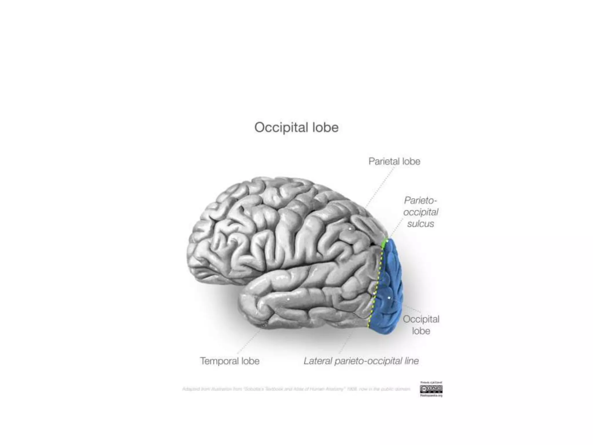
Occipital Lobe
Anatomy and Function
with clinical correlation
By Dr Srimant Pattnaik, DM PDT
Neuromedicine , BIN
Lesions in the occipital lobe
• Lesion of the primary visual area
• Lesion of the dorsal stream
• Lesion of ventral stream
Lesions of primary visual cortex
1. Field defects
2. Cortical blindness
3. Anton ‘ s syndrome
FIELD DEFECTS
• Binocular field defect usually localizes to or
beyond optic chiasm
• Retrochiasmal lesions are usually
homonymous hemianopia
• Bilatreal optic nerve or retinal disease can
produce bilateral field defect
• Retinal disease has ophthalmoscoic finding
• Optic nerve problem has acuity problem
left lower bank of calcarine sulcus
Left upper bank of calcarine sucus
case
• A patient recovering
from a right
hemipareisis, presented
to hospital with right
sided visual disturbance
Half moon or temporal crescent
syndrome
Cortical blindness
• Conceptualized as bilateral homonymous hemianopia.
Clinically blind.
• Due to blockage of bilateral PCA or distal Basilar artery
• Bilateral Area 17 involvement
• Central vision may be present (macular sparing):
reason for optokinetic nystagmus
• Pupilary reflex present
• Menace reflex ±
• VEP : no cortical potential
• EEG : loss of alpha rythm
• Visual imagination and visual imagery preserved
• Less complete bilateral lesion leave the patient
with varying degrees of visual perception
• There may be visual hallucination of either
complex or elementary type
• Recovery occurs in a regular fashion from cortical
blindness through visual agnosia and partially
impaired perceptual function to recovery
• After recovery , there may be p[roblems of visual
fatigue,fusion or fixation (3F’s)
Anton syndrome
• Visual Anosognosia : self unawareness of
visual deficit. Denies blindness
• Iinvolvement of primary visual area along with
visual association area
• Behaves as if he can see
• Confabulates. Has excuses for blindness
Inverse Anton Syndrome
• Also known as Blindism or Blindsight
• As such patient can not see but eye movements
are noticed in response to stimulus such as
flashes of light
• Due to nonconscious visual processing
• Pathways
– Retino tectal
– Retino tecto pulvino extrastriate
– Geniculoextrastriate
• Simulates hysterical blindness
Riddoch syndrome
• Cortical blindness but area V5 is spared
• Otherwise blind Patient can percieve moving
objects
• Hypothesis is of a direct connection between
LGB or Pulvinus to V5
Lesions of ventral stream
1. Visual object agnosia
2. Colour vision abnormality
3. Prosopagnosia
4. Pure Alexia
VISUAL OBJECT RECOGNITION
VISUAL STIMULI
PRIMARY SKETCH
VIEWER CENTERED REPRESENATION
OBJECT CENTERED REPRESENTATION
SEMANTIC CONCEPTUAL FIELD
VISUAL ANALYSIS
PHONOLOGICAL LEXICON
SEMANTIC-CONCEPTUAL SYSTEM
PRIMARY SKETCH-2D
VIEWER CENTERED SKETCH-2.5D
OBJECT CENTERED SKETCH-3D
MARR’S MODEL OF VISUAL
OBJECT RECOGNITION
Visual Object Agnosia
• Apperceptive Visual agnosia
– Perceptual categorization defect
• Associative visual agnosia
Apperceptive visual agnosia
VISUAL STIMULI
PRIMARY SKETCH
VIEWER CENTERED REPRESENATION
OBJECT CENTERED REPRESENTATION
SEMANTIC CONCEPTUAL FIELD
VISUAL ANALYSIS
PHONOLOGICAL LEXICON
Apperceptive visual agnosia
• Localization :
• bilateral lesion of calcarine cortex
• Occipitotemopral regions
• Pathology: mostly ischemic
• Most commonly seen during recovery from
cortical blindness
Clinical feature
• Pick out details of the object in fragments i.e
lines adges, intensity but fail to identify the
whole picture
• They can not name objects presented to them
or draw them or match them
• Mostly act as blind but avoid obstacles while
walking
Besdside tests
• Match test
• Figure copying test
Match test
Figure copying test
Perceptual categorization defect
VISUAL STIMULI
PRIMARY SKETCH
VIEWER CENTERED REPRESENATION
OBJECT CENTERED REPRESENTATION
SEMANTIC CONCEPTUAL FIELD
VISUAL ANALYSIS
PHONOLOGICAL LEXICON
Perceptual categorization defect:contd
Perceptual categorization defect:
contd
• Localization: right parietal lobe
• Patient can appreciate objects , match them
and and recognize them later as long as they
are presented in same conventional
orientation
tests
VOSP
Incomplete letter test
Silhouettes
BORB
Gollin figures
Unusual view test
Foreshortened match
silhouettes
Unusual view test
Gollin figures
Foreshortened match
Associative visual agnosia
VISUAL STIMULI
PRIMARY SKETCH
VIEWER CENTERED REPRESENATION
OBJECT CENTERED REPRESENTATION
SEMANTIC CONCEPTUAL FIELD
VISUAL ANALYSIS
PHONOLOGICAL LEXICON
SEMANTIC-CONCEPTUAL SYSTEM
PRIMARY SKETCH-2D
VIEWER CENTERED SKETCH-2.5D
OBJECT CENTERED SKETCH-3D
MARR’S MODEL OF VISUAL
OBJECT RECOGNITION
• Perception is stripped of meaning
• Patient can copy , match figures still can not
say what it is
• Can identify if object is presented by any other
modality
• Localized to bilateral posterior hemisphere
involving fusiform ot occipitotemporal gyri,
sometimes lingual gyrus and adjacent white
matter
Tests
• Questionnaire
• Sorting of stimuli
• Matching visually dissimilar objects according
to similar function
• Real and unreal objects
• Pyramids and palm trees test
Optic aphasia
VISUAL STIMULI
PRIMARY SKETCH
VIEWER CENTERED REPRESENATION
OBJECT CENTERED REPRESENTATION
SEMANTIC CONCEPTUAL FIELD
VISUAL ANALYSIS
PHONOLOGICAL LEXICON
• Optic aphasia or optic anomia, an
intermediate between agnosia and aphasia
• Localization: left occipitotemporal area
• Can not name object presented visually but
can recognise when given cue
• Preserved recpognition of the objects
differentiates frm associative agnosia
• Recognition with auditory or tactile cue
differentiates it from anomic aphasia
Face recognition
test
• Face identification from photographs
• Benton facial recognition test
Prosopagnosia
• Difficulty in recognising familiar faces
• Recognise faces from a singular feature like
glasses , beards etc or non visual cues like voice
or social context
• Apperceptive vs associative
• Localization
– B/L damage to fusiform & lingual gyri
– U/L Rt OP lesion
– B/L or U/L anterior temporal lesion
Causes of Prosopagnosia
• Congenital
– Hereditary : autosomal dominant inheritance
– Autistic spectrum disorder
• Acquired
– Stroke
– Herpes encephalitis
– Alzheimer
– Carbon monoxide poisoining
COLOUR STIMULI
COLOUR
DISCRIMINATION
COLOUR
CATEGORISATION
SEMANTIC CONCEPTUAL
FIELD
COLOR NAMING
ACHROMATOPSIA
COLOUR
AGNOSIA
COLOUR
AMNESIA
Cerebral Achromatopsia
• Localization : bilateral or nondominant inferior
occipito temporal lesions that damage lingual
and fusiform gyri sparing of calcarine cortex
COLOUR AGNOSIA
• Colour is stripped off its meaning
• Can see and discriminate though
• Can not point or paint
• Inferomedial aspect of dominant occipital anfd
temporal lobe
Colour Amnesia
• Defect in colour object association
• Can see, discriminate and point
• Can not paint
• Dominant hemisphere lesions
Colour Anomia
• Basically the optic aphasia for colours
• Defective naming only
ACHROMATOPSIA COLOUR
AGNOSIA
COLOUR
AMNESIA
COLOR
ANOMIA
ISHIHARA
CHART
COLOUR
PLATE
NAMING
POINTING TO
COLOUR
MATCHING
COLOUR
CONCEPTUAL
COLOUR
NAMING
COLOUR
PAINTING
simultanagnosia
• Inability to comprehend a complex visual
scene despite being able to comprehend
individual items
• Divided into
• Ventral
• Dorsal
SIMULTANAGNOSIA
DORSAL VENTRAL
LOCALIZATION B/L OCCIPITOPARIETAL LEFT OCCIPITOTEMPORAL
PATHOPHYSIOLOGY RESTRICTED VISUAL
ATTENTION
SLOW VISUAL PROCESSING
CLINICALLY BLIND NOT SO
LEVEL OF PERCEPTION LIMITED TO ONE OBJECT SEVERAL OBJECTS
PERCIEVED, ONE
RECOGNIZED
SCATTERED DOT
COUNTING
NOT ABLE TO DO ABLE TO DO
WORD READING CAN NOT READ WORDS READS BUT LETTER BY
LETTER
Tests of ventral Simultanagnosia
Mixed figure testing Cookie theft picture
Disorders of Dorsal stream
1. Balint Syndrome
2. Visual disorientation
3. Akinetopsia
4. Astereopsis
5. Visual extinction
Balint syndrome
• Occurs due to lesion in b/l parieto occipital
lesion in the convexity of the hemisphere
• Components
– Dorsal simultanagnosia
– Oculomotor apraxia
– Optic ataxia
Dorsal Simultanagnosia
• Due to restriction of visual attention
• Perception is limited to point of fixation
without the awareness of presence of other
parts
• Miss forest for trees.
• Functionally blind as they find it difficult to
move about
Oculomotor apraxia
• Not a true apraxia as the higher level motor
control is intact
• Also known as psychic paralysis of gaze
• Defective vision for action
• Deficit is a difficulty in shifting visual attention
to appropriate target
Optic ataxia
Optic ataxia
• Inability to reach a target under visual
guidance
• Defective action for vision
• No difficulty in touching body parts
• Tested by asking the patient to touch
examiner’s ear followed by touching own ear
Visual disorientation
• Error in judging the location and distance of
an object, and in judging the relative distance
between the two objects
• Bump into things while walking
• Difficulty in finding door handle
• Problem arises when things are placed at
different level
• Difficulty in reaching things becomes difficult,
mimics optic ataxia
• However, visual disorientation improves with
repeated attempts, differentiating from optic
ataxia
Astereopsis
• Loss of depth perception
• Depth is computed from binocular visual
information
• Retinal disparity is processed by the dorsal
stream
• Area 18 is primarily concerned with stereopsis
• Abnormality is seen due to bilateral damage,
more to the right side
astereopsis
akinetopsia
• Aquired defect of motion perception
• Sees moving people here and there
• Difficulty in pouring water from a cup to bottlr
as the water appears as ice
Visual extinction
Visual hallucination
• Positive visual phenomena in absence of external stimuli
• Simple formed hallucination
• Light dots, bars, lines , stars, fog, coloured senasation
• Mostly after occpital stroke: good recovery
• Psychiatric conditions
• Multicoloured wth spherical or circular pattern seen in
Occipital epileptic seizure
• Degenerative : AD,DLB
• Infective: CJD-Heidenhain variant
• Unformed hemifield hallucination post exercise : occiptal
lobe tumor
Peduncular Hallucinosis
• Charcot – Willebrand syndrome
• Vivid,colourful,formed hallucination of
p[eople and animal
• Usually hypnogogic
• Pseudohallucinosis as patient knows they are
unreal
• Also seen in midbrain lesion as a release
hallucination
Charles Bonnet Syndrome
• Seen in patients with visual loss due to
ophthalmological condition
• Well defined, organized and vivid scenes of
animals, flowers and people
• Also a pseudohallucination
• Can be abolished by repeated closing and
opening of eyes
• Deafferentiation of visual assdociation areas
leading to a form of phantom vision
Miscellaneous Positive visual
phenomena
• Metamorphopsia( distortions of shape)
• scintillating scotomas
• Teichopsia( irregular shapes)
• macropsia and micropsia
• palinopsia (perseveration of visual images)
• Visual allesthesia (spread of a visual image from a
normal to a hemianopic field)
• polyopia (duplication of objects)
• All these phenomena are disturbances of higher visual
perception rather than agnosias
Occipital lobe
Occipital lobe

Occipital lobe

  • 1.
    Occipital Lobe Anatomy andFunction with clinical correlation By Dr Srimant Pattnaik, DM PDT Neuromedicine , BIN
  • 10.
    Lesions in theoccipital lobe • Lesion of the primary visual area • Lesion of the dorsal stream • Lesion of ventral stream
  • 11.
    Lesions of primaryvisual cortex 1. Field defects 2. Cortical blindness 3. Anton ‘ s syndrome
  • 12.
  • 13.
    • Binocular fielddefect usually localizes to or beyond optic chiasm • Retrochiasmal lesions are usually homonymous hemianopia • Bilatreal optic nerve or retinal disease can produce bilateral field defect • Retinal disease has ophthalmoscoic finding • Optic nerve problem has acuity problem
  • 15.
    left lower bankof calcarine sulcus Left upper bank of calcarine sucus
  • 16.
    case • A patientrecovering from a right hemipareisis, presented to hospital with right sided visual disturbance
  • 18.
    Half moon ortemporal crescent syndrome
  • 19.
    Cortical blindness • Conceptualizedas bilateral homonymous hemianopia. Clinically blind. • Due to blockage of bilateral PCA or distal Basilar artery • Bilateral Area 17 involvement • Central vision may be present (macular sparing): reason for optokinetic nystagmus • Pupilary reflex present • Menace reflex ± • VEP : no cortical potential • EEG : loss of alpha rythm
  • 20.
    • Visual imaginationand visual imagery preserved • Less complete bilateral lesion leave the patient with varying degrees of visual perception • There may be visual hallucination of either complex or elementary type • Recovery occurs in a regular fashion from cortical blindness through visual agnosia and partially impaired perceptual function to recovery • After recovery , there may be p[roblems of visual fatigue,fusion or fixation (3F’s)
  • 21.
    Anton syndrome • VisualAnosognosia : self unawareness of visual deficit. Denies blindness • Iinvolvement of primary visual area along with visual association area • Behaves as if he can see • Confabulates. Has excuses for blindness
  • 22.
    Inverse Anton Syndrome •Also known as Blindism or Blindsight • As such patient can not see but eye movements are noticed in response to stimulus such as flashes of light • Due to nonconscious visual processing • Pathways – Retino tectal – Retino tecto pulvino extrastriate – Geniculoextrastriate • Simulates hysterical blindness
  • 23.
    Riddoch syndrome • Corticalblindness but area V5 is spared • Otherwise blind Patient can percieve moving objects • Hypothesis is of a direct connection between LGB or Pulvinus to V5
  • 24.
    Lesions of ventralstream 1. Visual object agnosia 2. Colour vision abnormality 3. Prosopagnosia 4. Pure Alexia
  • 25.
    VISUAL OBJECT RECOGNITION VISUALSTIMULI PRIMARY SKETCH VIEWER CENTERED REPRESENATION OBJECT CENTERED REPRESENTATION SEMANTIC CONCEPTUAL FIELD VISUAL ANALYSIS PHONOLOGICAL LEXICON
  • 26.
    SEMANTIC-CONCEPTUAL SYSTEM PRIMARY SKETCH-2D VIEWERCENTERED SKETCH-2.5D OBJECT CENTERED SKETCH-3D MARR’S MODEL OF VISUAL OBJECT RECOGNITION
  • 27.
    Visual Object Agnosia •Apperceptive Visual agnosia – Perceptual categorization defect • Associative visual agnosia
  • 28.
    Apperceptive visual agnosia VISUALSTIMULI PRIMARY SKETCH VIEWER CENTERED REPRESENATION OBJECT CENTERED REPRESENTATION SEMANTIC CONCEPTUAL FIELD VISUAL ANALYSIS PHONOLOGICAL LEXICON
  • 30.
    Apperceptive visual agnosia •Localization : • bilateral lesion of calcarine cortex • Occipitotemopral regions • Pathology: mostly ischemic • Most commonly seen during recovery from cortical blindness
  • 31.
    Clinical feature • Pickout details of the object in fragments i.e lines adges, intensity but fail to identify the whole picture • They can not name objects presented to them or draw them or match them • Mostly act as blind but avoid obstacles while walking
  • 32.
    Besdside tests • Matchtest • Figure copying test
  • 33.
  • 34.
  • 35.
    Perceptual categorization defect VISUALSTIMULI PRIMARY SKETCH VIEWER CENTERED REPRESENATION OBJECT CENTERED REPRESENTATION SEMANTIC CONCEPTUAL FIELD VISUAL ANALYSIS PHONOLOGICAL LEXICON
  • 36.
  • 37.
    Perceptual categorization defect: contd •Localization: right parietal lobe • Patient can appreciate objects , match them and and recognize them later as long as they are presented in same conventional orientation
  • 38.
    tests VOSP Incomplete letter test Silhouettes BORB Gollinfigures Unusual view test Foreshortened match
  • 40.
  • 41.
  • 42.
  • 43.
  • 44.
    Associative visual agnosia VISUALSTIMULI PRIMARY SKETCH VIEWER CENTERED REPRESENATION OBJECT CENTERED REPRESENTATION SEMANTIC CONCEPTUAL FIELD VISUAL ANALYSIS PHONOLOGICAL LEXICON
  • 45.
    SEMANTIC-CONCEPTUAL SYSTEM PRIMARY SKETCH-2D VIEWERCENTERED SKETCH-2.5D OBJECT CENTERED SKETCH-3D MARR’S MODEL OF VISUAL OBJECT RECOGNITION
  • 46.
    • Perception isstripped of meaning • Patient can copy , match figures still can not say what it is • Can identify if object is presented by any other modality • Localized to bilateral posterior hemisphere involving fusiform ot occipitotemporal gyri, sometimes lingual gyrus and adjacent white matter
  • 47.
    Tests • Questionnaire • Sortingof stimuli • Matching visually dissimilar objects according to similar function • Real and unreal objects • Pyramids and palm trees test
  • 51.
    Optic aphasia VISUAL STIMULI PRIMARYSKETCH VIEWER CENTERED REPRESENATION OBJECT CENTERED REPRESENTATION SEMANTIC CONCEPTUAL FIELD VISUAL ANALYSIS PHONOLOGICAL LEXICON
  • 52.
    • Optic aphasiaor optic anomia, an intermediate between agnosia and aphasia • Localization: left occipitotemporal area • Can not name object presented visually but can recognise when given cue • Preserved recpognition of the objects differentiates frm associative agnosia • Recognition with auditory or tactile cue differentiates it from anomic aphasia
  • 53.
  • 55.
    test • Face identificationfrom photographs • Benton facial recognition test
  • 56.
    Prosopagnosia • Difficulty inrecognising familiar faces • Recognise faces from a singular feature like glasses , beards etc or non visual cues like voice or social context • Apperceptive vs associative • Localization – B/L damage to fusiform & lingual gyri – U/L Rt OP lesion – B/L or U/L anterior temporal lesion
  • 57.
    Causes of Prosopagnosia •Congenital – Hereditary : autosomal dominant inheritance – Autistic spectrum disorder • Acquired – Stroke – Herpes encephalitis – Alzheimer – Carbon monoxide poisoining
  • 60.
  • 64.
    Cerebral Achromatopsia • Localization: bilateral or nondominant inferior occipito temporal lesions that damage lingual and fusiform gyri sparing of calcarine cortex
  • 65.
    COLOUR AGNOSIA • Colouris stripped off its meaning • Can see and discriminate though • Can not point or paint • Inferomedial aspect of dominant occipital anfd temporal lobe
  • 66.
    Colour Amnesia • Defectin colour object association • Can see, discriminate and point • Can not paint • Dominant hemisphere lesions
  • 67.
    Colour Anomia • Basicallythe optic aphasia for colours • Defective naming only
  • 68.
  • 69.
    simultanagnosia • Inability tocomprehend a complex visual scene despite being able to comprehend individual items • Divided into • Ventral • Dorsal
  • 70.
    SIMULTANAGNOSIA DORSAL VENTRAL LOCALIZATION B/LOCCIPITOPARIETAL LEFT OCCIPITOTEMPORAL PATHOPHYSIOLOGY RESTRICTED VISUAL ATTENTION SLOW VISUAL PROCESSING CLINICALLY BLIND NOT SO LEVEL OF PERCEPTION LIMITED TO ONE OBJECT SEVERAL OBJECTS PERCIEVED, ONE RECOGNIZED SCATTERED DOT COUNTING NOT ABLE TO DO ABLE TO DO WORD READING CAN NOT READ WORDS READS BUT LETTER BY LETTER
  • 71.
    Tests of ventralSimultanagnosia Mixed figure testing Cookie theft picture
  • 72.
    Disorders of Dorsalstream 1. Balint Syndrome 2. Visual disorientation 3. Akinetopsia 4. Astereopsis 5. Visual extinction
  • 73.
    Balint syndrome • Occursdue to lesion in b/l parieto occipital lesion in the convexity of the hemisphere • Components – Dorsal simultanagnosia – Oculomotor apraxia – Optic ataxia
  • 74.
    Dorsal Simultanagnosia • Dueto restriction of visual attention • Perception is limited to point of fixation without the awareness of presence of other parts • Miss forest for trees. • Functionally blind as they find it difficult to move about
  • 77.
    Oculomotor apraxia • Nota true apraxia as the higher level motor control is intact • Also known as psychic paralysis of gaze • Defective vision for action • Deficit is a difficulty in shifting visual attention to appropriate target
  • 78.
  • 79.
  • 80.
    • Inability toreach a target under visual guidance • Defective action for vision • No difficulty in touching body parts • Tested by asking the patient to touch examiner’s ear followed by touching own ear
  • 81.
    Visual disorientation • Errorin judging the location and distance of an object, and in judging the relative distance between the two objects • Bump into things while walking • Difficulty in finding door handle • Problem arises when things are placed at different level
  • 82.
    • Difficulty inreaching things becomes difficult, mimics optic ataxia • However, visual disorientation improves with repeated attempts, differentiating from optic ataxia
  • 83.
    Astereopsis • Loss ofdepth perception • Depth is computed from binocular visual information • Retinal disparity is processed by the dorsal stream • Area 18 is primarily concerned with stereopsis • Abnormality is seen due to bilateral damage, more to the right side
  • 84.
  • 85.
  • 86.
    • Aquired defectof motion perception • Sees moving people here and there • Difficulty in pouring water from a cup to bottlr as the water appears as ice
  • 87.
  • 88.
    Visual hallucination • Positivevisual phenomena in absence of external stimuli • Simple formed hallucination • Light dots, bars, lines , stars, fog, coloured senasation • Mostly after occpital stroke: good recovery • Psychiatric conditions • Multicoloured wth spherical or circular pattern seen in Occipital epileptic seizure • Degenerative : AD,DLB • Infective: CJD-Heidenhain variant • Unformed hemifield hallucination post exercise : occiptal lobe tumor
  • 89.
    Peduncular Hallucinosis • Charcot– Willebrand syndrome • Vivid,colourful,formed hallucination of p[eople and animal • Usually hypnogogic • Pseudohallucinosis as patient knows they are unreal • Also seen in midbrain lesion as a release hallucination
  • 90.
    Charles Bonnet Syndrome •Seen in patients with visual loss due to ophthalmological condition • Well defined, organized and vivid scenes of animals, flowers and people • Also a pseudohallucination • Can be abolished by repeated closing and opening of eyes • Deafferentiation of visual assdociation areas leading to a form of phantom vision
  • 91.
    Miscellaneous Positive visual phenomena •Metamorphopsia( distortions of shape) • scintillating scotomas • Teichopsia( irregular shapes) • macropsia and micropsia • palinopsia (perseveration of visual images) • Visual allesthesia (spread of a visual image from a normal to a hemianopic field) • polyopia (duplication of objects) • All these phenomena are disturbances of higher visual perception rather than agnosias

Editor's Notes

  • #3 Pyramidal in shape, 17 to 18% of weight
  • #6 A- amount of myelinated fibers, b – appearance of 6 cortical layers c; lesion based d; developmental e functional area f mri tactography
  • #7 Striate cortex : part of area 17 along lips of calcarine cortex,parastriate is 18. peristriate is 19
  • #20 Korsakoff amnestic defect, akinetic mute stagemay be acco pained,Adams
  • #23 Usual pathway is retino geniculo striate pathway
  • #28 Lissauer made this clasification around 1890, which still is used. By 30 he died
  • #32 How to exclude anomia in this patient
  • #34 Match test
  • #54 Skin conductance response
  • #81 Touching body parts does not require visual guidance