Cranial Nerve II and Visual Pathways.pptxAtemJoshua
Definition of visual field
Normal vision
What are the causes of visual field defect?
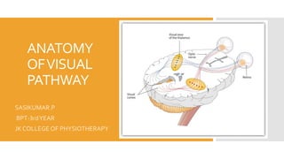
Anatomy of Visual pathways
Optic nerve
Optic chiasm
Retrochiasm
Visual Reflex and Visual field deficits.
Cranial Nerve II and Visual Pathways.pptxAtemJoshua
Definition of visual field
Normal vision
What are the causes of visual field defect?
Anatomy of Visual pathways
Optic nerve
Optic chiasm
Retrochiasm
Visual Reflex and Visual field deficits.
he sense organs — eyes, ears, tongue, skin, and nose — help to protect the body. The human sense organs contain receptors that relay information through sensory neurons to the appropriate places within the nervous system.
Each sense organ contains different receptors.
General receptors are found throughout the body because they are present in skin, visceral organs (visceral meaning in the abdominal cavity), muscles, and joints.
Special receptors include chemoreceptors (chemical receptors) found in the mouth and nose, photoreceptors (light receptors) found in the eyes, and mechanoreceptors found in the ears.
Human eye is a sense organ that responses to light and allows vision. Eyeball is placed in bony orbit in the skull and protected by eyelids. Eyeball is made up of three layers; Fibrous tunic (cornea and sclera), Vascular tunic (choroid, ciliary body and iris) and Retina. There are six extra ocular muscles to control movement of each eye. Optic nerve for its co-ordination with the brain. Blood is supplied to eye by the branches of internal carotid artery.
TEST BANK for Operations Management, 14th Edition by William J. Stevenson, Ve...kevinkariuki227
TEST BANK for Operations Management, 14th Edition by William J. Stevenson, Verified Chapters 1 - 19, Complete Newest Version.pdf
TEST BANK for Operations Management, 14th Edition by William J. Stevenson, Verified Chapters 1 - 19, Complete Newest Version.pdf
More Related Content
Similar to ANATOMY AND PHYSIOLOGY OF VISUAL PATHWAY
he sense organs — eyes, ears, tongue, skin, and nose — help to protect the body. The human sense organs contain receptors that relay information through sensory neurons to the appropriate places within the nervous system.
Each sense organ contains different receptors.
General receptors are found throughout the body because they are present in skin, visceral organs (visceral meaning in the abdominal cavity), muscles, and joints.
Special receptors include chemoreceptors (chemical receptors) found in the mouth and nose, photoreceptors (light receptors) found in the eyes, and mechanoreceptors found in the ears.
Human eye is a sense organ that responses to light and allows vision. Eyeball is placed in bony orbit in the skull and protected by eyelids. Eyeball is made up of three layers; Fibrous tunic (cornea and sclera), Vascular tunic (choroid, ciliary body and iris) and Retina. There are six extra ocular muscles to control movement of each eye. Optic nerve for its co-ordination with the brain. Blood is supplied to eye by the branches of internal carotid artery.
TEST BANK for Operations Management, 14th Edition by William J. Stevenson, Ve...kevinkariuki227
TEST BANK for Operations Management, 14th Edition by William J. Stevenson, Verified Chapters 1 - 19, Complete Newest Version.pdf
TEST BANK for Operations Management, 14th Edition by William J. Stevenson, Verified Chapters 1 - 19, Complete Newest Version.pdf
Flu Vaccine Alert in Bangalore Karnatakaaddon Scans
As flu season approaches, health officials in Bangalore, Karnataka, are urging residents to get their flu vaccinations. The seasonal flu, while common, can lead to severe health complications, particularly for vulnerable populations such as young children, the elderly, and those with underlying health conditions.
Dr. Vidisha Kumari, a leading epidemiologist in Bangalore, emphasizes the importance of getting vaccinated. "The flu vaccine is our best defense against the influenza virus. It not only protects individuals but also helps prevent the spread of the virus in our communities," he says.
This year, the flu season is expected to coincide with a potential increase in other respiratory illnesses. The Karnataka Health Department has launched an awareness campaign highlighting the significance of flu vaccinations. They have set up multiple vaccination centers across Bangalore, making it convenient for residents to receive their shots.
To encourage widespread vaccination, the government is also collaborating with local schools, workplaces, and community centers to facilitate vaccination drives. Special attention is being given to ensuring that the vaccine is accessible to all, including marginalized communities who may have limited access to healthcare.
Residents are reminded that the flu vaccine is safe and effective. Common side effects are mild and may include soreness at the injection site, mild fever, or muscle aches. These side effects are generally short-lived and far less severe than the flu itself.
Healthcare providers are also stressing the importance of continuing COVID-19 precautions. Wearing masks, practicing good hand hygiene, and maintaining social distancing are still crucial, especially in crowded places.
Protect yourself and your loved ones by getting vaccinated. Together, we can help keep Bangalore healthy and safe this flu season. For more information on vaccination centers and schedules, residents can visit the Karnataka Health Department’s official website or follow their social media pages.
Stay informed, stay safe, and get your flu shot today!
Prix Galien International 2024 Forum ProgramLevi Shapiro
June 20, 2024, Prix Galien International and Jerusalem Ethics Forum in ROME. Detailed agenda including panels:
- ADVANCES IN CARDIOLOGY: A NEW PARADIGM IS COMING
- WOMEN’S HEALTH: FERTILITY PRESERVATION
- WHAT’S NEW IN THE TREATMENT OF INFECTIOUS,
ONCOLOGICAL AND INFLAMMATORY SKIN DISEASES?
- ARTIFICIAL INTELLIGENCE AND ETHICS
- GENE THERAPY
- BEYOND BORDERS: GLOBAL INITIATIVES FOR DEMOCRATIZING LIFE SCIENCE TECHNOLOGIES AND PROMOTING ACCESS TO HEALTHCARE
- ETHICAL CHALLENGES IN LIFE SCIENCES
- Prix Galien International Awards Ceremony
These lecture slides, by Dr Sidra Arshad, offer a quick overview of physiological basis of a normal electrocardiogram.
Learning objectives:
1. Define an electrocardiogram (ECG) and electrocardiography
2. Describe how dipoles generated by the heart produce the waveforms of the ECG
3. Describe the components of a normal electrocardiogram of a typical bipolar leads (limb II)
4. Differentiate between intervals and segments
5. Enlist some common indications for obtaining an ECG
Study Resources:
1. Chapter 11, Guyton and Hall Textbook of Medical Physiology, 14th edition
2. Chapter 9, Human Physiology - From Cells to Systems, Lauralee Sherwood, 9th edition
3. Chapter 29, Ganong’s Review of Medical Physiology, 26th edition
4. Electrocardiogram, StatPearls - https://www.ncbi.nlm.nih.gov/books/NBK549803/
5. ECG in Medical Practice by ABM Abdullah, 4th edition
6. ECG Basics, http://www.nataliescasebook.com/tag/e-c-g-basics
Tom Selleck Health: A Comprehensive Look at the Iconic Actor’s Wellness Journeygreendigital
Tom Selleck, an enduring figure in Hollywood. has captivated audiences for decades with his rugged charm, iconic moustache. and memorable roles in television and film. From his breakout role as Thomas Magnum in Magnum P.I. to his current portrayal of Frank Reagan in Blue Bloods. Selleck's career has spanned over 50 years. But beyond his professional achievements. fans have often been curious about Tom Selleck Health. especially as he has aged in the public eye.
Follow us on: Pinterest
Introduction
Many have been interested in Tom Selleck health. not only because of his enduring presence on screen but also because of the challenges. and lifestyle choices he has faced and made over the years. This article delves into the various aspects of Tom Selleck health. exploring his fitness regimen, diet, mental health. and the challenges he has encountered as he ages. We'll look at how he maintains his well-being. the health issues he has faced, and his approach to ageing .
Early Life and Career
Childhood and Athletic Beginnings
Tom Selleck was born on January 29, 1945, in Detroit, Michigan, and grew up in Sherman Oaks, California. From an early age, he was involved in sports, particularly basketball. which played a significant role in his physical development. His athletic pursuits continued into college. where he attended the University of Southern California (USC) on a basketball scholarship. This early involvement in sports laid a strong foundation for his physical health and disciplined lifestyle.
Transition to Acting
Selleck's transition from an athlete to an actor came with its physical demands. His first significant role in "Magnum P.I." required him to perform various stunts and maintain a fit appearance. This role, which he played from 1980 to 1988. necessitated a rigorous fitness routine to meet the show's demands. setting the stage for his long-term commitment to health and wellness.
Fitness Regimen
Workout Routine
Tom Selleck health and fitness regimen has evolved. adapting to his changing roles and age. During his "Magnum, P.I." days. Selleck's workouts were intense and focused on building and maintaining muscle mass. His routine included weightlifting, cardiovascular exercises. and specific training for the stunts he performed on the show.
Selleck adjusted his fitness routine as he aged to suit his body's needs. Today, his workouts focus on maintaining flexibility, strength, and cardiovascular health. He incorporates low-impact exercises such as swimming, walking, and light weightlifting. This balanced approach helps him stay fit without putting undue strain on his joints and muscles.
Importance of Flexibility and Mobility
In recent years, Selleck has emphasized the importance of flexibility and mobility in his fitness regimen. Understanding the natural decline in muscle mass and joint flexibility with age. he includes stretching and yoga in his routine. These practices help prevent injuries, improve posture, and maintain mobilit
Pulmonary Thromboembolism - etilogy, types, medical- Surgical and nursing man...VarunMahajani
Disruption of blood supply to lung alveoli due to blockage of one or more pulmonary blood vessels is called as Pulmonary thromboembolism. In this presentation we will discuss its causes, types and its management in depth.
Couples presenting to the infertility clinic- Do they really have infertility...Sujoy Dasgupta
Dr Sujoy Dasgupta presented the study on "Couples presenting to the infertility clinic- Do they really have infertility? – The unexplored stories of non-consummation" in the 13th Congress of the Asia Pacific Initiative on Reproduction (ASPIRE 2024) at Manila on 24 May, 2024.
3. INTRODUCTION :
Visual pathway or optic pathway is
the nervous pathway that transmits impulses
from retina visual center in cerebral cortex.
In binocular vision, the light rays
from temporal(outer) half of visual field fall
upon the nasal part of corresponding retina.
The rays from nasal (inner) half of visual field
fall upon the temporal part of retina.
4.
5. BASIC'SOF
EYE
The eye is a special orang of the sense of sight.
The adult human eyeball is a hollow,spherical
stucture,24mm in diameter and situated in the
orbital cavity.
Only 1/6th of the eyeball is avisible outside.
Layer's of eyeball
Outer fibrous layer - Sclera and cornea.
Middle layer - Choroid,Ciliary body and
Iris.
Inner nervous layer - Retina.
6.
7. VISUAL
RECEPTORS
Rods and cones which are present in the retina
of eye form the visual receptors.
Fibers from the visual receptors synapse with
dendrites of bipolar cells of inner nuclear layer
of the retina.
8.
9. ORDEROF
NEURONS
FIRST ORDER NEURONS
First order neurons (primary neurons) are bipolar cells
in the retina. Axons from the bipolar cells synapse with
dendrites of ganglionic cells.
SECOND ORDER NEURONS
Second order neurons (secondary neurons) are the
ganglionic cells in ganglionic cell layer of retina, Axons
of the ganglionic cells form optic nerve. Optic nerve
leaves the eye and terminates in lateral geniculate
body.
THIRD ORDER NEURONS
Third order neurons are in the lateral geniculate body.
Fibers arising from here, reach the visual cortex.
13. Optic nerve
Optic nerve is the 2nd cranial nerve.
It is purely sensory and responsible for vision.
It is also called the nerve of sight.
Optic nerve is 4cm in length.
Optic nerve is formed by the axons of
ganglionic cells.
Optic nerve leaves the eye through optic disk.
The fibers from temporal part of retina are in
lateral part of the nerve and carry the impulses
from nasal half of visual field of same eye.
The fibers from nasal part of retina are in
medial part of the nerve and carry the impulses
from temporal half of visual field of same eye.
14.
15. OPTIC
CHIASMA
Medial fibers of each optic nerve cross the
midline and join the uncrossed lateral fibers of
opposite side, to form the optic tract.
This area of crossing of the optic nerve fibers is
called optic chiasma.
16.
17. OPTIC TRACT
Optic tract is formed by uncrossed fibers of
optic nerve on the same side and crossed fibers
of optic nerve from the opposite side.
All the fibers of optic tract run backward,
outward and towards the cerebral peduncle.
While reaching the peduncle, the fibers pass
between tuber cinereum and anterior
perforated substance.
The fibers turn around the peduncle to reach
the lateral geniculate body in thalamus.
18. . Due to crossing of medial fibers in optic
chiasma, the left optic tract carries impulses
from temporal part of left retina and nasal part
of right retina (i.e.it is responsible for vision in
nasal half of left visual field and temporal half
of right visual field)
The right optic tract contains fibers from nasal
half of left retina and temporal half of right
retina (i.e. It is responsible for vision in
temporal half of left visual field and nasal half
of right visual field.)
19.
20. LATERAL
GENICULATE
BODY
Majority of the fibers of optic tract terminate
in lateral geniculate body, which forms the
subcortical center for visual sensation. From
here, the geniculocalcarine tract or optic
radiation arises.This tract is the last relay of
visual pathway.
Superior colliculus: It is concerned with
reflex movements of eyeballs and head, in
response to optic stimulus
Pretectal nucleus: It is concerned with light
reflexes
Supraoptic nucleus of hypothalamus: It is
concerned with the retinal control of
pituitary in animals. But in human, it does
not play any important role.
21.
22. OPTIC
RADIATION
Fibers from lateral geniculate body pass
through internal capsule and form optic
radiation.
The fibers between lateral geniculate body
and visual cortex are also called
geniculocalcarine fibers. Optic radiation ends in
visual cortex
23.
24. VISUAL
CORTEX
Primary cortical center for vision is called
visual cortex, which is located on the medial
surface of occipital lobe.
It forms the walls and lips of calcarine fissure
in medial surface of occipital lobe.
The peripheral retinal representation occupies
the anterior part of visual cortex.
Macular representation occupies the posterior
part of visual cortex near occipital pole.
25. Areas ofVisual Cortex and their Function
Primary visual area (area 17), which is
concerned with the perception of visual
impulses.
Secondary visual area or visual association
area (area 18), which is concerned with the
interpretation of visual impulses.
Occipital eye field (area 19), which is
concerned with the movement of eyes.
26.
27. APPLIED
ANATOMY
EFFECTS OF LESION INVISUAL PATHWAY:
Injury to any part of optic pathway causes
visual defect and the nature of defect
depends upon the location and extent of
injury.
ANOPIA:
Anopiais the loss of vision in one visual
field.
Hemianopia:
Loss of vision in one half of visual field.
Hemianopia is classified into two types:
Homonymous hemianopia .
Heteronymous hemianopia.
28. Homonymous hemianopia:
Homonymous hemianopsia (or
homonymous hemianopia, HH) is a field loss
deficit in the same halves of the visual field of
each eye.
This condition most commonly results from
stroke for adults, or tumors/lesions for
patients under the age of 18.
Heteronymous hemianopia:
A heteronymous hemianopsia is the loss of
half of the visual field on different sides in
both eyes.
It is separated into two categories:
Binasal hemianopsia – the loss of the
fields surrounding the nose.
Bitemporal hemianopsia – the loss of the
fields closest to the temples.
32. Refractive error
(i.e. inadequate
focussing on the
retina, e.g. hyper-
metropia,myopia)
can be overcome
by testing reading
acuity through a
pinhole.This
concentrates a
thin beam of vision
on the macula.
33. Visual fields
Gross testing by CONFRONTATION. Compare the
patient’s fields of vision by advancing a
moving finger or, more accurately, a red 5 mm pin
from the extreme periphery towards the fixation
point.This maps out ‘cone’ vision. A 2 mm pin will
define central feld defects which may only
manifest as a loss of colour perception.
In the temporal portion of the visual field the
physiological blind spot may be detected. A 2 mm
object should disappear here.