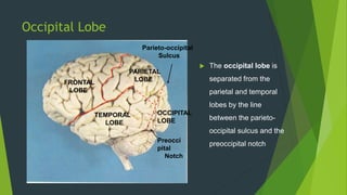
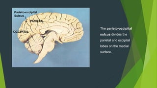
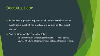
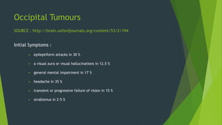
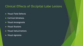
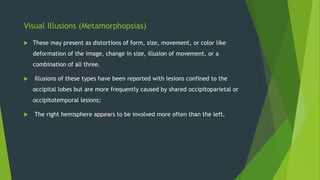
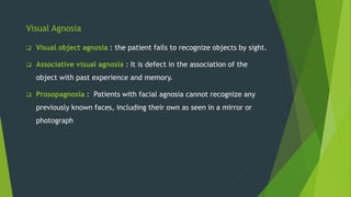
Occipital Tumours
Occipital Lobe
OCCIPITAL
LOBE
FRONTAL
LOBE
PARIETAL
LOBE
TEMPORAL
LOBE
Preocci
pital
Notch
Parieto-occipital
Sulcus
 The occipital lobe is
separated from the
parietal and temporal
lobes by the line
between the parieto-
occipital sulcus and the
preoccipital notch
Parieto-occipital
Sulcus
OCCIPITAL
PARIETAL
The parieto-occipital
sulcus divides the
parietal and occipital
lobes on the medial
surface.
Occipital Lobe
 is the visual processing center of the mammalian brain
containing most of the anatomical region of the visual
cortex.
 Subdivisions of the occipital lobe :
• V1 (Primary visual cortex; Brodmann area 17; Striate cortex)
• V2, V3, V4, V5, V6 ( Secondary visual cortex, extrastriate regions)
Functions :
 V1 & V2 - function to segregate info to other areas.,
receives primary visual impressions Color/Form/Motion/Size
and illumination.
 V3, V3A, V4, V5- Visual association areas- Recognition and
identification of objects, storage of visual memories, it
functions in more complex visual recognition and
perception, revisualization, visual association and spatial
orientation.
Occipital Tumours
Initial Symptoms :
 epileptiform attacks in 30 %
 a visual aura or visual hallucinations in 12.5 %
 general mental impairment in 17 %
 headache in 35 %
 transient or progressive failure of vision in 15 %
 strabismus in 2·5 %
SOURCE : http://brain.oxfordjournals.org/content/53/2/194
On Examination :
 Mental changes, 60 %
 Contralateral homonymous defects of the visual fields, 94 %
 Papilloedema or optic atrophy, 70 %
 Inequality of the pupils, 35 %
 Ocular pareses, usually of the external recti, 30 %
 Nystagmus and nystagmoid jerkings, 35 %
 Disturbances of speech functions, 30 %
 Minor degrees of motor disturbance usually in the contra-lateral limbs, 90 %
Clinical Effects of Occipital Lobe Lesions
 Visual Field Defects
 Cortical blindness
 Visual Anosognosia
 Visual Illusions
 Visual hallucinations
 Visual Agnosias
Visual Field Defects
 The most familiar clinical abnormality resulting from a lesion of one
occipital lobe, is a contralateral homonymous hemianopia
 With a neoplastic lesion that eventually involves the entire striate region,
the field defect may extend from the periphery toward the center
 Loss of color vision (hemiachromatopsia) often precedes loss of black and
white.
 Destruction of only part of the striate cortex on one side yields characteristic
field defects that accurately indicate the loci of the lesion.
Cortical Blindness
 With bilateral lesions of the occipital lobes (destruction of area 17 of
both hemispheres), may have complete “cortical” blindness.
 The degree of blindness may be equivalent to that which follows severing of
the optic nerves.
 The pupillary light reflexes are preserved because they depend upon visual
fibers that terminate in the midbrain, but reflex closure of the eyelids to
threat or bright light may not be preserved
Visual Anosognosia (Anton Syndrome )
 The main characteristic of this disorder is the denial of blindness by a patient
who obviously cannot see.
 The patient acts as though he could see, and in attempting to walk, collides
with objects, even to the point of injury.
 The lesions in cases of negation of blindness extend beyond the striate cortex
to involve the visual association areas.
Visual Illusions (Metamorphopsias)
 These may present as distortions of form, size, movement, or color like
deformation of the image, change in size, illusion of movement, or a
combination of all three.
 Illusions of these types have been reported with lesions confined to the
occipital lobes but are more frequently caused by shared occipitoparietal or
occipitotemporal lesions;
 The right hemisphere appears to be involved more often than the left.
Visual Hallucinations
 These phenomena may be elementary or complex, and both types have
sensory as well as cognitive aspects.
 Elementary (or unformed) hallucinations : include flashes of light,
colors, luminous points, stars, multiple lights (like candles), and
geometric forms (circles, squares, and hexagons). They may be stationary
or moving (zigzag, oscillations, vibrations, or pulsations).
 Complex hallucinations : include objects, persons, or animals and
infrequently, more complete scenes that are indicative of lesions in the
visual association areas or their connections with the temporal lobes.
Visual Agnosia
 Visual object agnosia : the patient fails to recognize objects by sight.
 Associative visual agnosia : It is defect in the association of the
object with past experience and memory.
 Prosopagnosia : Patients with facial agnosia cannot recognize any
previously known faces, including their own as seen in a mirror or
photograph

Opthalmic tumours

  • 1.
  • 2.
    Occipital Lobe OCCIPITAL LOBE FRONTAL LOBE PARIETAL LOBE TEMPORAL LOBE Preocci pital Notch Parieto-occipital Sulcus  Theoccipital lobe is separated from the parietal and temporal lobes by the line between the parieto- occipital sulcus and the preoccipital notch
  • 3.
    Parieto-occipital Sulcus OCCIPITAL PARIETAL The parieto-occipital sulcus dividesthe parietal and occipital lobes on the medial surface.
  • 4.
    Occipital Lobe  isthe visual processing center of the mammalian brain containing most of the anatomical region of the visual cortex.  Subdivisions of the occipital lobe : • V1 (Primary visual cortex; Brodmann area 17; Striate cortex) • V2, V3, V4, V5, V6 ( Secondary visual cortex, extrastriate regions)
  • 5.
    Functions :  V1& V2 - function to segregate info to other areas., receives primary visual impressions Color/Form/Motion/Size and illumination.  V3, V3A, V4, V5- Visual association areas- Recognition and identification of objects, storage of visual memories, it functions in more complex visual recognition and perception, revisualization, visual association and spatial orientation.
  • 6.
    Occipital Tumours Initial Symptoms:  epileptiform attacks in 30 %  a visual aura or visual hallucinations in 12.5 %  general mental impairment in 17 %  headache in 35 %  transient or progressive failure of vision in 15 %  strabismus in 2·5 % SOURCE : http://brain.oxfordjournals.org/content/53/2/194
  • 7.
    On Examination : Mental changes, 60 %  Contralateral homonymous defects of the visual fields, 94 %  Papilloedema or optic atrophy, 70 %  Inequality of the pupils, 35 %  Ocular pareses, usually of the external recti, 30 %  Nystagmus and nystagmoid jerkings, 35 %  Disturbances of speech functions, 30 %  Minor degrees of motor disturbance usually in the contra-lateral limbs, 90 %
  • 8.
    Clinical Effects ofOccipital Lobe Lesions  Visual Field Defects  Cortical blindness  Visual Anosognosia  Visual Illusions  Visual hallucinations  Visual Agnosias
  • 9.
    Visual Field Defects The most familiar clinical abnormality resulting from a lesion of one occipital lobe, is a contralateral homonymous hemianopia  With a neoplastic lesion that eventually involves the entire striate region, the field defect may extend from the periphery toward the center  Loss of color vision (hemiachromatopsia) often precedes loss of black and white.  Destruction of only part of the striate cortex on one side yields characteristic field defects that accurately indicate the loci of the lesion.
  • 10.
    Cortical Blindness  Withbilateral lesions of the occipital lobes (destruction of area 17 of both hemispheres), may have complete “cortical” blindness.  The degree of blindness may be equivalent to that which follows severing of the optic nerves.  The pupillary light reflexes are preserved because they depend upon visual fibers that terminate in the midbrain, but reflex closure of the eyelids to threat or bright light may not be preserved
  • 11.
    Visual Anosognosia (AntonSyndrome )  The main characteristic of this disorder is the denial of blindness by a patient who obviously cannot see.  The patient acts as though he could see, and in attempting to walk, collides with objects, even to the point of injury.  The lesions in cases of negation of blindness extend beyond the striate cortex to involve the visual association areas.
  • 12.
    Visual Illusions (Metamorphopsias) These may present as distortions of form, size, movement, or color like deformation of the image, change in size, illusion of movement, or a combination of all three.  Illusions of these types have been reported with lesions confined to the occipital lobes but are more frequently caused by shared occipitoparietal or occipitotemporal lesions;  The right hemisphere appears to be involved more often than the left.
  • 13.
    Visual Hallucinations  Thesephenomena may be elementary or complex, and both types have sensory as well as cognitive aspects.  Elementary (or unformed) hallucinations : include flashes of light, colors, luminous points, stars, multiple lights (like candles), and geometric forms (circles, squares, and hexagons). They may be stationary or moving (zigzag, oscillations, vibrations, or pulsations).  Complex hallucinations : include objects, persons, or animals and infrequently, more complete scenes that are indicative of lesions in the visual association areas or their connections with the temporal lobes.
  • 14.
    Visual Agnosia  Visualobject agnosia : the patient fails to recognize objects by sight.  Associative visual agnosia : It is defect in the association of the object with past experience and memory.  Prosopagnosia : Patients with facial agnosia cannot recognize any previously known faces, including their own as seen in a mirror or photograph

Editor's Notes

  • #5 V = Vision ; V2,V3,V4,V5,V5 = Broadman areas.