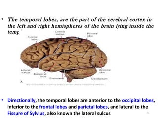
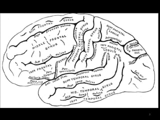
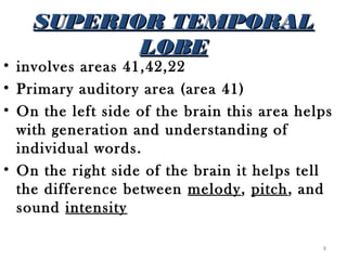
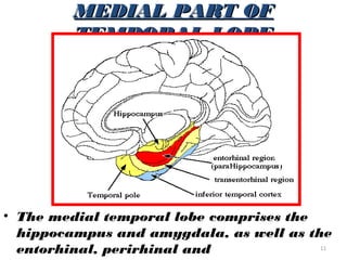
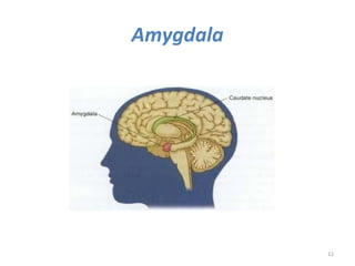
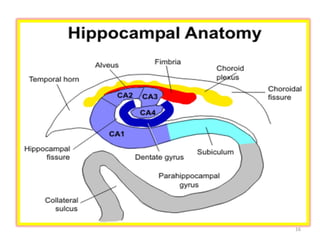
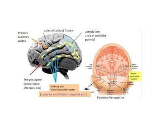
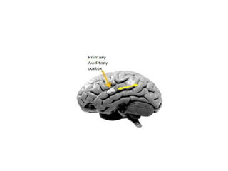
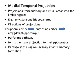
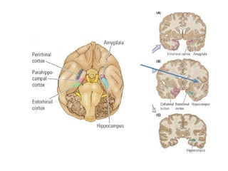
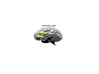
TEMPORAL
  LOBE

  PRESENTOR:- DR VIRINCHI
                SHARMA
        MODERATOR:-
              Dr. V S RAJ
                       1
0VERVIEW
• ANATOMY(parts)

• FUNCTIONAL   AREAS

• LOOPS & PATHWAYS

• FUNCTIONS

• DISORDERS            2
• The Temporal lobes are one of the most
  heterogeneous regions of the Neocortex.  
• Input for the Limbic and Para-Limbic cortex.  
• This is the lobe that is credited with being the seat
  of human para-Psychological and Psychic
  abilities!  
• The temporal lobe, Is considered in its broadest
  context, links the present sensory and emotional
  experience of our world


                                                    3
M Y
      A T O
    N
  A OF             B E
              L LO
          R A
      P O
T E M                4
• The temporal lobes, are the part of the cerebral cortex in
  the left and right hemispheres of the brain lying inside the
  temples.


• Directionally, the temporal lobes are anterior to the occipital lobes,
  inferior to the frontal lobes and parietal lobes, and lateral to the
  Fissure of Sylvius, also known the lateral sulcus                   5
• SUPERIOR AND INFERIOR
  TEMPORAL SULCI DIVIDE
  TEMPORAL LOBE INTO 3 LOBES

• SUPERIOR TEMPORAL LOBE

• MIDDLE TEMPORAL LOBE

• INFERIOR TEMPORAL LOBE


                           6
7
SUPERIOR TEMPORAL
           LOBE
• involves areas 41,42,22
• Primary auditory area (area 41)
• On the left side of the brain this area helps
  with generation and understanding of
  individual words.
• On the right side of the brain it helps tell
  the difference between melody, pitch, and
  sound intensity

                                             8
MIDDLE TEMPORAL LOBE

The region encompasses most of the lateral
  temporal cortex, a region believed to play a
  part in auditory processing and language.
• Language function is left lateralized in most
  individuals.
• Brodmann area 21.


                                            9
INFERIOR TEMPORAL
           LOBE
• It refers to a subdivision of the
  cytoarchitecturally defined temporal
  region of cerebral cortex. In the human
  it corresponds approximately to the
  inferior temporal gyrus.
• Brodmann area 20.
• The region encompasses most of the
  ventral temporal cortex, a region
  believed to play a part in high-level
  visual processing and recognition    10
MEDIAL PART OF
         TEMPORAL LOBE


• The medial temporal lobe comprises the
  hippocampus and amygdala, as well as the
  entorhinal, perirhinal and             11
Amygdala


           12
Amygdala
• The fear & flight response
• Inputs: The association areas of visual, auditory, and
  somato sensory cortices are the main inputs to the
  amygdala.
• Outputs: The main outputs of the amygdala are to the
  hypothalamus and brainstem autonomic centers,
  including the vagal nuclei and the sympathetic
  neurons.
• The amygdala is also involved with mood and the
  conscious emotional response to an event
• The amygdala is also extensively interconnected with
  frontal cortex, medio dorsal thalamus, and the medial
  striatum.                                            13
• The deep group, which
  includes the lateral, basal,
  accessory basal nucleic
• Func: collects input from
  sensory cortex.


• The more dorsal group,
  which includes the central &
  medial nuclei
• Func: receives projections
  from the deep group and
  sends the signal out to
  autonomic centers.


                                 14
• The amygdala is the heart of the emotional
  system. It processes and interprets all sensory
  data.
• It modulates the flow of emotional
  information between the cerebral cortex and
  the hypothalamus, and in doing that, it
  modulates autonomic, endocrine, and
  affective responses.
• Lesions in amygdala lead to-- agitation,
  irritability, anxiety, mood disorders,
  paranoia, and psychosis.

                                                15
16
The Hippocampus
• The hippocampus is a scrolled structure
  located in the medial temporal lobe.
• The hippocampus can be divided into at least
  five different areas.
• The dentate gyrus is the dense dark layer of
  cells at the "tip" of the hippocampus. Areas
  CA3 and CA1 are more diffuse; the small
  CA2 is hard to distinguish between them.
  (CA stands for cornu ammonis, from its ram's
  horn shape.)
• The subiculum sits at the base of the
  hippocampus, and is continuous with
  entorhinal cortex, which is part of the  17
18
FUNCTIONAL
  AREAS OF
TEMPORAL LOBE


                19
• AUDITORY – primary & Association

• OLFACTORY - primary & Association

• VISUAL (Recognition & association)

• MEMORY

• EMOTIONAL & SOCIAL

• Link past and present sensory and
  emotional experiences into a         20
LANGUAGE &
               COMPREHENSION
• LANGUAGE             AREAS
• A specialized integration area called Wernicke's area is
  found in the posterior temporal lobe of one hemisphere
  (usually the left). Commonly called the "speech area, "
  Wernicke's area surrounds and encompasses part of the
  auditory association area.
• AFFECTIVE           LANGUAGE             AREAS
• Affective language areas involved in the nonverbal
  emotional components of language - appear to be present
  in the hemisphere opposite Brocas's and Wernickes's
  areas.
• These "mirror images" allow the lilt of tone of our voice
  and our gestures to express our emotions when we speak,
  and permit us to comprehend the emotional content of
  what we hear.                                            21
• Lesions to this area result in aprosodia, a condition in
AUDITORY SENSES
• PRIMARY AUDITORY AREA (area 41)
   Essential to detect changes in
  frequency , & to know the direction
  from which sounds originate.

• AUDITORY ASSOCIATION AREA
  (area 42)

• HIGHER AUDITORY ASSOCIATION      22
23
VISUAL SENSES
• Processing of our
  recognition of objects
  occurs in a path on the
  lower, dorsal stream in
  the temporal lobe; here
  you find areas sensitive to
  faces vs. objects,
• Area MT (right) performs
  processing on motion.
  Subjects without an area
  MT describe seeing
  motion as discontinuous
                                24
  pictures – eg.having to
OLFACTORY SENSES


The rightmost green
spots are the location in
cortex where smell is
processed.


                            25
GUSTATORY SENSES
• The sensation of
  taste is processed in
  insular cortex


                          26
connections of the Temporal Lobes

•   Five main types:
•   Hierarchical sensory pathway
•   Dorsal auditory pathway
•   Polymodal pathway
•   Medial (mesial) temporal pathway
•   Frontal lobe projection
• Hierarchical sensory pathway
• connections from
• primary(sensory neuron) and secondary auditory
                                   and visual
  cortical


•       through the lateral temporal cortex


•          terminate in the temporal pole
•   visual travels        inferior temporal gyrus
•
•  auditory travels        e suprior temporal gyrus
•  Major destinations:
•  amygdala and hippocampus
•  This results in the integration of information into:
   memory, retrieval of stored information,
   emotional tone
• Ultimate effect
 stimulus recognition
• The familiar conscious experience of knowing,
   assimilating, and feeling
• Dorsal auditory pathway

• Forms important functional connections with
  the posterior parietal cortex
• Enables location of sounds in space
• Promotes orienting and initiation of
  movements relative to sound location
Polymodal Pathway
• connections emerging from the auditory and
  visual hierarchical pathways
• Directed towards the neurons enfolded within
  the superior temporal sulcus
• Polymodal nature of neurons
• Assigns stimuli to specific category of classes,
  linked to and can be retrieved by memory
• Medial Temporal Projection
• Projections from auditory and visual areas into the
  limbic regions
• E.g., amygdala and hippocampus
• Directions of projections
Peripheral cortex       entorhinalcortex
  amygdala/hippocampus
• Perforant pathway
• forms the main projection to thehippocampus
• Damage in this region severely affects memory
  formation
• Frontal-lobe Projection

• Neurons from the temporal lobe have strong
  connections with the frontal lobe
• Posterior temporal cortex
• Projects to the dorsolateral prefrontal cortex
• anterior temporal cortex
• Projects to the orbital frontal cortex
• Damage leads to terrible life decisions
DISORDERS OF
TEMPORAL LOBE

                39
8 principal symptoms of
     temporal lobe damage:
• Disturbance of auditory sensation and
  perception
• Disturbance of selective attention of
  auditory and visual input
• Disorders of visual perception
• Impaired organization and categorization of
  verbal material
• Disturbance of language comprehension
• Impaired long-term memory
• Altered personality and affective behaviour
• Altered sexual behaviour                40
Manifestations of temporal lobe
                lesions
• Disorders of auditory perception:
   – Lesions of the left superior temporal gyrus produce
     problems of speech perception with difficulty in
     discriminating speech and the temporal order of sounds is
     impaired.

• Lesions of the right superior temporal gyrus can
  produce disorders of perception of music with inability to
  discriminate melodies and produce prosody

• The inferior temporal cortex is responsible for visual
  perception and lesions produce inability to recognise faces,
  called prosopagnosia.

• There may be disturbance of visual and auditory input
  selection. This presents as impairment of short term memory,
                                                            41
disorders of memory
• The medial and inferior temporal cortex and hippocampus are
  responsible for memory.
• There is complete anterograde amnesia following bilateral
  removal of medial temporal lobes, including hippocampus &
  amygdala.
• There is difficulty recalling information.
• The left side is responsible for verbal material and the right for
  non-verbal memory such as faces, tunes and drawings.


                                                                 42
• temporal lobe personality. There is
  egocentricity, pedantic speech, perseveration
  of speech, paranoia, religious preoccupations
  and a tendency to aggressive outbursts,
  especially after right temporal lobectomy.
• temporal lobe lesions can present with visual
  field defects in the form of superior quadrant
  loss, sometimes called the "pie in the sky
  defect."


• Stroke normally reduces libido but temporal   43
APHASIA
• Any disturbance in the comprehension or
  expression of language caused by a brain
  lesion.
• NONFLUENT APHASIA, i.e. in lesion to
  Broca's area results in slow speech, difficulty
  in choosing words, or use of words that only
  approximate the correct word.e.g., a person
  may say "tssair" when asked to identify a
  picture of a chair.
• A lesion to Wernicke's area may result in
  FLUENT APHASIA, in which a person speaks
  normally, and sometimes excessively, but uses
  jargon and invented words, that make little 44
Kluver-Bucy syndrome
     Kluver-Bucy syndrome results due to a bilateral
 destruction of the amygdaloid body and inferior
 temporal cortex.
 It is characterized by
      Visual agnosia,
      Placidity,
       Hypermetamorphosis,
         Hyperorality
  Hypersexuality.
 causes: cerebral trauma; infections including
 herpes and other encephalitides; Alzheimer's
 disease and other dementias; Niemann-Pick disease 45
 and cerebrovascular disease.
Diseases of the Hippocampus
• The hippocampus is
  particularly vulnerable to
  several disease processes,
  including ischemia, which
  is any obstruction of blood
  flow or oxygen deprivation,
  Alzheimer's disease,
  and epilepsy.
• These diseases selectively
  attack CA1, which
  effectively cuts through the
  hippocampal circuit.


                                 46
TEMPORAL LOBE EPILEPSY
TEMPORAL LOBE EPILEPSY (TLE) WAS DEFINED IN 1985 BY
THE INTERNATIONAL LEAGUE AGAINST EPILEPSY (ILAE) AS
A CONDITION CHARACTERIZED BY RECURRENT
UNPROVOKED SEIZURES ORIGINATING FROM THE MEDIAL
OR LATERAL TEMPORAL LOBE.


LATERAL TEMPORAL LOBE EPILEPSY : ARISES IN THE
NEOCORTEX
MESIAL TEMPORAL LOBE EPILEPSY:HIPPOCAMPUS,
PARAHIPPOCAMPAL GYRUS AMYGDALA
                                                      47
cAuSES
•   Hereditary
•   Febrile sezuires
•   Brain tumors
•   spinal meningitis,
•    encephalitis,
•    head injury or blood vessel malformations
•   Trauma
•   congenital brain malformations
•   Alcohol withdrawl
•   Sleep deprivation
                                                 48
SYMPTOM
•                  S
    Seizures usually simple partial but can be complex
    partial seizures also
•   Duration is one to two minutes
•   Auras
•   Motionless staring
•   Anxiety
•   Emergent past memories
•   Spiritual religious experience

• Most common type comprising 70percent of
  seizures with 1.5%of population world wide
                                                         49
TREATMEnT
• Antiepilectic drugs


• Surgical procedures


                         50
cVA---TEMPORAL LOBE
•   Middle cerebral artery in farct:
    – Aphasia or non-dominant hemisphere findings
      depending on the side.
    – “Partial” middle cerebral artery syndromes, almost
      always of embolic origin, may include a) sensorimotor
      paresis with little aphasia b) conduction aphasia c)
      Wernicke’s aphasia without hemiparesis.
    – Wernicke's aphasia, caused most often by occlusion of
      the lower division of the MCA bifurcation or one of its
      branches. Patients with Wernicke's aphasia vocalize
      smoothly and with expression, but they demonstrate
      paraphasias or speech with distorted phonetic structure,
      word substitution, and additional prefixes and suffixes.

    – The infarct responsible for a classic Wernicke's aphasia
      includes the dominant posterior temporal, inferior
      parietal, and lateral temporo-occipital regions.
                                                             51
• Posterior cerebral artery syndrome:
TEMPORAL LOBE ABScESS
• Brain abscess is a
  newly formed cavity in
  brain tissue, filled with
  pus.
• Mastoiditis causes
  temporal lobe abscess.
• Hematogenous
  abscesses are often
  multiple.

                              52
TEMPORAL LOBE
          TUMORS
• As many as 50 to 55 percent of patients
  with temporal lobe tumors experience
  psychiatric, behavioral, or personality
  changes

• Psychopathology related to temporal lobe
  tumors can be ictal, that is, seizure
  associated, or interictal, completely
  unrelated to seizure activity.

• Patients with tumors of the temporal lobe
  who have temporal lobe seizures often
  have seizure-associated schizophrenia-like 53
SYMPTOMS INCLUDE

• Auditory hallucinations and atypical
  dream-like episodes, depersonalization,
  blanking-out spells, and dazed feelings
• Patients with temporal lobe seizures may
  present with depression and frontal lobe–
  like apathy and irritability,
• Or with features suggesting hypomania or
  mania,
• Personality changes commonly occur and
  may be one of the earliest indications of
  an undiagnosed temporal lobe tumor          54
Thank you

 Thank u…….

              55

Temporal lobe ppt

  • 1.
    TEMPORAL LOBE PRESENTOR:- DR VIRINCHI SHARMA MODERATOR:- Dr. V S RAJ 1
  • 2.
    0VERVIEW • ANATOMY(parts) • FUNCTIONAL AREAS • LOOPS & PATHWAYS • FUNCTIONS • DISORDERS 2
  • 3.
    • The Temporallobes are one of the most heterogeneous regions of the Neocortex.   • Input for the Limbic and Para-Limbic cortex.   • This is the lobe that is credited with being the seat of human para-Psychological and Psychic abilities!   • The temporal lobe, Is considered in its broadest context, links the present sensory and emotional experience of our world 3
  • 4.
    M Y A T O N A OF B E L LO R A P O T E M 4
  • 5.
    • The temporallobes, are the part of the cerebral cortex in the left and right hemispheres of the brain lying inside the temples. • Directionally, the temporal lobes are anterior to the occipital lobes, inferior to the frontal lobes and parietal lobes, and lateral to the Fissure of Sylvius, also known the lateral sulcus 5
  • 6.
    • SUPERIOR ANDINFERIOR TEMPORAL SULCI DIVIDE TEMPORAL LOBE INTO 3 LOBES • SUPERIOR TEMPORAL LOBE • MIDDLE TEMPORAL LOBE • INFERIOR TEMPORAL LOBE 6
  • 7.
  • 8.
    SUPERIOR TEMPORAL LOBE • involves areas 41,42,22 • Primary auditory area (area 41) • On the left side of the brain this area helps with generation and understanding of individual words. • On the right side of the brain it helps tell the difference between melody, pitch, and sound intensity 8
  • 9.
    MIDDLE TEMPORAL LOBE Theregion encompasses most of the lateral temporal cortex, a region believed to play a part in auditory processing and language. • Language function is left lateralized in most individuals. • Brodmann area 21. 9
  • 10.
    INFERIOR TEMPORAL LOBE • It refers to a subdivision of the cytoarchitecturally defined temporal region of cerebral cortex. In the human it corresponds approximately to the inferior temporal gyrus. • Brodmann area 20. • The region encompasses most of the ventral temporal cortex, a region believed to play a part in high-level visual processing and recognition 10
  • 11.
    MEDIAL PART OF TEMPORAL LOBE • The medial temporal lobe comprises the hippocampus and amygdala, as well as the entorhinal, perirhinal and 11
  • 12.
  • 13.
    Amygdala • The fear& flight response • Inputs: The association areas of visual, auditory, and somato sensory cortices are the main inputs to the amygdala. • Outputs: The main outputs of the amygdala are to the hypothalamus and brainstem autonomic centers, including the vagal nuclei and the sympathetic neurons. • The amygdala is also involved with mood and the conscious emotional response to an event • The amygdala is also extensively interconnected with frontal cortex, medio dorsal thalamus, and the medial striatum. 13
  • 14.
    • The deepgroup, which includes the lateral, basal, accessory basal nucleic • Func: collects input from sensory cortex. • The more dorsal group, which includes the central & medial nuclei • Func: receives projections from the deep group and sends the signal out to autonomic centers. 14
  • 15.
    • The amygdalais the heart of the emotional system. It processes and interprets all sensory data. • It modulates the flow of emotional information between the cerebral cortex and the hypothalamus, and in doing that, it modulates autonomic, endocrine, and affective responses. • Lesions in amygdala lead to-- agitation, irritability, anxiety, mood disorders, paranoia, and psychosis. 15
  • 16.
  • 17.
    The Hippocampus • Thehippocampus is a scrolled structure located in the medial temporal lobe. • The hippocampus can be divided into at least five different areas. • The dentate gyrus is the dense dark layer of cells at the "tip" of the hippocampus. Areas CA3 and CA1 are more diffuse; the small CA2 is hard to distinguish between them. (CA stands for cornu ammonis, from its ram's horn shape.) • The subiculum sits at the base of the hippocampus, and is continuous with entorhinal cortex, which is part of the 17
  • 18.
  • 19.
    FUNCTIONAL AREASOF TEMPORAL LOBE 19
  • 20.
    • AUDITORY –primary & Association • OLFACTORY - primary & Association • VISUAL (Recognition & association) • MEMORY • EMOTIONAL & SOCIAL • Link past and present sensory and emotional experiences into a 20
  • 21.
    LANGUAGE & COMPREHENSION • LANGUAGE AREAS • A specialized integration area called Wernicke's area is found in the posterior temporal lobe of one hemisphere (usually the left). Commonly called the "speech area, " Wernicke's area surrounds and encompasses part of the auditory association area. • AFFECTIVE LANGUAGE AREAS • Affective language areas involved in the nonverbal emotional components of language - appear to be present in the hemisphere opposite Brocas's and Wernickes's areas. • These "mirror images" allow the lilt of tone of our voice and our gestures to express our emotions when we speak, and permit us to comprehend the emotional content of what we hear. 21 • Lesions to this area result in aprosodia, a condition in
  • 22.
    AUDITORY SENSES • PRIMARYAUDITORY AREA (area 41) Essential to detect changes in frequency , & to know the direction from which sounds originate. • AUDITORY ASSOCIATION AREA (area 42) • HIGHER AUDITORY ASSOCIATION 22
  • 23.
  • 24.
    VISUAL SENSES • Processingof our recognition of objects occurs in a path on the lower, dorsal stream in the temporal lobe; here you find areas sensitive to faces vs. objects, • Area MT (right) performs processing on motion. Subjects without an area MT describe seeing motion as discontinuous 24 pictures – eg.having to
  • 25.
    OLFACTORY SENSES The rightmostgreen spots are the location in cortex where smell is processed. 25
  • 26.
    GUSTATORY SENSES • Thesensation of taste is processed in insular cortex 26
  • 27.
    connections of theTemporal Lobes • Five main types: • Hierarchical sensory pathway • Dorsal auditory pathway • Polymodal pathway • Medial (mesial) temporal pathway • Frontal lobe projection
  • 28.
    • Hierarchical sensorypathway • connections from • primary(sensory neuron) and secondary auditory and visual cortical • through the lateral temporal cortex • terminate in the temporal pole
  • 30.
    visual travels inferior temporal gyrus • • auditory travels e suprior temporal gyrus • Major destinations: • amygdala and hippocampus • This results in the integration of information into: memory, retrieval of stored information, emotional tone • Ultimate effect stimulus recognition • The familiar conscious experience of knowing, assimilating, and feeling
  • 31.
    • Dorsal auditorypathway • Forms important functional connections with the posterior parietal cortex • Enables location of sounds in space • Promotes orienting and initiation of movements relative to sound location
  • 33.
    Polymodal Pathway • connectionsemerging from the auditory and visual hierarchical pathways • Directed towards the neurons enfolded within the superior temporal sulcus • Polymodal nature of neurons • Assigns stimuli to specific category of classes, linked to and can be retrieved by memory
  • 35.
    • Medial TemporalProjection • Projections from auditory and visual areas into the limbic regions • E.g., amygdala and hippocampus • Directions of projections Peripheral cortex entorhinalcortex amygdala/hippocampus • Perforant pathway • forms the main projection to thehippocampus • Damage in this region severely affects memory formation
  • 37.
    • Frontal-lobe Projection •Neurons from the temporal lobe have strong connections with the frontal lobe • Posterior temporal cortex • Projects to the dorsolateral prefrontal cortex • anterior temporal cortex • Projects to the orbital frontal cortex • Damage leads to terrible life decisions
  • 39.
  • 40.
    8 principal symptomsof temporal lobe damage: • Disturbance of auditory sensation and perception • Disturbance of selective attention of auditory and visual input • Disorders of visual perception • Impaired organization and categorization of verbal material • Disturbance of language comprehension • Impaired long-term memory • Altered personality and affective behaviour • Altered sexual behaviour 40
  • 41.
    Manifestations of temporallobe lesions • Disorders of auditory perception: – Lesions of the left superior temporal gyrus produce problems of speech perception with difficulty in discriminating speech and the temporal order of sounds is impaired. • Lesions of the right superior temporal gyrus can produce disorders of perception of music with inability to discriminate melodies and produce prosody • The inferior temporal cortex is responsible for visual perception and lesions produce inability to recognise faces, called prosopagnosia. • There may be disturbance of visual and auditory input selection. This presents as impairment of short term memory, 41
  • 42.
    disorders of memory •The medial and inferior temporal cortex and hippocampus are responsible for memory. • There is complete anterograde amnesia following bilateral removal of medial temporal lobes, including hippocampus & amygdala. • There is difficulty recalling information. • The left side is responsible for verbal material and the right for non-verbal memory such as faces, tunes and drawings. 42
  • 43.
    • temporal lobepersonality. There is egocentricity, pedantic speech, perseveration of speech, paranoia, religious preoccupations and a tendency to aggressive outbursts, especially after right temporal lobectomy. • temporal lobe lesions can present with visual field defects in the form of superior quadrant loss, sometimes called the "pie in the sky defect." • Stroke normally reduces libido but temporal 43
  • 44.
    APHASIA • Any disturbancein the comprehension or expression of language caused by a brain lesion. • NONFLUENT APHASIA, i.e. in lesion to Broca's area results in slow speech, difficulty in choosing words, or use of words that only approximate the correct word.e.g., a person may say "tssair" when asked to identify a picture of a chair. • A lesion to Wernicke's area may result in FLUENT APHASIA, in which a person speaks normally, and sometimes excessively, but uses jargon and invented words, that make little 44
  • 45.
    Kluver-Bucy syndrome  Kluver-Bucy syndrome results due to a bilateral destruction of the amygdaloid body and inferior temporal cortex.  It is characterized by Visual agnosia, Placidity, Hypermetamorphosis, Hyperorality Hypersexuality.  causes: cerebral trauma; infections including herpes and other encephalitides; Alzheimer's disease and other dementias; Niemann-Pick disease 45 and cerebrovascular disease.
  • 46.
    Diseases of theHippocampus • The hippocampus is particularly vulnerable to several disease processes, including ischemia, which is any obstruction of blood flow or oxygen deprivation, Alzheimer's disease, and epilepsy. • These diseases selectively attack CA1, which effectively cuts through the hippocampal circuit. 46
  • 47.
    TEMPORAL LOBE EPILEPSY TEMPORALLOBE EPILEPSY (TLE) WAS DEFINED IN 1985 BY THE INTERNATIONAL LEAGUE AGAINST EPILEPSY (ILAE) AS A CONDITION CHARACTERIZED BY RECURRENT UNPROVOKED SEIZURES ORIGINATING FROM THE MEDIAL OR LATERAL TEMPORAL LOBE. LATERAL TEMPORAL LOBE EPILEPSY : ARISES IN THE NEOCORTEX MESIAL TEMPORAL LOBE EPILEPSY:HIPPOCAMPUS, PARAHIPPOCAMPAL GYRUS AMYGDALA 47
  • 48.
    cAuSES • Hereditary • Febrile sezuires • Brain tumors • spinal meningitis, • encephalitis, • head injury or blood vessel malformations • Trauma • congenital brain malformations • Alcohol withdrawl • Sleep deprivation 48
  • 49.
    SYMPTOM • S Seizures usually simple partial but can be complex partial seizures also • Duration is one to two minutes • Auras • Motionless staring • Anxiety • Emergent past memories • Spiritual religious experience • Most common type comprising 70percent of seizures with 1.5%of population world wide 49
  • 50.
  • 51.
    cVA---TEMPORAL LOBE • Middle cerebral artery in farct: – Aphasia or non-dominant hemisphere findings depending on the side. – “Partial” middle cerebral artery syndromes, almost always of embolic origin, may include a) sensorimotor paresis with little aphasia b) conduction aphasia c) Wernicke’s aphasia without hemiparesis. – Wernicke's aphasia, caused most often by occlusion of the lower division of the MCA bifurcation or one of its branches. Patients with Wernicke's aphasia vocalize smoothly and with expression, but they demonstrate paraphasias or speech with distorted phonetic structure, word substitution, and additional prefixes and suffixes. – The infarct responsible for a classic Wernicke's aphasia includes the dominant posterior temporal, inferior parietal, and lateral temporo-occipital regions. 51 • Posterior cerebral artery syndrome:
  • 52.
    TEMPORAL LOBE ABScESS •Brain abscess is a newly formed cavity in brain tissue, filled with pus. • Mastoiditis causes temporal lobe abscess. • Hematogenous abscesses are often multiple. 52
  • 53.
    TEMPORAL LOBE TUMORS • As many as 50 to 55 percent of patients with temporal lobe tumors experience psychiatric, behavioral, or personality changes • Psychopathology related to temporal lobe tumors can be ictal, that is, seizure associated, or interictal, completely unrelated to seizure activity. • Patients with tumors of the temporal lobe who have temporal lobe seizures often have seizure-associated schizophrenia-like 53
  • 54.
    SYMPTOMS INCLUDE • Auditoryhallucinations and atypical dream-like episodes, depersonalization, blanking-out spells, and dazed feelings • Patients with temporal lobe seizures may present with depression and frontal lobe– like apathy and irritability, • Or with features suggesting hypomania or mania, • Personality changes commonly occur and may be one of the earliest indications of an undiagnosed temporal lobe tumor 54
  • 55.
    Thank you Thanku……. 55