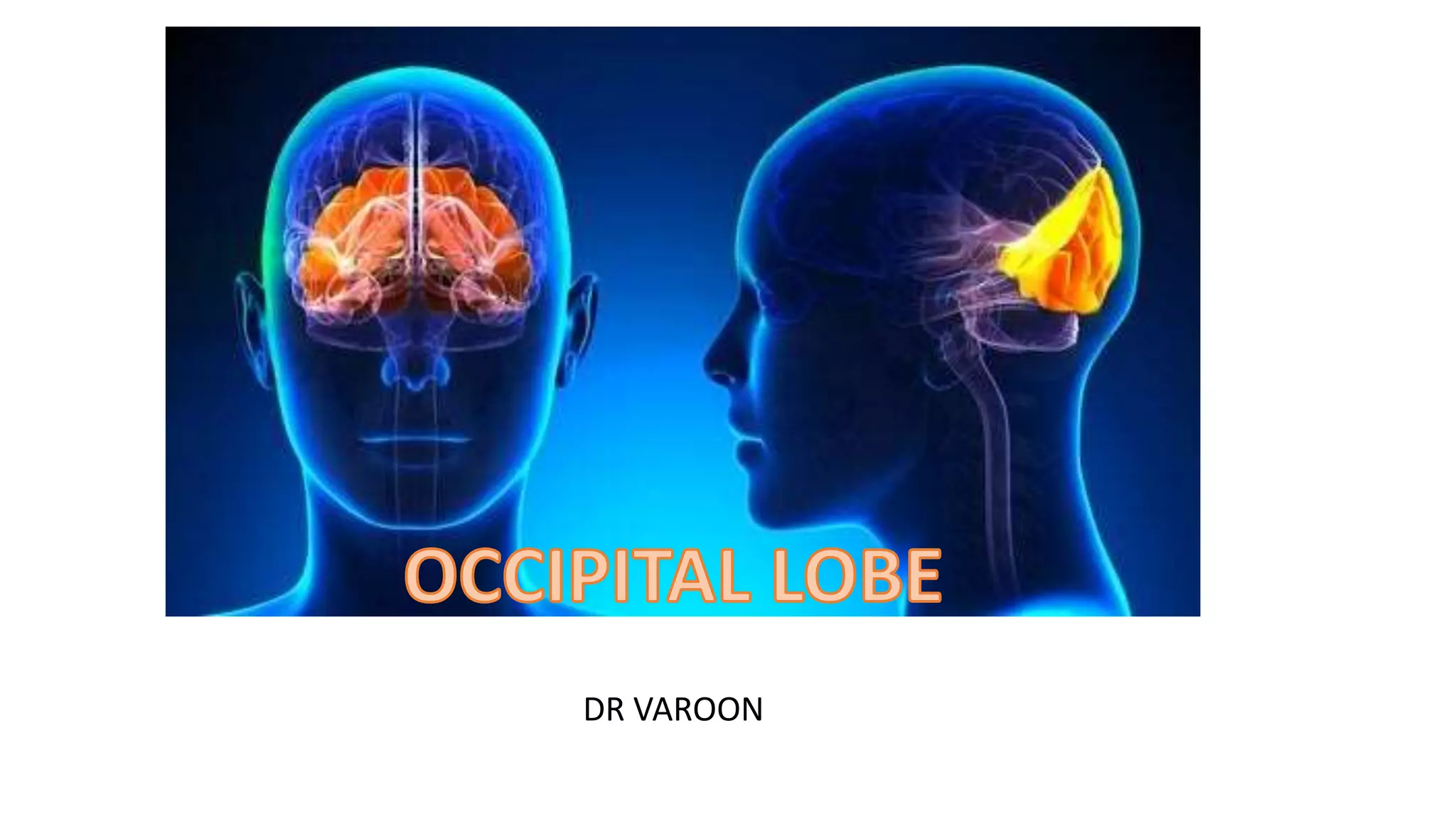
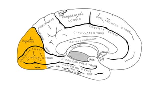
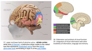
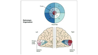
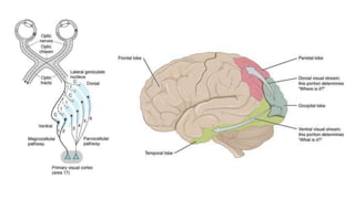
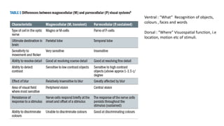
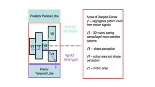
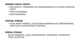
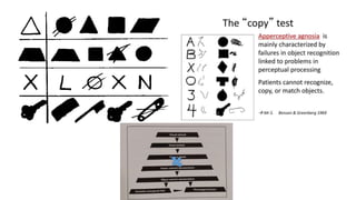
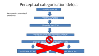
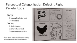
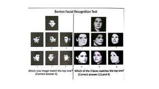
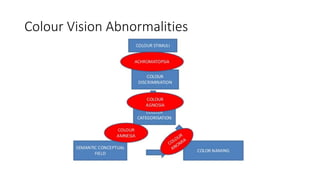
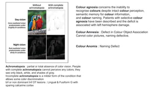
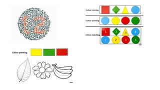
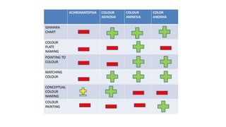
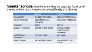
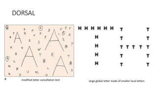
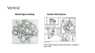
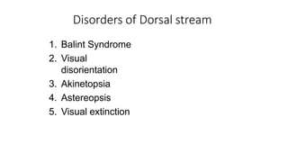
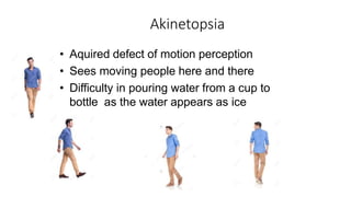
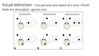
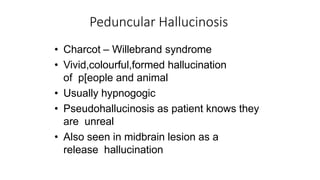
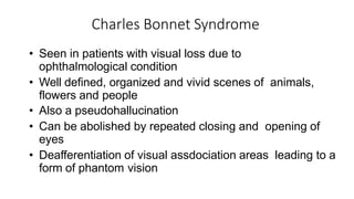
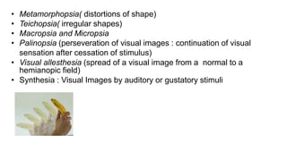
DR VAROON
18 : Elaboration and synthesis of visual function
19 : Integration of Visual Information with other
modalities of information, language and memory.
17 : upper and lower bank of calcarine sulcus : striate cortex :
white stria of Gennari, a distinctive stripe visible to the naked
eye that represents myelinated axons from the lateral
geniculate body terminating in layer 4 of the gray matter.
Ventral : “What” Recognition of objects,
colours , faces and words
Dorsal : “Where” Visuospatial function, i.e
location, motion etc of stimuli.
LESIONS OF OCCIPITAL LOBE AND
EXAMINATION
PRIMARY VISUAL CORTEX
• FIELD DEFECTS : HEMIANOPIA, B/L QUADRANTANOPIA, ALTITUDINAL HEMIFIELD
DEFECT
• CORTICAL BLINDNESS
• ANTON SYNDROME
VENTRAL STREAM
• VISUAL OBJECT AGNOSIA , COLOUR VISION ABNORMALITIES, PROSOPAGNOSIA,
PURE ALEXIA ( ALEXIA WITHOUT AGRAPHIA)
DORSAL STREAM
BALINT SYNDROME ; VISUAL DISORIENTATION ; AKINETOPSIA ; ASTEREOPSIS ;
VISUAL EXTINCTION
Primary Visual Cortex
CORTICAL BLINDNESS
• MACULAR SPARING : SHIFT IN OCULAR FIXATION ; DUAL BLOOD
SUPPLY ( MCA + PCA) ; macular representation ( large cortical)
• Occipital cortex
• Cerebral blindness : Circuits, Double hemianopia ; adrenal
leukodystrophy
Anton-Babinski syndrome (Anton syndrome or ABS)
Visual anosognosia (denial of loss of vision) associated with
confabulation (defined as the emergence of memories of events
and experiences which never took place) in the setting of obvious
visual loss and cortical blindness
Field
Defects
b/l altitudinal defect
VENTRAL STREAM
• Agnosia : Perceptual Disorder where
sensations are preserved but the
ability to recognize a stimulus or
identify its meaning is lost
Visual Agnosia
• Apperceptive visual agnosia :
• Abnormality in visual perception and discriminative process, despite
the absence of elementary visual deficits.
• These people are unable to recognize objects, draw, or copy a figure.
• They cannot perceive correct forms of the object, although knowledge
of the object is intact.
• Typically associated with lesions to the parietal, occipital cortex
TESTS : MATCHING ; COPYING
Perceptual categorization defect
VISUAL STIMULI
PRIMARY SKETCH
VIEWER CENTERED REPRESENATION
OBJECT CENTERED REPRESENTATION
SEMANTIC CONCEPTUAL
FIELD
VISUAL ANALYSIS
PHONOLOGICAL
LEXICON
Recognize in conventional
orientation
Perceptual Categorization Defect : Right
Parietal Lobe
Visual object and space perception battery
Birmingham object recognition battery
• Associative visual agnosia :
• Difficulty with understanding the meaning of what they are seeing.
• They can draw or copy but do not know what they have drawn.
• They correctly perceive the form and know the object when tested with
verbal or tactile information, but cannot identify the object.
• They are unable to link the fully perceived visual stimulus to prior
experience to help them recognize the stimulus.
• Usually associated with damage to the bilateral inferior occipitotemporal
Question : Picture of object, animal or tool … ask properties , function..
Sorting : according to category
Match visually dissimilar object with same function. Eg Keys, glass.
• Optic aphasia is a disconnection syndrome ( Language—Vision) in
which patients are unable to name visually presented objects but
have no difficulty in naming those objects on tactile or verbal
presentation. Left Occipitotemporal
• Prosopagnosia : Inability to identify familiar faces . Fusiform Gyrus.
– B/L damage to fusiform & lingual gyri
– U/L Rt OP lesion
– B/L or U/L anterior temporal lesion
1. Informal test : face identification
2. Benton Facial Recognition Test ( Formal )
Colour Vision Abnormalities
Achromatopsia : partial or total absence of color vision. People
with complete achromatopsia cannot perceive any colors; they
see only black, white, and shades of gray.
Incomplete achromatopsia is a milder form of the condition that
allows some color discrimination
b/l or non dominant Inf OT lesions : Lingual & Fusiform G with
sparing calcarine cortex
Colour agnosia concerns the inability to
recognise colours despite intact colour perception,
semantic memory for colour information,
and colour naming. Patients with selective colour
agnosia have been described and the deficit is
associated with left hemisphere damage.
Colour Amnesia : Defect in Colour Object Association
Cannot color pictures, naming defective.
Colour Anomia : Naming Defect
Simultanagnosia : inability to synthesize separate features of
the visual field into a meaningful whole(Totality of a Scene)
DORSAL
large global letter made of smaller local letters
modified letter cancellation test
Ventral
DORSAL STREAM
Disorders of Dorsal stream
1. Balint Syndrome
2. Visual
disorientation
3. Akinetopsia
4. Astereopsis
5. Visual extinction
Balint syndrome
• Occurs due to lesion in b/l parieto occipital lesion in the
convexity of the hemisphere
• Components
• Dorsal simultanagnosia
• Oculomotor apraxia : An inability to visualize more than one object in
the visual field at a time (psychic paralysis of gaze or visual inattention
• Optic ataxia : failure to reach an object under visual guidance.
(Misreaching).
• Test : touch examiner`s ear --- own ear
Visual disorientation : Egocentric topographic
disorientation
• Error in judging the location and distance of an object, and
in judging the relative distance between the two objects
• Bump into things while walking , Difficulty in finding door
handle
• Problem arises when things are placed at different level.
Test : Two Objects at 15cm distance ---half meter from
eyes--- ask near / far ; Dot at the center of the circle
• Visual disorientation improves with repeated attempts,
differentiating from optic ataxia
Astereopsis
• Loss of depth perception
• Depth is computed from binocular
visual information
• Retinal disparity is processed by the
dorsal stream
• Area 18 is primarily concerned with
stereopsis
• Abnormality is seen due to bilateral
damage, more to the right side
Akinetopsia
• Aquired defect of motion perception
• Sees moving people here and there
• Difficulty in pouring water from a cup to
bottle as the water appears as ice
Visual extinction : Can perceive one object at a time. If both
fields are stimulated , ignores one
Other visual phenomenas
Peduncular Hallucinosis
• Charcot – Willebrand syndrome
• Vivid,colourful,formed hallucination
of p[eople and animal
• Usually hypnogogic
• Pseudohallucinosis as patient knows they
are unreal
• Also seen in midbrain lesion as a
release hallucination
Charles Bonnet Syndrome
• Seen in patients with visual loss due to
ophthalmological condition
• Well defined, organized and vivid scenes of animals,
flowers and people
• Also a pseudohallucination
• Can be abolished by repeated closing and opening of
eyes
• Deafferentiation of visual assdociation areas leading to a
form of phantom vision
• Metamorphopsia( distortions of shape)
• Teichopsia( irregular shapes)
• Macropsia and Micropsia
• Palinopsia (perseveration of visual images : continuation of visual
sensation after cessation of stimulus)
• Visual allesthesia (spread of a visual image from a normal to a
hemianopic field)
• Synthesia : Visual Images by auditory or gustatory stimuli

Occipital lobe

  • 1.
  • 3.
    18 : Elaborationand synthesis of visual function 19 : Integration of Visual Information with other modalities of information, language and memory. 17 : upper and lower bank of calcarine sulcus : striate cortex : white stria of Gennari, a distinctive stripe visible to the naked eye that represents myelinated axons from the lateral geniculate body terminating in layer 4 of the gray matter.
  • 6.
    Ventral : “What”Recognition of objects, colours , faces and words Dorsal : “Where” Visuospatial function, i.e location, motion etc of stimuli.
  • 8.
    LESIONS OF OCCIPITALLOBE AND EXAMINATION
  • 9.
    PRIMARY VISUAL CORTEX •FIELD DEFECTS : HEMIANOPIA, B/L QUADRANTANOPIA, ALTITUDINAL HEMIFIELD DEFECT • CORTICAL BLINDNESS • ANTON SYNDROME VENTRAL STREAM • VISUAL OBJECT AGNOSIA , COLOUR VISION ABNORMALITIES, PROSOPAGNOSIA, PURE ALEXIA ( ALEXIA WITHOUT AGRAPHIA) DORSAL STREAM BALINT SYNDROME ; VISUAL DISORIENTATION ; AKINETOPSIA ; ASTEREOPSIS ; VISUAL EXTINCTION
  • 10.
  • 11.
    CORTICAL BLINDNESS • MACULARSPARING : SHIFT IN OCULAR FIXATION ; DUAL BLOOD SUPPLY ( MCA + PCA) ; macular representation ( large cortical) • Occipital cortex • Cerebral blindness : Circuits, Double hemianopia ; adrenal leukodystrophy Anton-Babinski syndrome (Anton syndrome or ABS) Visual anosognosia (denial of loss of vision) associated with confabulation (defined as the emergence of memories of events and experiences which never took place) in the setting of obvious visual loss and cortical blindness
  • 12.
  • 13.
  • 14.
    • Agnosia :Perceptual Disorder where sensations are preserved but the ability to recognize a stimulus or identify its meaning is lost
  • 15.
    Visual Agnosia • Apperceptivevisual agnosia : • Abnormality in visual perception and discriminative process, despite the absence of elementary visual deficits. • These people are unable to recognize objects, draw, or copy a figure. • They cannot perceive correct forms of the object, although knowledge of the object is intact. • Typically associated with lesions to the parietal, occipital cortex TESTS : MATCHING ; COPYING
  • 17.
    Perceptual categorization defect VISUALSTIMULI PRIMARY SKETCH VIEWER CENTERED REPRESENATION OBJECT CENTERED REPRESENTATION SEMANTIC CONCEPTUAL FIELD VISUAL ANALYSIS PHONOLOGICAL LEXICON Recognize in conventional orientation
  • 18.
    Perceptual Categorization Defect: Right Parietal Lobe Visual object and space perception battery Birmingham object recognition battery
  • 19.
    • Associative visualagnosia : • Difficulty with understanding the meaning of what they are seeing. • They can draw or copy but do not know what they have drawn. • They correctly perceive the form and know the object when tested with verbal or tactile information, but cannot identify the object. • They are unable to link the fully perceived visual stimulus to prior experience to help them recognize the stimulus. • Usually associated with damage to the bilateral inferior occipitotemporal
  • 20.
    Question : Pictureof object, animal or tool … ask properties , function.. Sorting : according to category Match visually dissimilar object with same function. Eg Keys, glass.
  • 22.
    • Optic aphasiais a disconnection syndrome ( Language—Vision) in which patients are unable to name visually presented objects but have no difficulty in naming those objects on tactile or verbal presentation. Left Occipitotemporal • Prosopagnosia : Inability to identify familiar faces . Fusiform Gyrus. – B/L damage to fusiform & lingual gyri – U/L Rt OP lesion – B/L or U/L anterior temporal lesion 1. Informal test : face identification 2. Benton Facial Recognition Test ( Formal )
  • 25.
  • 26.
    Achromatopsia : partialor total absence of color vision. People with complete achromatopsia cannot perceive any colors; they see only black, white, and shades of gray. Incomplete achromatopsia is a milder form of the condition that allows some color discrimination b/l or non dominant Inf OT lesions : Lingual & Fusiform G with sparing calcarine cortex Colour agnosia concerns the inability to recognise colours despite intact colour perception, semantic memory for colour information, and colour naming. Patients with selective colour agnosia have been described and the deficit is associated with left hemisphere damage. Colour Amnesia : Defect in Colour Object Association Cannot color pictures, naming defective. Colour Anomia : Naming Defect
  • 29.
    Simultanagnosia : inabilityto synthesize separate features of the visual field into a meaningful whole(Totality of a Scene)
  • 30.
    DORSAL large global lettermade of smaller local letters modified letter cancellation test
  • 31.
  • 32.
  • 33.
    Disorders of Dorsalstream 1. Balint Syndrome 2. Visual disorientation 3. Akinetopsia 4. Astereopsis 5. Visual extinction
  • 34.
    Balint syndrome • Occursdue to lesion in b/l parieto occipital lesion in the convexity of the hemisphere • Components • Dorsal simultanagnosia • Oculomotor apraxia : An inability to visualize more than one object in the visual field at a time (psychic paralysis of gaze or visual inattention • Optic ataxia : failure to reach an object under visual guidance. (Misreaching). • Test : touch examiner`s ear --- own ear
  • 35.
    Visual disorientation :Egocentric topographic disorientation • Error in judging the location and distance of an object, and in judging the relative distance between the two objects • Bump into things while walking , Difficulty in finding door handle • Problem arises when things are placed at different level. Test : Two Objects at 15cm distance ---half meter from eyes--- ask near / far ; Dot at the center of the circle • Visual disorientation improves with repeated attempts, differentiating from optic ataxia
  • 36.
    Astereopsis • Loss ofdepth perception • Depth is computed from binocular visual information • Retinal disparity is processed by the dorsal stream • Area 18 is primarily concerned with stereopsis • Abnormality is seen due to bilateral damage, more to the right side
  • 37.
    Akinetopsia • Aquired defectof motion perception • Sees moving people here and there • Difficulty in pouring water from a cup to bottle as the water appears as ice
  • 38.
    Visual extinction :Can perceive one object at a time. If both fields are stimulated , ignores one
  • 39.
  • 40.
    Peduncular Hallucinosis • Charcot– Willebrand syndrome • Vivid,colourful,formed hallucination of p[eople and animal • Usually hypnogogic • Pseudohallucinosis as patient knows they are unreal • Also seen in midbrain lesion as a release hallucination
  • 41.
    Charles Bonnet Syndrome •Seen in patients with visual loss due to ophthalmological condition • Well defined, organized and vivid scenes of animals, flowers and people • Also a pseudohallucination • Can be abolished by repeated closing and opening of eyes • Deafferentiation of visual assdociation areas leading to a form of phantom vision
  • 42.
    • Metamorphopsia( distortionsof shape) • Teichopsia( irregular shapes) • Macropsia and Micropsia • Palinopsia (perseveration of visual images : continuation of visual sensation after cessation of stimulus) • Visual allesthesia (spread of a visual image from a normal to a hemianopic field) • Synthesia : Visual Images by auditory or gustatory stimuli

Editor's Notes

  • #12 Point fixation shifted on periphery.