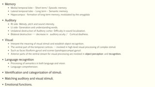
The temporal lobe is involved in several important functions:
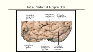
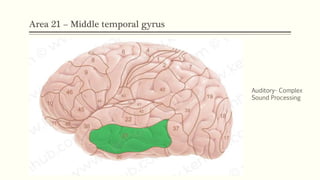
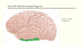
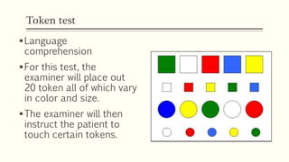
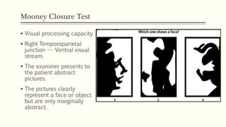
1) It processes auditory and visual information through distinct cortical areas.
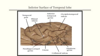
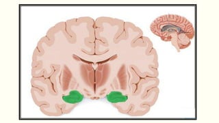
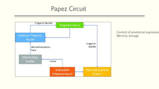
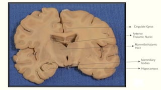
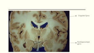
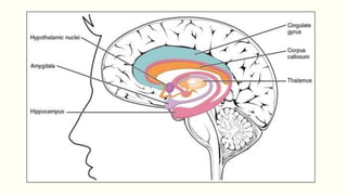
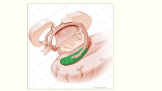
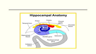
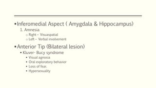
2) The medial temporal lobe structures including the hippocampus and amygdala are critical for forming memories and regulating emotions.
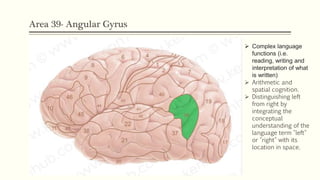
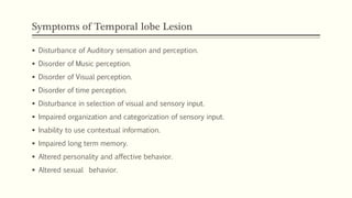
3) Disorders of the temporal lobe can cause problems with memory, language processing, perception and personality changes depending on the area affected.


![References
Localization in Clinical Neurology, Paul W. Brazis 7th edi (SAE).
Bradley 8th edition.
J. A. Kiernan, "Anatomy of the Temporal Lobe", Epilepsy Research and Treatment,
vol. 2012, Article ID 176157, 12 pages, 2012. https://doi.org/10.1155/2012/176157
Patel A, Biso GMNR, Fowler JB. Neuroanatomy, Temporal Lobe. [Updated 2021 Jul
31].
Bajada CJ, Haroon HA, Azadbakht H, Parker GJM, Lambon Ralph MA, Cloutman LL.
The tract terminations in the temporal lobe: Their location and associated
functions. Cortex. 2017 Dec;97:277-290.](https://image.slidesharecdn.com/anatomylocalizationoftemporallobe-220309015038/85/Anatomy-Function-of-Temporal-lobe-44-320.jpg)


![DUAL AND TRIPLE ANTITHROMBOTIC THERAPY FOR SECONDARY STROKE [Autosaved].pptx](https://cdn.slidesharecdn.com/ss_thumbnails/dualandtripleantithrombotictherapyforsecondarystrokeautosaved-230904113552-c3502b37-thumbnail.jpg?width=640&height=640&fit=bounds)


![CASE_PRESENTATION_ON_subdural_hematoma(SDH)[1 FINAL PPT]-1.pptx](https://cdn.slidesharecdn.com/ss_thumbnails/casepresentationonsubduralhematomasdh1finalppt-1-260129172522-d405d375-thumbnail.jpg?width=640&height=640&fit=bounds)


