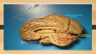
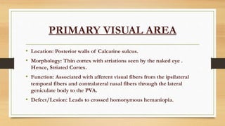
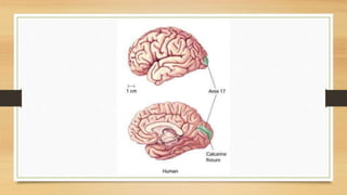
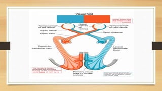
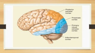
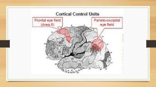
MEHWISH ASLAM RAHEEMI
OCCIPITAL LOBE
Posterior most of the brain lobes,
associated primarily with vision.
GYRI & SULCI
1)No distinguishable gyri.
2)Sulci present.i.e Parieto-occipital sulcus separating the
occipital lobe from parietal lobe and the calcarine sulcus
(medial surface) extending from the posterior part of corpus
callosum up to the occipital pole.
3)Parieto-occipital sulcus makes an acute angle with the
calcarine sulcus.
AREAS
•PRIMARY VISUAL AREA
•SECONDARY VISUAL AREA
PRIMARY VISUAL AREA
• Location: Posterior walls of Calcarine sulcus.
• Morphology: Thin cortex with striations seen by the naked eye .
Hence, Striated Cortex.
• Function: Associated with afferent visual fibers from the ipsilateral
temporal fibers and contralateral nasal fibers through the lateral
geniculate body to the PVA.
• Defect/Lesion: Leads to crossed homonymous hemaniopia.
SECONDARY VISUAL CORTEX
• Location: around the Primary visual cortex
medially and laterally.
• Function: Associates vision with past experience .
Helps in Appreciation & Recognition.
• Defect/Lesion : Disability to recognize Objects.
OCCIPITAL EYEFIELD
• Depends on stimulus.
• Involuntary.
• Reflex tracking movement
• Difference b/w Frontal & Occipital eye field: The formal is
independent of stimulus , voluntary scanning movement.
THANK YOU!

Occipital Lobe

  • 1.
    MEHWISH ASLAM RAHEEMI OCCIPITALLOBE Posterior most of the brain lobes, associated primarily with vision.
  • 3.
    GYRI & SULCI 1)Nodistinguishable gyri. 2)Sulci present.i.e Parieto-occipital sulcus separating the occipital lobe from parietal lobe and the calcarine sulcus (medial surface) extending from the posterior part of corpus callosum up to the occipital pole. 3)Parieto-occipital sulcus makes an acute angle with the calcarine sulcus.
  • 5.
  • 6.
    PRIMARY VISUAL AREA •Location: Posterior walls of Calcarine sulcus. • Morphology: Thin cortex with striations seen by the naked eye . Hence, Striated Cortex. • Function: Associated with afferent visual fibers from the ipsilateral temporal fibers and contralateral nasal fibers through the lateral geniculate body to the PVA. • Defect/Lesion: Leads to crossed homonymous hemaniopia.
  • 9.
    SECONDARY VISUAL CORTEX •Location: around the Primary visual cortex medially and laterally. • Function: Associates vision with past experience . Helps in Appreciation & Recognition. • Defect/Lesion : Disability to recognize Objects.
  • 11.
    OCCIPITAL EYEFIELD • Dependson stimulus. • Involuntary. • Reflex tracking movement • Difference b/w Frontal & Occipital eye field: The formal is independent of stimulus , voluntary scanning movement.
  • 13.