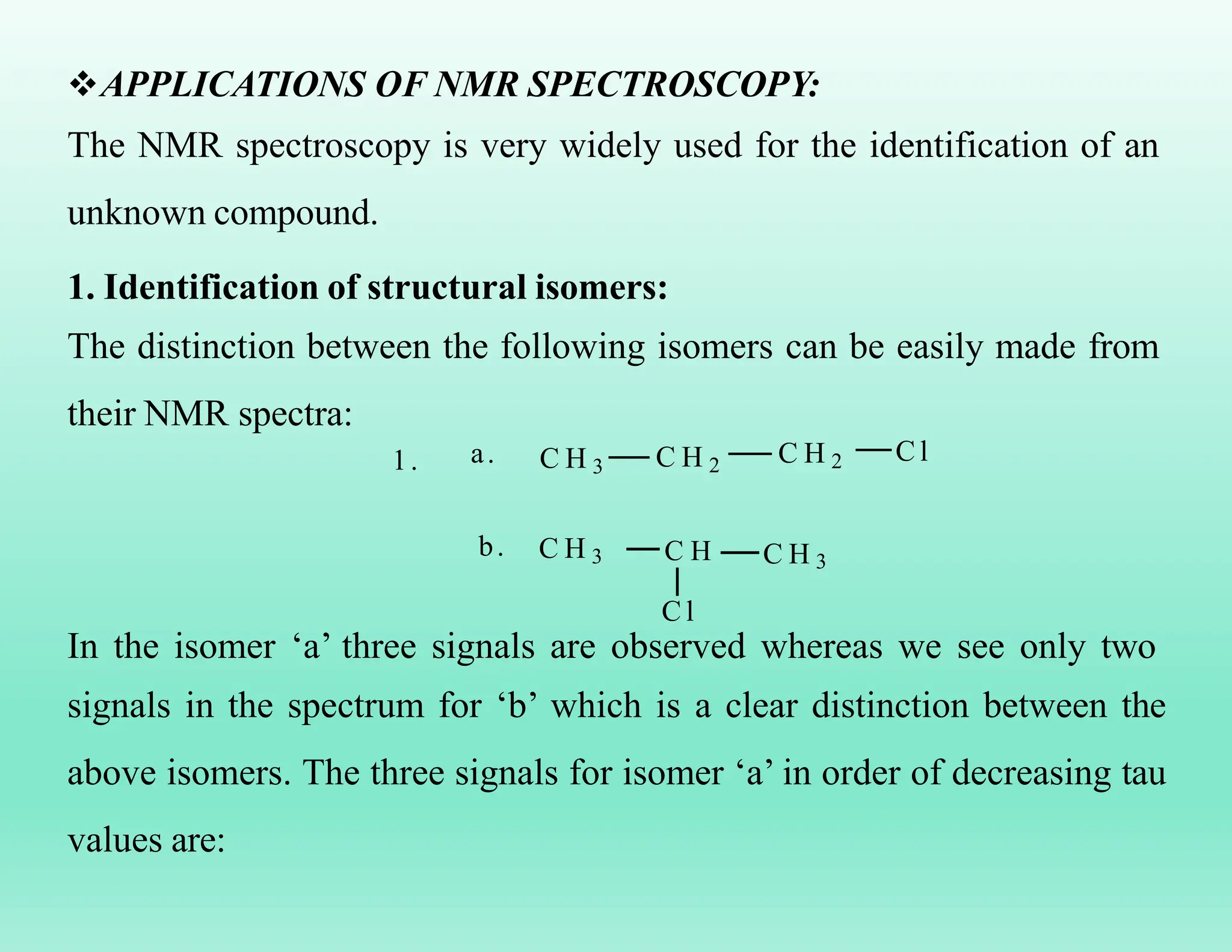
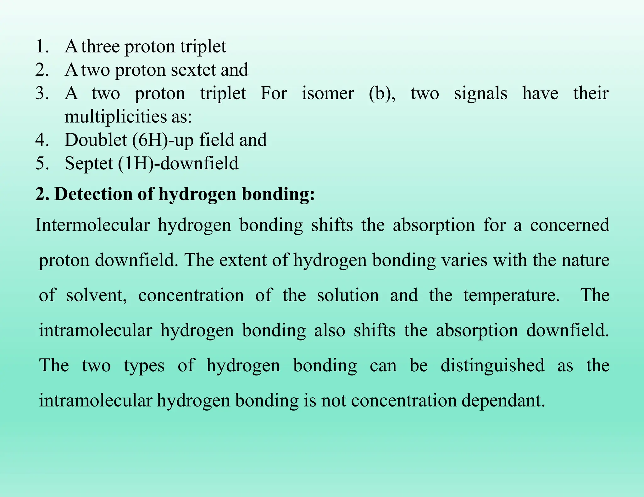
SlideShare a Scribd company logo
1 of 38
NMR SPECTROSCOPY
Mrs. Poonam Sunil Aher (M.Pharm, PhD)
Assistant Professor
Sanjivani College of Pharmaceutical Education and Research
(Autonomous),
Kopargaon, Ahmednagar-423603 (M.S.), INDIA
Mobile: +91-9689942854
LEARNING OUTCOMES
After studying this Chapter, you shall be able to:
To understand the basic concept of NMR.
To know about NMR active and inactive nuclei.
To know about the conditions of a nuclei to be called an
NMR active nuclei.
To know the types of relaxation processes a nuclei
undergoes.
To know when a nuclei is called shielded and deshielded.
INTRODUCTION:
The concept of NMR was represented at first in 1946 by two groups of
eminent physicist; Black Hensen and Packard at Stanford University
detected a signal from the Protons of water and Parcell, Torrey and
pound at Havard University observed a signal from the photons in
Paraffin wax. Black and Parcell were jointly awarded a Nobel Prize for
Physics in 1952 for this discovery.
NMR spectroscopy involvers transition of a nucleus from one spin state
to other with the resultant absorption of electromagnetic radiation by spin
active nuclei (having nuclei spin not equal to zero) when they are placed
in magnetic field. Nuclear magnetic resonance spectroscopy related to
the nuclei and only one type of nucleus at a particular timeline.
1H or C13, F19 when the frequency of the rotating magnetic field and that
of the processing nucleus (Lamar Frequency) become equal, they are
said to be in resonance absorption or emission of energy by the nucleus
can be obtained. Plot of the peak intensities versus the frequencies of
objection (represented by δ or τ) establish an NMR spectrum.
The 1H nucleus is most commonly studied by NMR spectroscopy
because of its high natural abundance (99.98%) and the fact that it is
present in the majority of organic compounds, the PMR or 1H NMR
spectrum provides information about the number of different types of
protons and also the chemical environment of each of them.
TWO TYPES NMR SPECTROSCOPY
1.1H NMR
2.13C NMR
PROTON MAGNETIC RESONANCE (1H NMR) SPECTROSCOPY
1HNMR or PMR spectroscopy is the most widely applicable Nuclear
Magnetic Resonance spectroscopy for the structural determination of
various organic compounds but the other NMR spectroscopic methods
like C13 and P31 NMR spectroscopy; F19 spectroscopy can also be helpful
in the structural determination of the compounds.
Spin active nuclei:
All those nuclei which having the full integer or half integer nuclear spin
value are known as spin active nuclei. With the help of the number of the
electron/proton and neutron in the various nuclei, the spin active or
inactive nature for them can be defined as:
e-/p Neutron (n) Nuclear spin
(I)
Nuclei Examples
Even Even 0 Inactive 8O16
Odd Odd Full integer Active 7N14, 6C12
Even Odd Full integer Active 6C13
Odd Even Half integer Active 15P31, 1H1
Phenomena of energy absorption (Resonance & relaxation
phenomena):
In the absence of external magnetic field H0 the nuclear spin are
randomly oriented, However when the sample is placed in an external
magnetic field then the nuclei (proton) with the spin +half (1/2) are
aligned with the applied field in that lower energy α- spin stagehand the
nuclei with the spin -1/2 are aligned against to the external magnetic field
in the higher energy β- spin state that can be represented as:
-spinstate
(I=-1/2)
E
-spinstate
(I=+1/2)
H0
Randomarrangementofspin
activenucleiintheabsanceofH0
H0
Orientation of spinning nuclei in absence and presence of external
magnetic field
α and β spin state depends on the
strength of external magnetic field
H0 according to the equation ∆E = 2µh0 during the PMR spectroscopy,
and then it can be represented as:
 -spin state
E
 - spin state
E
H 0
Energy states of nuclear spin
Thus according to above spin states resonance phenomena may be
defined as the transition of spin active nuclei from α-spin state to the β-
spin state by the absorption of Rf radiation while the phenomena of
returning the spin active nuclei from high energy β- spin state to the low
energy α-spin state is known as relaxation phenomena. Both the
resonance and relaxation phenomena can be represented as:
 -spin state
Resonance
Phenomena
Relaxation
Phenomena
-spin state
Resonance and Relaxation phenomenon
NUCLEAR SHIELDING AND DESHIELDING:
Electron surrounding the spin active nuclei can also generate their own
magnetic field which is called as induced magnetic field, that oppose the
applied magnetic field in the region of the nucleus and these e- which
generate their induced magnetic field are known as diamagnetic e- and
this effect on the nucleus by these e- is known as diamagnetic shielding
Nucleus
Induced magnetic
field by the e-
H0
Shielding and deshielding of a nucleus
The external magnetic field is uniform over the entire molecule and
therefore cannot differentiate to the different types of the proton.
However the induced magnetic field generated by the e- around the
nucleus is not uniform, this situation makes the different spin active
nuclei 9proton) to be non-equivalent. Thus each proton in the different
electronic environment show slightly different magnetic field due to the
circulation of e- in the neighboring bond.
Thus the effective magnetic field for the different spin active nuclei can
be calculated through the following equations:
H effect = H0 – H induced
Form the above equation the shielded and deshielded proton concept can
be given as:
CHEMICALLY EQUIVALENT & NON- EQUIVALENT
PROTONS:
Those protons present in the sample of spin active nuclei which having
same chemical environment are known as chemically equivalent protons.
The entire chemically equivalent protons appear as a single signal in the
PMR spectrum.
On the other hand those protons present in the sample of spin active
nuclei which having different chemical environment are known as
chemically non-equivalent proton. Chemically non-equivalent proton
represents the different signal in the PMR spectrum.
B r
H
H
C C
1 .
a
C H 3
b
c
I n a m o l e c u l e r e v e a l s t h a t t h r r e d i f f e r e n t t y p e s o f p r o t o n s , i n d i c a t e d b y t h e l e t t e r s a , b , c .
2 .
C H 3
H
H
C C
a
C H 3
b
b
3 . C H 3 - C H C l - C - C l
a b
c
a
T h e r e a r e t w o t y p e s o f p r o t o n s . S i x m e t h y l p r o t o n s o n t h e L H S a r e e q u i v a l e n t . T h e p r o t o n s o n R H S a r e
a g a i n e q u i v a l e n t
d
H
H
P r o t o n s m a r k e d c a n d d a p p e a r t o b e e q u i v a l e n t , b u t t h e y a r e n o a c t u a l y s o . T h u s i t h a s f o u r t y p e s o f p r o t o n s .
4 .
2
C H 2
a
C H 2
a
C H 2
C H a
a C H 2
a C H 2
a
I n t h i s c a s e a l l t h e p r o t o n s a r e e q u i v a l e n t g i v i n g r i s e t o o n l y o n e P M R s i g n a l s
5 . C H 3
6 .
T h e r e a r e t w o t y p e s o f p r o t o n s , b e n z e n e r i n g p r o t o n s a n d t h e m e t h y l p r o t o n s . T w o s i g n a l s w i l l b e o b s e r v e d .
b
H
a c
C H 3 - C - C H O
H b
T h e r e a r e t h r e e k i n d o f p r o t o n s g i v i n g r i s e t o t h r e e s i g n a l s .
CHEMICAL SHIFT :
Chemical shift it expresses the difference in the resonance frequency of a
given proton compared to that of the methyl protons of TMS, under the
experimental conditions. In practice, this difference is divided by the
operating radio-frequency of the instrument and the chemical shift δ is
expressed, downfield from TMS, as per the given equation.
For example, if the observed shift from TMS is 200 Hz and the operating
frequency of the instrument id 100 MHz, then the chemical shift δ is
given by the following expression:
This frequency ratio (2.0 x 10⁻⁶) is
multiplied by 10-6 in order to obtain an easily handled number (2.0 x
10-6 x 106 ppm) and consequently the chemical shift δ is expressed as
part per million (106) of the operating frequency.
Thus,
Chemicalshift =
s- T
M
S(Hz)
Operatingfriquency
x 106
T M S
S h i e l d e d r e g i o n
o r
U p f i e l d r e g i o n
D o w n f i e l d r e g i o n
D e s h i e l d e d r e g i o n
o r
C h e m i c a l s h i f t
 p p m
Chemical shift region
Internal standard for NMR spectroscopy:
That compound which is used as a reference standard to represent the
NMR/PMR signal of the compounds is known as internal standard for
the NMR or PMR spectroscopy.
In case of PMR spectroscopy Tetramethylsilane (TMS) used as a internal
standard due to the following reasons:
1. Due to the more shielded nature of the proton of TMS in compare to
the protons of most of the organic compound.
2. It is chemically inert and miscible with large range of solvent.
3. It does not take part in intermolecular association with the sample.
4. Due to the volatile nature of TMS.
The internal standard for some other type of the NMR spectroscopic
methods can be given as:
F19 NMR – CFCl3
N15 NMR – NH4NO3
C13 NMR – TMS
SPIN-SPIN SPLITTING AND COUPLING CONSTANTS:
The coupling interaction between two or more protons, most often
through the bond, results in splitting of the spectral lines. This is called
spin-spin coupling. It is related to the possible combinations of the spin
orientations of the neighboring protons. The phenomena of splitting the
signal of any particular type of proton by the spin orientation of the non-
equivalent proton present adjacent to it is known as spin-spin coupling
phenomena.
Exa
mpl
e:
a
b
CH3CHCl2
According to the NMR spectroscopy the signal of two different type of
the proton present in this compound can be give as:
T M S
a
b
O

According to the spin-spin coupling phenomena the splitting of the
signal of Hb proton & Ha proton according to the spin orientation of non-
equivalent adjacent proton can be given as:
1.Spin orientation of Ha for the splitting of
the signal of Hb :
T M S
b
H o )
(

2. Spin orientation of Hb for the splitting of the signal of Ha
T M S
H o )
(
a
Couplingconstant(J):
The distance between the centers of the two adjacent peaks in a multiplet
is usually constant and is called the coupling constant. The value of
coupling constant is independent of the external field. It is measured
Hertz (Hz) or in cps (cycle per second). It is denote by the letter J. In
other words, we can say the value of J remains the same whatever the
applied field. The value of J generally lies between 0 and 20 Hertz (Hz).
Always the value of coupling constant being same for the protons which
causing the splitting each other signal.
Now, let us consider a compound:
a
CH2
b
CH
In this compound two signals are expected in the NMR spectrum. Under
the influence of two equivalent proton a, the signal for proton b will
appear as a triplet. The distance between any two adjacent peaks in a
multiplet will be exactly the same. The spin-spin coupling is given
below:
T M S
J
J
spin-spin coupling
It may be clearly noted that the value of coupling constant depends on the number
of covalent bonds through which protons may interact and also upon the structural
relationship between the coupled protons. Various types of the coupling may be given
as:
Geminal coupling:
Such type of the spin-spin coupling phenomena in which two chemically
non-equivalent protons present at the same c atom causing the splitting
of each other signal will be called as Germinal coupling or J2coupling.
C C
a
H
H
b
H
c
d
CH3 Geminal coupling
J2 coupling
Vicinal coupling-
Such type of the coupling phenomena in which two chemically non-
equivalent protons present at the adjacent carleon atom causing the
splitting of each other signal are known as vicinal coupling or J3
coupling.
J 3 c o u p l i n g
C C
H
H
C C
H C l
C
a
H
a
H
c
H
Long range coupling:
Such type of the coupling phenomena in which two chemically non-
equivalent protons causing the splitting of each other signal being
separated by more than three covalent bonds are known as multi
range/long range coupling. The probability of this type of the coupling
phenomena in the organic compounds being very low.
L o n g r a n g e c o u p l i n g
a
H
APPLICATIONS OF NMR SPECTROSCOPY:
The NMR spectroscopy is very widely used for the identification of an
unknown compound.
1. Identification of structural isomers:
The distinction between the following isomers can be easily made from
their NMR spectra:
1. C H 3 C H 2
C l
a. C H 2
C H 3 C H C H 3
b.
C l
In the isomer ‘a’ three signals are observed whereas we see only two
signals in the spectrum for ‘b’ which is a clear distinction between the
above isomers. The three signals for isomer ‘a’ in order of decreasing tau
values are:
1. Athree proton triplet
2. Atwo proton sextet and
3. A two proton triplet For isomer (b), two signals have their
multiplicities as:
4. Doublet (6H)-up field and
5. Septet (1H)-downfield
2. Detection of hydrogen bonding:
Intermolecular hydrogen bonding shifts the absorption for a concerned
proton downfield. The extent of hydrogen bonding varies with the nature
of solvent, concentration of the solution and the temperature. The
intramolecular hydrogen bonding also shifts the absorption downfield.
The two types of hydrogen bonding can be distinguished as the
intramolecular hydrogen bonding is not concentration dependant.
3. Distinction between Cis-Trans Isomers and conformers:
The is and trans isomers of a compound can be easily distinguished as
the concerned protons have different values of the chemical shift as well
as the coupling constants. Similarly, the various conformation of a
compound, the axial and equatorial positions of the proton or group
carrying a proton can be distinguished from their different values of the
coupling.
4. Detection of some double bond character due to resonance:
In some compounds, the molecule acquires a little double bond character
due to resonance. Due to this, two signals can be expected for apparently
equivalent protons, It is due to the hindered rotation which changes the
geometry of the molecules. Consider N, N- dimethyl formamide.
It can be written in the following resonating structures:
• For structure (a), two signals (singlets) should be
expected with peak areas 6:1 as the two methyls are
exactly equivalent.
• In structure (b), the presence of double bond
restricts rotation and now the two methyl groups
remain no longer equivalent (Geometrical
isomers). For this structure, two signals, appear for
two methyl groups.
CH3
3
CH3
O
..
C N
a
CH
a
b H
O
C N
a
CH3
b
c
H
a
b
INTERPRETATION OF PMR SPECTRA OF SIMPLE ORGANIC
MOLECULES:
NMR interpretation plays a important role in molecular identifications.
As interpreting NMR spectra, the structure of an unknown compound, as
well as known structures, can be assigned by several factors such as
chemical shift, spin multiplicity, coupling constants, and integration. For
the interpreting of the NMR spectra, the following points may be noted:
1. Molecular formula is determined by chemical analysis such as
elementary analysis.
2. Double-bond equivalent or Degree of Unsaturation: It is calculated
by a simple equation to estimate the number of the multiple bonds and
rings. It assumes that oxygen (O) and sulfur (S) are ignored and halogen
(Cl, Br) and nitrogen is replaced by CH. The resulting empirical formula is CxHy
Double Bond Equivalent (DBE) =
3. Structure fragmentation is determined by
(2x + 2) - y
2
chemical shift, spin
multiplicity, integral (peak area), and coupling constant(1J, 2J)
4. Molecular skeleton is built up using 2-dimensional NMR
spectroscopy.
5. Relative configuration is predicted by coupling constant (3J).
EXAMPLES:
1. Ethyl bromide (CH3CH2Br):
In this compound (Ethyl bromide) containing two set of equivalent
protons ‘a’ and ‘b’ types. The signal due to equivalent protons a will be
under the influence of neighboring proton b.
will be under the influence of
three equivalent proton a whose
spin possibilities with respect to
the applied field can be given as-
The following peaks can be identified in the spectrum
(a) Triplet, δ 1.7, 3H (b) Quartet, δ 3.4, 2H
The proton at δ 1.7 is given by the three methyl protons which are
magnetically equivalent and are coupled with the two methylene protons
to give a shielding. While the quartet at δ 3.4 is from the two equivalent
methylene protons which are coupled with the three methylene protons
to produce a downfield quartet as a result of deshielding influence of
bromine.
2. Interpretation of PMR spectra of ethanol:
The 1H NMR spectrum of ethanol (Figure 1.9) shows the methyl peak
has been split into three peaks (triplet) and the methylene peak has been
split into four peaks (quartet). This occurs because there is a small
interaction between the two groups of protons. The space between the
peaks of the methyl triplet is equal to the space between the peaks of the
methylene quartet. This spacing is measured in Hertz and is called the
coupling constant, J.
PMR Spectrum of Ethyl alcohol
In the above PMR spectrum of ethyl alcohol the following observation
takes place:
•A triplet centered at 1.18 δ, equivalent to 3H, represents the CH3 (a)
proton.
•Asinglet at 4.51 δ, equivalent to 1H, exhibit the OH (c) proton.
•A quartet centered at 3.63 δ, equivalent to 2H indicates the CH2 (b)
proton.
NUCLEAR MAGNETIC RESONANCE SPECTROSCOPY(NMR)

More Related Content

What's hot

Nmr spectroscopy
Nmr spectroscopyNmr spectroscopy
Nmr spectroscopyAsma Ashraf
 
13 C NMR Spectroscopy with examples by Dr Anthony Crasto
13 C NMR Spectroscopy with examples by Dr Anthony Crasto13 C NMR Spectroscopy with examples by Dr Anthony Crasto
13 C NMR Spectroscopy with examples by Dr Anthony CrastoAnthony Melvin Crasto Ph.D
 
Spin spin coupling and decoupling
Spin spin coupling and decouplingSpin spin coupling and decoupling
Spin spin coupling and decouplingAKSHAY KUMAR
 
Solvents and solvent effect in UV - Vis Spectroscopy, By Dr. Umesh Kumar sh...
Solvents and  solvent effect in UV -  Vis Spectroscopy, By Dr. Umesh Kumar sh...Solvents and  solvent effect in UV -  Vis Spectroscopy, By Dr. Umesh Kumar sh...
Solvents and solvent effect in UV - Vis Spectroscopy, By Dr. Umesh Kumar sh...Dr. UMESH KUMAR SHARMA
 
Liquid chromatography nuclear magnetic resonance spectroscopy
Liquid chromatography  nuclear magnetic resonance spectroscopyLiquid chromatography  nuclear magnetic resonance spectroscopy
Liquid chromatography nuclear magnetic resonance spectroscopySanket Bhatshankar
 
Nuclear magnetic resonance (NMR) spectroscopy
Nuclear magnetic resonance (NMR) spectroscopyNuclear magnetic resonance (NMR) spectroscopy
Nuclear magnetic resonance (NMR) spectroscopyVK VIKRAM VARMA
 
13 C NMR Spectroscopy by Dr Anthony Melvin Crasto
13 C NMR Spectroscopy by Dr Anthony Melvin Crasto13 C NMR Spectroscopy by Dr Anthony Melvin Crasto
13 C NMR Spectroscopy by Dr Anthony Melvin CrastoAnthony Melvin Crasto Ph.D
 
IR interpretation and sample handling
 IR interpretation and sample handling  IR interpretation and sample handling
IR interpretation and sample handling Afzaye Rasul
 
NMR spectroscopy- Spin-lattice & spin-spin relaxation, signal splitting & sig...
NMR spectroscopy- Spin-lattice & spin-spin relaxation, signal splitting & sig...NMR spectroscopy- Spin-lattice & spin-spin relaxation, signal splitting & sig...
NMR spectroscopy- Spin-lattice & spin-spin relaxation, signal splitting & sig...AYESHA NAZEER
 
Instrumentation of uv spectroscopy
Instrumentation of uv spectroscopyInstrumentation of uv spectroscopy
Instrumentation of uv spectroscopyArabinda Changmai
 
C-13 NMR Spectroscopy
C-13 NMR SpectroscopyC-13 NMR Spectroscopy
C-13 NMR SpectroscopyMANISHSAHU106
 
NMR SPECTROSCOPY
NMR SPECTROSCOPYNMR SPECTROSCOPY
NMR SPECTROSCOPYPV. Viji
 
NMR SPECTROSCOPY
NMR SPECTROSCOPYNMR SPECTROSCOPY
NMR SPECTROSCOPYVidyaNani
 
Fragmentation rules mass spectroscopy
Fragmentation rules mass spectroscopyFragmentation rules mass spectroscopy
Fragmentation rules mass spectroscopySanthosh Kalakar dj
 

What's hot (20)

Nmr spectroscopy
Nmr spectroscopyNmr spectroscopy
Nmr spectroscopy
 
Coupling constant
Coupling constantCoupling constant
Coupling constant
 
Double resonance
Double resonanceDouble resonance
Double resonance
 
2D NMR.pptx
2D NMR.pptx2D NMR.pptx
2D NMR.pptx
 
13 C NMR Spectroscopy with examples by Dr Anthony Crasto
13 C NMR Spectroscopy with examples by Dr Anthony Crasto13 C NMR Spectroscopy with examples by Dr Anthony Crasto
13 C NMR Spectroscopy with examples by Dr Anthony Crasto
 
Spin spin coupling and decoupling
Spin spin coupling and decouplingSpin spin coupling and decoupling
Spin spin coupling and decoupling
 
Solvents and solvent effect in UV - Vis Spectroscopy, By Dr. Umesh Kumar sh...
Solvents and  solvent effect in UV -  Vis Spectroscopy, By Dr. Umesh Kumar sh...Solvents and  solvent effect in UV -  Vis Spectroscopy, By Dr. Umesh Kumar sh...
Solvents and solvent effect in UV - Vis Spectroscopy, By Dr. Umesh Kumar sh...
 
Liquid chromatography nuclear magnetic resonance spectroscopy
Liquid chromatography  nuclear magnetic resonance spectroscopyLiquid chromatography  nuclear magnetic resonance spectroscopy
Liquid chromatography nuclear magnetic resonance spectroscopy
 
Nuclear magnetic resonance (NMR) spectroscopy
Nuclear magnetic resonance (NMR) spectroscopyNuclear magnetic resonance (NMR) spectroscopy
Nuclear magnetic resonance (NMR) spectroscopy
 
13 C NMR Spectroscopy by Dr Anthony Melvin Crasto
13 C NMR Spectroscopy by Dr Anthony Melvin Crasto13 C NMR Spectroscopy by Dr Anthony Melvin Crasto
13 C NMR Spectroscopy by Dr Anthony Melvin Crasto
 
IR interpretation and sample handling
 IR interpretation and sample handling  IR interpretation and sample handling
IR interpretation and sample handling
 
NMR spectroscopy- Spin-lattice & spin-spin relaxation, signal splitting & sig...
NMR spectroscopy- Spin-lattice & spin-spin relaxation, signal splitting & sig...NMR spectroscopy- Spin-lattice & spin-spin relaxation, signal splitting & sig...
NMR spectroscopy- Spin-lattice & spin-spin relaxation, signal splitting & sig...
 
Instrumentation of uv spectroscopy
Instrumentation of uv spectroscopyInstrumentation of uv spectroscopy
Instrumentation of uv spectroscopy
 
C-13 NMR Spectroscopy
C-13 NMR SpectroscopyC-13 NMR Spectroscopy
C-13 NMR Spectroscopy
 
NMR Spectroscopy
NMR SpectroscopyNMR Spectroscopy
NMR Spectroscopy
 
NMR SPECTROSCOPY
NMR SPECTROSCOPYNMR SPECTROSCOPY
NMR SPECTROSCOPY
 
C13-NMR
C13-NMRC13-NMR
C13-NMR
 
NMR SPECTROSCOPY
NMR SPECTROSCOPYNMR SPECTROSCOPY
NMR SPECTROSCOPY
 
Fragmentation rules mass spectroscopy
Fragmentation rules mass spectroscopyFragmentation rules mass spectroscopy
Fragmentation rules mass spectroscopy
 
Nmr 2
Nmr 2Nmr 2
Nmr 2
 

Similar to NUCLEAR MAGNETIC RESONANCE SPECTROSCOPY(NMR) (20)

CHE-504 Lecture 3 Basics of NMR Spectroscopy by Dr. Charu C. Pant.pdf
CHE-504 Lecture 3 Basics of NMR Spectroscopy by Dr. Charu C. Pant.pdfCHE-504 Lecture 3 Basics of NMR Spectroscopy by Dr. Charu C. Pant.pdf
CHE-504 Lecture 3 Basics of NMR Spectroscopy by Dr. Charu C. Pant.pdf
 
Basics of NMR Spectroscopy
Basics of NMR SpectroscopyBasics of NMR Spectroscopy
Basics of NMR Spectroscopy
 
NMR spectroscopy by G Shashikanth
NMR spectroscopy by G ShashikanthNMR spectroscopy by G Shashikanth
NMR spectroscopy by G Shashikanth
 
Proton NMR
Proton NMRProton NMR
Proton NMR
 
NMR spectroscopy.ppt instrumentation, principle
NMR spectroscopy.ppt instrumentation, principleNMR spectroscopy.ppt instrumentation, principle
NMR spectroscopy.ppt instrumentation, principle
 
2D NMR & PROBLEMS BASED ON IT
2D NMR & PROBLEMS BASED ON IT2D NMR & PROBLEMS BASED ON IT
2D NMR & PROBLEMS BASED ON IT
 
NMR
NMRNMR
NMR
 
NEPHAR 407 NMR_19.pdf
NEPHAR 407 NMR_19.pdfNEPHAR 407 NMR_19.pdf
NEPHAR 407 NMR_19.pdf
 
Nmr spectroscopy by dr. pramod r. padole
Nmr spectroscopy by dr. pramod r. padoleNmr spectroscopy by dr. pramod r. padole
Nmr spectroscopy by dr. pramod r. padole
 
Nmr theory
Nmr theoryNmr theory
Nmr theory
 
Parts per million
Parts per millionParts per million
Parts per million
 
spectroscopy nmr for basic principles nmr
spectroscopy nmr for basic principles nmrspectroscopy nmr for basic principles nmr
spectroscopy nmr for basic principles nmr
 
NMR
NMRNMR
NMR
 
Introduction To Proton NMR and Interpretation
Introduction To Proton NMR and InterpretationIntroduction To Proton NMR and Interpretation
Introduction To Proton NMR and Interpretation
 
NMR - KRISHNAN
NMR - KRISHNANNMR - KRISHNAN
NMR - KRISHNAN
 
Nmr spectroscopy
Nmr spectroscopyNmr spectroscopy
Nmr spectroscopy
 
NMR
NMRNMR
NMR
 
NMR 7..pptx
NMR 7..pptxNMR 7..pptx
NMR 7..pptx
 
Nmr final
Nmr finalNmr final
Nmr final
 
Proton nmr by Bhushan Chavan
Proton nmr by Bhushan ChavanProton nmr by Bhushan Chavan
Proton nmr by Bhushan Chavan
 

More from Poonam Aher Patil

Gravimetric Analysis (Gravimetric Titration)
Gravimetric Analysis (Gravimetric Titration)Gravimetric Analysis (Gravimetric Titration)
Gravimetric Analysis (Gravimetric Titration)Poonam Aher Patil
 
THERMAL ANALYSIS (DIFFERENTIAL THERMAL ANALYSIS AND DSC)
THERMAL ANALYSIS (DIFFERENTIAL THERMAL ANALYSIS AND DSC)THERMAL ANALYSIS (DIFFERENTIAL THERMAL ANALYSIS AND DSC)
THERMAL ANALYSIS (DIFFERENTIAL THERMAL ANALYSIS AND DSC)Poonam Aher Patil
 
Differential scanning Colorimetry (DSC).
Differential scanning Colorimetry (DSC).Differential scanning Colorimetry (DSC).
Differential scanning Colorimetry (DSC).Poonam Aher Patil
 
Quenching of fluorescence Spectroscopy PPT
Quenching of fluorescence Spectroscopy PPTQuenching of fluorescence Spectroscopy PPT
Quenching of fluorescence Spectroscopy PPTPoonam Aher Patil
 
HIGH PERFORMANCE THIN LAYER CHROMATOGRAPHY
HIGH PERFORMANCE THIN LAYER CHROMATOGRAPHYHIGH PERFORMANCE THIN LAYER CHROMATOGRAPHY
HIGH PERFORMANCE THIN LAYER CHROMATOGRAPHYPoonam Aher Patil
 
Non Aqueous titration: Definition, Principle and Application
Non Aqueous titration: Definition, Principle and ApplicationNon Aqueous titration: Definition, Principle and Application
Non Aqueous titration: Definition, Principle and ApplicationPoonam Aher Patil
 
COMPLEXOMETRIC TITRATION OR CHEALATOMETRIC TITRATION
COMPLEXOMETRIC TITRATION OR CHEALATOMETRIC TITRATIONCOMPLEXOMETRIC TITRATION OR CHEALATOMETRIC TITRATION
COMPLEXOMETRIC TITRATION OR CHEALATOMETRIC TITRATIONPoonam Aher Patil
 
THEORIES OF GAS CHROMATOGRAPHY: RATE THEORY AND PLATE THEORY
THEORIES OF GAS CHROMATOGRAPHY: RATE THEORY AND PLATE THEORYTHEORIES OF GAS CHROMATOGRAPHY: RATE THEORY AND PLATE THEORY
THEORIES OF GAS CHROMATOGRAPHY: RATE THEORY AND PLATE THEORYPoonam Aher Patil
 
Thin layer chromatography ( Horizontal)
Thin layer chromatography  ( Horizontal)Thin layer chromatography  ( Horizontal)
Thin layer chromatography ( Horizontal)Poonam Aher Patil
 
Paper Chromatography or Paper partition chromatography
Paper Chromatography or Paper partition chromatographyPaper Chromatography or Paper partition chromatography
Paper Chromatography or Paper partition chromatographyPoonam Aher Patil
 
Gel Filtration or Permeation Chromatography
Gel Filtration or Permeation ChromatographyGel Filtration or Permeation Chromatography
Gel Filtration or Permeation ChromatographyPoonam Aher Patil
 
Introduction of Accuracy and Precision
Introduction of Accuracy and PrecisionIntroduction of Accuracy and Precision
Introduction of Accuracy and PrecisionPoonam Aher Patil
 
introduction of glasswares.pptx
introduction of glasswares.pptxintroduction of glasswares.pptx
introduction of glasswares.pptxPoonam Aher Patil
 
Methods of expressing Concentrations
Methods of expressing ConcentrationsMethods of expressing Concentrations
Methods of expressing ConcentrationsPoonam Aher Patil
 
Different techniques of analysis
Different techniques of analysisDifferent techniques of analysis
Different techniques of analysisPoonam Aher Patil
 

More from Poonam Aher Patil (20)

Gravimetric Analysis (Gravimetric Titration)
Gravimetric Analysis (Gravimetric Titration)Gravimetric Analysis (Gravimetric Titration)
Gravimetric Analysis (Gravimetric Titration)
 
THERMAL ANALYSIS (DIFFERENTIAL THERMAL ANALYSIS AND DSC)
THERMAL ANALYSIS (DIFFERENTIAL THERMAL ANALYSIS AND DSC)THERMAL ANALYSIS (DIFFERENTIAL THERMAL ANALYSIS AND DSC)
THERMAL ANALYSIS (DIFFERENTIAL THERMAL ANALYSIS AND DSC)
 
Differential scanning Colorimetry (DSC).
Differential scanning Colorimetry (DSC).Differential scanning Colorimetry (DSC).
Differential scanning Colorimetry (DSC).
 
Quenching of fluorescence Spectroscopy PPT
Quenching of fluorescence Spectroscopy PPTQuenching of fluorescence Spectroscopy PPT
Quenching of fluorescence Spectroscopy PPT
 
HIGH PERFORMANCE THIN LAYER CHROMATOGRAPHY
HIGH PERFORMANCE THIN LAYER CHROMATOGRAPHYHIGH PERFORMANCE THIN LAYER CHROMATOGRAPHY
HIGH PERFORMANCE THIN LAYER CHROMATOGRAPHY
 
Non Aqueous titration: Definition, Principle and Application
Non Aqueous titration: Definition, Principle and ApplicationNon Aqueous titration: Definition, Principle and Application
Non Aqueous titration: Definition, Principle and Application
 
COMPLEXOMETRIC TITRATION OR CHEALATOMETRIC TITRATION
COMPLEXOMETRIC TITRATION OR CHEALATOMETRIC TITRATIONCOMPLEXOMETRIC TITRATION OR CHEALATOMETRIC TITRATION
COMPLEXOMETRIC TITRATION OR CHEALATOMETRIC TITRATION
 
THEORIES OF GAS CHROMATOGRAPHY: RATE THEORY AND PLATE THEORY
THEORIES OF GAS CHROMATOGRAPHY: RATE THEORY AND PLATE THEORYTHEORIES OF GAS CHROMATOGRAPHY: RATE THEORY AND PLATE THEORY
THEORIES OF GAS CHROMATOGRAPHY: RATE THEORY AND PLATE THEORY
 
Thin layer chromatography ( Horizontal)
Thin layer chromatography  ( Horizontal)Thin layer chromatography  ( Horizontal)
Thin layer chromatography ( Horizontal)
 
Paper Chromatography or Paper partition chromatography
Paper Chromatography or Paper partition chromatographyPaper Chromatography or Paper partition chromatography
Paper Chromatography or Paper partition chromatography
 
Gel Filtration or Permeation Chromatography
Gel Filtration or Permeation ChromatographyGel Filtration or Permeation Chromatography
Gel Filtration or Permeation Chromatography
 
flouroscence.pptx
flouroscence.pptxflouroscence.pptx
flouroscence.pptx
 
flamephotometry.pptx
flamephotometry.pptxflamephotometry.pptx
flamephotometry.pptx
 
UV Visible spectroscopy
UV Visible spectroscopyUV Visible spectroscopy
UV Visible spectroscopy
 
Introduction of Accuracy and Precision
Introduction of Accuracy and PrecisionIntroduction of Accuracy and Precision
Introduction of Accuracy and Precision
 
reagents.pptx
reagents.pptxreagents.pptx
reagents.pptx
 
Pharmacopoeia.pptx
Pharmacopoeia.pptxPharmacopoeia.pptx
Pharmacopoeia.pptx
 
introduction of glasswares.pptx
introduction of glasswares.pptxintroduction of glasswares.pptx
introduction of glasswares.pptx
 
Methods of expressing Concentrations
Methods of expressing ConcentrationsMethods of expressing Concentrations
Methods of expressing Concentrations
 
Different techniques of analysis
Different techniques of analysisDifferent techniques of analysis
Different techniques of analysis
 

Recently uploaded

PossibleEoarcheanRecordsoftheGeomagneticFieldPreservedintheIsuaSupracrustalBe...
PossibleEoarcheanRecordsoftheGeomagneticFieldPreservedintheIsuaSupracrustalBe...PossibleEoarcheanRecordsoftheGeomagneticFieldPreservedintheIsuaSupracrustalBe...
PossibleEoarcheanRecordsoftheGeomagneticFieldPreservedintheIsuaSupracrustalBe...Sérgio Sacani
 
The Black hole shadow in Modified Gravity
The Black hole shadow in Modified GravityThe Black hole shadow in Modified Gravity
The Black hole shadow in Modified GravitySubhadipsau21168
 
Artificial Intelligence In Microbiology by Dr. Prince C P
Artificial Intelligence In Microbiology by Dr. Prince C PArtificial Intelligence In Microbiology by Dr. Prince C P
Artificial Intelligence In Microbiology by Dr. Prince C PPRINCE C P
 
Unlocking the Potential: Deep dive into ocean of Ceramic Magnets.pptx
Unlocking  the Potential: Deep dive into ocean of Ceramic Magnets.pptxUnlocking  the Potential: Deep dive into ocean of Ceramic Magnets.pptx
Unlocking the Potential: Deep dive into ocean of Ceramic Magnets.pptxanandsmhk
 
A relative description on Sonoporation.pdf
A relative description on Sonoporation.pdfA relative description on Sonoporation.pdf
A relative description on Sonoporation.pdfnehabiju2046
 
Biopesticide (2).pptx .This slides helps to know the different types of biop...
Biopesticide (2).pptx  .This slides helps to know the different types of biop...Biopesticide (2).pptx  .This slides helps to know the different types of biop...
Biopesticide (2).pptx .This slides helps to know the different types of biop...RohitNehra6
 
TOPIC 8 Temperature and Heat.pdf physics
TOPIC 8 Temperature and Heat.pdf physicsTOPIC 8 Temperature and Heat.pdf physics
TOPIC 8 Temperature and Heat.pdf physicsssuserddc89b
 
STERILITY TESTING OF PHARMACEUTICALS ppt by DR.C.P.PRINCE
STERILITY TESTING OF PHARMACEUTICALS ppt by DR.C.P.PRINCESTERILITY TESTING OF PHARMACEUTICALS ppt by DR.C.P.PRINCE
STERILITY TESTING OF PHARMACEUTICALS ppt by DR.C.P.PRINCEPRINCE C P
 
BIOETHICS IN RECOMBINANT DNA TECHNOLOGY.
BIOETHICS IN RECOMBINANT DNA TECHNOLOGY.BIOETHICS IN RECOMBINANT DNA TECHNOLOGY.
BIOETHICS IN RECOMBINANT DNA TECHNOLOGY.PraveenaKalaiselvan1
 
Call Girls in Mayapuri Delhi 💯Call Us 🔝9953322196🔝 💯Escort.
Call Girls in Mayapuri Delhi 💯Call Us 🔝9953322196🔝 💯Escort.Call Girls in Mayapuri Delhi 💯Call Us 🔝9953322196🔝 💯Escort.
Call Girls in Mayapuri Delhi 💯Call Us 🔝9953322196🔝 💯Escort.aasikanpl
 
Work, Energy and Power for class 10 ICSE Physics
Work, Energy and Power for class 10 ICSE PhysicsWork, Energy and Power for class 10 ICSE Physics
Work, Energy and Power for class 10 ICSE Physicsvishikhakeshava1
 
Animal Communication- Auditory and Visual.pptx
Animal Communication- Auditory and Visual.pptxAnimal Communication- Auditory and Visual.pptx
Animal Communication- Auditory and Visual.pptxUmerFayaz5
 
SOLUBLE PATTERN RECOGNITION RECEPTORS.pptx
SOLUBLE PATTERN RECOGNITION RECEPTORS.pptxSOLUBLE PATTERN RECOGNITION RECEPTORS.pptx
SOLUBLE PATTERN RECOGNITION RECEPTORS.pptxkessiyaTpeter
 
Boyles law module in the grade 10 science
Boyles law module in the grade 10 scienceBoyles law module in the grade 10 science
Boyles law module in the grade 10 sciencefloriejanemacaya1
 
Call Girls in Munirka Delhi 💯Call Us 🔝9953322196🔝 💯Escort.
Call Girls in Munirka Delhi 💯Call Us 🔝9953322196🔝 💯Escort.Call Girls in Munirka Delhi 💯Call Us 🔝9953322196🔝 💯Escort.
Call Girls in Munirka Delhi 💯Call Us 🔝9953322196🔝 💯Escort.aasikanpl
 
zoogeography of pakistan.pptx fauna of Pakistan
zoogeography of pakistan.pptx fauna of Pakistanzoogeography of pakistan.pptx fauna of Pakistan
zoogeography of pakistan.pptx fauna of Pakistanzohaibmir069
 
Genomic DNA And Complementary DNA Libraries construction.
Genomic DNA And Complementary DNA Libraries construction.Genomic DNA And Complementary DNA Libraries construction.
Genomic DNA And Complementary DNA Libraries construction.k64182334
 
Call Us ≽ 9953322196 ≼ Call Girls In Mukherjee Nagar(Delhi) |
Call Us ≽ 9953322196 ≼ Call Girls In Mukherjee Nagar(Delhi) |Call Us ≽ 9953322196 ≼ Call Girls In Mukherjee Nagar(Delhi) |
Call Us ≽ 9953322196 ≼ Call Girls In Mukherjee Nagar(Delhi) |aasikanpl
 

Recently uploaded (20)

PossibleEoarcheanRecordsoftheGeomagneticFieldPreservedintheIsuaSupracrustalBe...
PossibleEoarcheanRecordsoftheGeomagneticFieldPreservedintheIsuaSupracrustalBe...PossibleEoarcheanRecordsoftheGeomagneticFieldPreservedintheIsuaSupracrustalBe...
PossibleEoarcheanRecordsoftheGeomagneticFieldPreservedintheIsuaSupracrustalBe...
 
The Black hole shadow in Modified Gravity
The Black hole shadow in Modified GravityThe Black hole shadow in Modified Gravity
The Black hole shadow in Modified Gravity
 
Artificial Intelligence In Microbiology by Dr. Prince C P
Artificial Intelligence In Microbiology by Dr. Prince C PArtificial Intelligence In Microbiology by Dr. Prince C P
Artificial Intelligence In Microbiology by Dr. Prince C P
 
Unlocking the Potential: Deep dive into ocean of Ceramic Magnets.pptx
Unlocking  the Potential: Deep dive into ocean of Ceramic Magnets.pptxUnlocking  the Potential: Deep dive into ocean of Ceramic Magnets.pptx
Unlocking the Potential: Deep dive into ocean of Ceramic Magnets.pptx
 
A relative description on Sonoporation.pdf
A relative description on Sonoporation.pdfA relative description on Sonoporation.pdf
A relative description on Sonoporation.pdf
 
Biopesticide (2).pptx .This slides helps to know the different types of biop...
Biopesticide (2).pptx  .This slides helps to know the different types of biop...Biopesticide (2).pptx  .This slides helps to know the different types of biop...
Biopesticide (2).pptx .This slides helps to know the different types of biop...
 
TOPIC 8 Temperature and Heat.pdf physics
TOPIC 8 Temperature and Heat.pdf physicsTOPIC 8 Temperature and Heat.pdf physics
TOPIC 8 Temperature and Heat.pdf physics
 
Engler and Prantl system of classification in plant taxonomy
Engler and Prantl system of classification in plant taxonomyEngler and Prantl system of classification in plant taxonomy
Engler and Prantl system of classification in plant taxonomy
 
STERILITY TESTING OF PHARMACEUTICALS ppt by DR.C.P.PRINCE
STERILITY TESTING OF PHARMACEUTICALS ppt by DR.C.P.PRINCESTERILITY TESTING OF PHARMACEUTICALS ppt by DR.C.P.PRINCE
STERILITY TESTING OF PHARMACEUTICALS ppt by DR.C.P.PRINCE
 
BIOETHICS IN RECOMBINANT DNA TECHNOLOGY.
BIOETHICS IN RECOMBINANT DNA TECHNOLOGY.BIOETHICS IN RECOMBINANT DNA TECHNOLOGY.
BIOETHICS IN RECOMBINANT DNA TECHNOLOGY.
 
Call Girls in Mayapuri Delhi 💯Call Us 🔝9953322196🔝 💯Escort.
Call Girls in Mayapuri Delhi 💯Call Us 🔝9953322196🔝 💯Escort.Call Girls in Mayapuri Delhi 💯Call Us 🔝9953322196🔝 💯Escort.
Call Girls in Mayapuri Delhi 💯Call Us 🔝9953322196🔝 💯Escort.
 
Work, Energy and Power for class 10 ICSE Physics
Work, Energy and Power for class 10 ICSE PhysicsWork, Energy and Power for class 10 ICSE Physics
Work, Energy and Power for class 10 ICSE Physics
 
Animal Communication- Auditory and Visual.pptx
Animal Communication- Auditory and Visual.pptxAnimal Communication- Auditory and Visual.pptx
Animal Communication- Auditory and Visual.pptx
 
SOLUBLE PATTERN RECOGNITION RECEPTORS.pptx
SOLUBLE PATTERN RECOGNITION RECEPTORS.pptxSOLUBLE PATTERN RECOGNITION RECEPTORS.pptx
SOLUBLE PATTERN RECOGNITION RECEPTORS.pptx
 
Boyles law module in the grade 10 science
Boyles law module in the grade 10 scienceBoyles law module in the grade 10 science
Boyles law module in the grade 10 science
 
The Philosophy of Science
The Philosophy of ScienceThe Philosophy of Science
The Philosophy of Science
 
Call Girls in Munirka Delhi 💯Call Us 🔝9953322196🔝 💯Escort.
Call Girls in Munirka Delhi 💯Call Us 🔝9953322196🔝 💯Escort.Call Girls in Munirka Delhi 💯Call Us 🔝9953322196🔝 💯Escort.
Call Girls in Munirka Delhi 💯Call Us 🔝9953322196🔝 💯Escort.
 
zoogeography of pakistan.pptx fauna of Pakistan
zoogeography of pakistan.pptx fauna of Pakistanzoogeography of pakistan.pptx fauna of Pakistan
zoogeography of pakistan.pptx fauna of Pakistan
 
Genomic DNA And Complementary DNA Libraries construction.
Genomic DNA And Complementary DNA Libraries construction.Genomic DNA And Complementary DNA Libraries construction.
Genomic DNA And Complementary DNA Libraries construction.
 
Call Us ≽ 9953322196 ≼ Call Girls In Mukherjee Nagar(Delhi) |
Call Us ≽ 9953322196 ≼ Call Girls In Mukherjee Nagar(Delhi) |Call Us ≽ 9953322196 ≼ Call Girls In Mukherjee Nagar(Delhi) |
Call Us ≽ 9953322196 ≼ Call Girls In Mukherjee Nagar(Delhi) |
 

NUCLEAR MAGNETIC RESONANCE SPECTROSCOPY(NMR)

  • 1. NMR SPECTROSCOPY Mrs. Poonam Sunil Aher (M.Pharm, PhD) Assistant Professor Sanjivani College of Pharmaceutical Education and Research (Autonomous), Kopargaon, Ahmednagar-423603 (M.S.), INDIA Mobile: +91-9689942854
  • 2. LEARNING OUTCOMES After studying this Chapter, you shall be able to: To understand the basic concept of NMR. To know about NMR active and inactive nuclei. To know about the conditions of a nuclei to be called an NMR active nuclei. To know the types of relaxation processes a nuclei undergoes. To know when a nuclei is called shielded and deshielded.
  • 3. INTRODUCTION: The concept of NMR was represented at first in 1946 by two groups of eminent physicist; Black Hensen and Packard at Stanford University detected a signal from the Protons of water and Parcell, Torrey and pound at Havard University observed a signal from the photons in Paraffin wax. Black and Parcell were jointly awarded a Nobel Prize for Physics in 1952 for this discovery. NMR spectroscopy involvers transition of a nucleus from one spin state to other with the resultant absorption of electromagnetic radiation by spin active nuclei (having nuclei spin not equal to zero) when they are placed in magnetic field. Nuclear magnetic resonance spectroscopy related to the nuclei and only one type of nucleus at a particular timeline.
  • 4. 1H or C13, F19 when the frequency of the rotating magnetic field and that of the processing nucleus (Lamar Frequency) become equal, they are said to be in resonance absorption or emission of energy by the nucleus can be obtained. Plot of the peak intensities versus the frequencies of objection (represented by δ or τ) establish an NMR spectrum. The 1H nucleus is most commonly studied by NMR spectroscopy because of its high natural abundance (99.98%) and the fact that it is present in the majority of organic compounds, the PMR or 1H NMR spectrum provides information about the number of different types of protons and also the chemical environment of each of them.
  • 5. TWO TYPES NMR SPECTROSCOPY 1.1H NMR 2.13C NMR
  • 6. PROTON MAGNETIC RESONANCE (1H NMR) SPECTROSCOPY 1HNMR or PMR spectroscopy is the most widely applicable Nuclear Magnetic Resonance spectroscopy for the structural determination of various organic compounds but the other NMR spectroscopic methods like C13 and P31 NMR spectroscopy; F19 spectroscopy can also be helpful in the structural determination of the compounds. Spin active nuclei: All those nuclei which having the full integer or half integer nuclear spin value are known as spin active nuclei. With the help of the number of the electron/proton and neutron in the various nuclei, the spin active or inactive nature for them can be defined as:
  • 7. e-/p Neutron (n) Nuclear spin (I) Nuclei Examples Even Even 0 Inactive 8O16 Odd Odd Full integer Active 7N14, 6C12 Even Odd Full integer Active 6C13 Odd Even Half integer Active 15P31, 1H1 Phenomena of energy absorption (Resonance & relaxation phenomena): In the absence of external magnetic field H0 the nuclear spin are randomly oriented, However when the sample is placed in an external magnetic field then the nuclei (proton) with the spin +half (1/2) are aligned with the applied field in that lower energy α- spin stagehand the
  • 8. nuclei with the spin -1/2 are aligned against to the external magnetic field in the higher energy β- spin state that can be represented as: -spinstate (I=-1/2) E -spinstate (I=+1/2) H0 Randomarrangementofspin activenucleiintheabsanceofH0 H0 Orientation of spinning nuclei in absence and presence of external magnetic field
  • 9. α and β spin state depends on the strength of external magnetic field H0 according to the equation ∆E = 2µh0 during the PMR spectroscopy, and then it can be represented as:  -spin state E  - spin state E H 0 Energy states of nuclear spin
  • 10. Thus according to above spin states resonance phenomena may be defined as the transition of spin active nuclei from α-spin state to the β- spin state by the absorption of Rf radiation while the phenomena of returning the spin active nuclei from high energy β- spin state to the low energy α-spin state is known as relaxation phenomena. Both the resonance and relaxation phenomena can be represented as:  -spin state Resonance Phenomena Relaxation Phenomena -spin state Resonance and Relaxation phenomenon
  • 11. NUCLEAR SHIELDING AND DESHIELDING: Electron surrounding the spin active nuclei can also generate their own magnetic field which is called as induced magnetic field, that oppose the applied magnetic field in the region of the nucleus and these e- which generate their induced magnetic field are known as diamagnetic e- and this effect on the nucleus by these e- is known as diamagnetic shielding Nucleus Induced magnetic field by the e- H0 Shielding and deshielding of a nucleus
  • 12. The external magnetic field is uniform over the entire molecule and therefore cannot differentiate to the different types of the proton. However the induced magnetic field generated by the e- around the nucleus is not uniform, this situation makes the different spin active nuclei 9proton) to be non-equivalent. Thus each proton in the different electronic environment show slightly different magnetic field due to the circulation of e- in the neighboring bond. Thus the effective magnetic field for the different spin active nuclei can be calculated through the following equations: H effect = H0 – H induced Form the above equation the shielded and deshielded proton concept can be given as:
  • 13. CHEMICALLY EQUIVALENT & NON- EQUIVALENT PROTONS: Those protons present in the sample of spin active nuclei which having same chemical environment are known as chemically equivalent protons. The entire chemically equivalent protons appear as a single signal in the PMR spectrum. On the other hand those protons present in the sample of spin active nuclei which having different chemical environment are known as chemically non-equivalent proton. Chemically non-equivalent proton represents the different signal in the PMR spectrum.
  • 14. B r H H C C 1 . a C H 3 b c I n a m o l e c u l e r e v e a l s t h a t t h r r e d i f f e r e n t t y p e s o f p r o t o n s , i n d i c a t e d b y t h e l e t t e r s a , b , c . 2 . C H 3 H H C C a C H 3 b b 3 . C H 3 - C H C l - C - C l a b c a T h e r e a r e t w o t y p e s o f p r o t o n s . S i x m e t h y l p r o t o n s o n t h e L H S a r e e q u i v a l e n t . T h e p r o t o n s o n R H S a r e a g a i n e q u i v a l e n t d H H P r o t o n s m a r k e d c a n d d a p p e a r t o b e e q u i v a l e n t , b u t t h e y a r e n o a c t u a l y s o . T h u s i t h a s f o u r t y p e s o f p r o t o n s . 4 . 2 C H 2 a C H 2 a C H 2 C H a a C H 2 a C H 2 a I n t h i s c a s e a l l t h e p r o t o n s a r e e q u i v a l e n t g i v i n g r i s e t o o n l y o n e P M R s i g n a l s 5 . C H 3 6 . T h e r e a r e t w o t y p e s o f p r o t o n s , b e n z e n e r i n g p r o t o n s a n d t h e m e t h y l p r o t o n s . T w o s i g n a l s w i l l b e o b s e r v e d . b H a c C H 3 - C - C H O H b T h e r e a r e t h r e e k i n d o f p r o t o n s g i v i n g r i s e t o t h r e e s i g n a l s .
  • 15. CHEMICAL SHIFT : Chemical shift it expresses the difference in the resonance frequency of a given proton compared to that of the methyl protons of TMS, under the experimental conditions. In practice, this difference is divided by the operating radio-frequency of the instrument and the chemical shift δ is expressed, downfield from TMS, as per the given equation. For example, if the observed shift from TMS is 200 Hz and the operating frequency of the instrument id 100 MHz, then the chemical shift δ is given by the following expression:
  • 16. This frequency ratio (2.0 x 10⁻⁶) is multiplied by 10-6 in order to obtain an easily handled number (2.0 x 10-6 x 106 ppm) and consequently the chemical shift δ is expressed as part per million (106) of the operating frequency. Thus, Chemicalshift = s- T M S(Hz) Operatingfriquency x 106
  • 17. T M S S h i e l d e d r e g i o n o r U p f i e l d r e g i o n D o w n f i e l d r e g i o n D e s h i e l d e d r e g i o n o r C h e m i c a l s h i f t  p p m Chemical shift region
  • 18. Internal standard for NMR spectroscopy: That compound which is used as a reference standard to represent the NMR/PMR signal of the compounds is known as internal standard for the NMR or PMR spectroscopy. In case of PMR spectroscopy Tetramethylsilane (TMS) used as a internal standard due to the following reasons: 1. Due to the more shielded nature of the proton of TMS in compare to the protons of most of the organic compound. 2. It is chemically inert and miscible with large range of solvent. 3. It does not take part in intermolecular association with the sample. 4. Due to the volatile nature of TMS.
  • 19. The internal standard for some other type of the NMR spectroscopic methods can be given as: F19 NMR – CFCl3 N15 NMR – NH4NO3 C13 NMR – TMS SPIN-SPIN SPLITTING AND COUPLING CONSTANTS: The coupling interaction between two or more protons, most often through the bond, results in splitting of the spectral lines. This is called spin-spin coupling. It is related to the possible combinations of the spin orientations of the neighboring protons. The phenomena of splitting the signal of any particular type of proton by the spin orientation of the non- equivalent proton present adjacent to it is known as spin-spin coupling phenomena.
  • 20. Exa mpl e: a b CH3CHCl2 According to the NMR spectroscopy the signal of two different type of the proton present in this compound can be give as: T M S a b O  According to the spin-spin coupling phenomena the splitting of the signal of Hb proton & Ha proton according to the spin orientation of non- equivalent adjacent proton can be given as:
  • 21. 1.Spin orientation of Ha for the splitting of the signal of Hb : T M S b H o ) ( 
  • 22. 2. Spin orientation of Hb for the splitting of the signal of Ha T M S H o ) ( a
  • 23. Couplingconstant(J): The distance between the centers of the two adjacent peaks in a multiplet is usually constant and is called the coupling constant. The value of coupling constant is independent of the external field. It is measured Hertz (Hz) or in cps (cycle per second). It is denote by the letter J. In other words, we can say the value of J remains the same whatever the applied field. The value of J generally lies between 0 and 20 Hertz (Hz). Always the value of coupling constant being same for the protons which causing the splitting each other signal. Now, let us consider a compound: a CH2 b CH
  • 24. In this compound two signals are expected in the NMR spectrum. Under the influence of two equivalent proton a, the signal for proton b will appear as a triplet. The distance between any two adjacent peaks in a multiplet will be exactly the same. The spin-spin coupling is given below: T M S J J spin-spin coupling
  • 25. It may be clearly noted that the value of coupling constant depends on the number of covalent bonds through which protons may interact and also upon the structural relationship between the coupled protons. Various types of the coupling may be given as: Geminal coupling: Such type of the spin-spin coupling phenomena in which two chemically non-equivalent protons present at the same c atom causing the splitting of each other signal will be called as Germinal coupling or J2coupling. C C a H H b H c d CH3 Geminal coupling J2 coupling
  • 26. Vicinal coupling- Such type of the coupling phenomena in which two chemically non- equivalent protons present at the adjacent carleon atom causing the splitting of each other signal are known as vicinal coupling or J3 coupling. J 3 c o u p l i n g C C H H
  • 27. C C H C l C a H a H c H Long range coupling: Such type of the coupling phenomena in which two chemically non- equivalent protons causing the splitting of each other signal being separated by more than three covalent bonds are known as multi range/long range coupling. The probability of this type of the coupling phenomena in the organic compounds being very low. L o n g r a n g e c o u p l i n g a H
  • 28. APPLICATIONS OF NMR SPECTROSCOPY: The NMR spectroscopy is very widely used for the identification of an unknown compound. 1. Identification of structural isomers: The distinction between the following isomers can be easily made from their NMR spectra: 1. C H 3 C H 2 C l a. C H 2 C H 3 C H C H 3 b. C l In the isomer ‘a’ three signals are observed whereas we see only two signals in the spectrum for ‘b’ which is a clear distinction between the above isomers. The three signals for isomer ‘a’ in order of decreasing tau values are:
  • 29. 1. Athree proton triplet 2. Atwo proton sextet and 3. A two proton triplet For isomer (b), two signals have their multiplicities as: 4. Doublet (6H)-up field and 5. Septet (1H)-downfield 2. Detection of hydrogen bonding: Intermolecular hydrogen bonding shifts the absorption for a concerned proton downfield. The extent of hydrogen bonding varies with the nature of solvent, concentration of the solution and the temperature. The intramolecular hydrogen bonding also shifts the absorption downfield. The two types of hydrogen bonding can be distinguished as the intramolecular hydrogen bonding is not concentration dependant.
  • 30. 3. Distinction between Cis-Trans Isomers and conformers: The is and trans isomers of a compound can be easily distinguished as the concerned protons have different values of the chemical shift as well as the coupling constants. Similarly, the various conformation of a compound, the axial and equatorial positions of the proton or group carrying a proton can be distinguished from their different values of the coupling. 4. Detection of some double bond character due to resonance: In some compounds, the molecule acquires a little double bond character due to resonance. Due to this, two signals can be expected for apparently equivalent protons, It is due to the hindered rotation which changes the geometry of the molecules. Consider N, N- dimethyl formamide.
  • 31. It can be written in the following resonating structures: • For structure (a), two signals (singlets) should be expected with peak areas 6:1 as the two methyls are exactly equivalent. • In structure (b), the presence of double bond restricts rotation and now the two methyl groups remain no longer equivalent (Geometrical isomers). For this structure, two signals, appear for two methyl groups. CH3 3 CH3 O .. C N a CH a b H O C N a CH3 b c H a b
  • 32. INTERPRETATION OF PMR SPECTRA OF SIMPLE ORGANIC MOLECULES: NMR interpretation plays a important role in molecular identifications. As interpreting NMR spectra, the structure of an unknown compound, as well as known structures, can be assigned by several factors such as chemical shift, spin multiplicity, coupling constants, and integration. For the interpreting of the NMR spectra, the following points may be noted: 1. Molecular formula is determined by chemical analysis such as elementary analysis. 2. Double-bond equivalent or Degree of Unsaturation: It is calculated by a simple equation to estimate the number of the multiple bonds and rings. It assumes that oxygen (O) and sulfur (S) are ignored and halogen
  • 33. (Cl, Br) and nitrogen is replaced by CH. The resulting empirical formula is CxHy Double Bond Equivalent (DBE) = 3. Structure fragmentation is determined by (2x + 2) - y 2 chemical shift, spin multiplicity, integral (peak area), and coupling constant(1J, 2J) 4. Molecular skeleton is built up using 2-dimensional NMR spectroscopy. 5. Relative configuration is predicted by coupling constant (3J). EXAMPLES: 1. Ethyl bromide (CH3CH2Br): In this compound (Ethyl bromide) containing two set of equivalent protons ‘a’ and ‘b’ types. The signal due to equivalent protons a will be under the influence of neighboring proton b.
  • 34. will be under the influence of three equivalent proton a whose spin possibilities with respect to the applied field can be given as-
  • 35. The following peaks can be identified in the spectrum (a) Triplet, δ 1.7, 3H (b) Quartet, δ 3.4, 2H The proton at δ 1.7 is given by the three methyl protons which are magnetically equivalent and are coupled with the two methylene protons to give a shielding. While the quartet at δ 3.4 is from the two equivalent methylene protons which are coupled with the three methylene protons to produce a downfield quartet as a result of deshielding influence of bromine.
  • 36. 2. Interpretation of PMR spectra of ethanol: The 1H NMR spectrum of ethanol (Figure 1.9) shows the methyl peak has been split into three peaks (triplet) and the methylene peak has been split into four peaks (quartet). This occurs because there is a small interaction between the two groups of protons. The space between the peaks of the methyl triplet is equal to the space between the peaks of the methylene quartet. This spacing is measured in Hertz and is called the coupling constant, J.
  • 37. PMR Spectrum of Ethyl alcohol In the above PMR spectrum of ethyl alcohol the following observation takes place: •A triplet centered at 1.18 δ, equivalent to 3H, represents the CH3 (a) proton. •Asinglet at 4.51 δ, equivalent to 1H, exhibit the OH (c) proton. •A quartet centered at 3.63 δ, equivalent to 2H indicates the CH2 (b) proton.