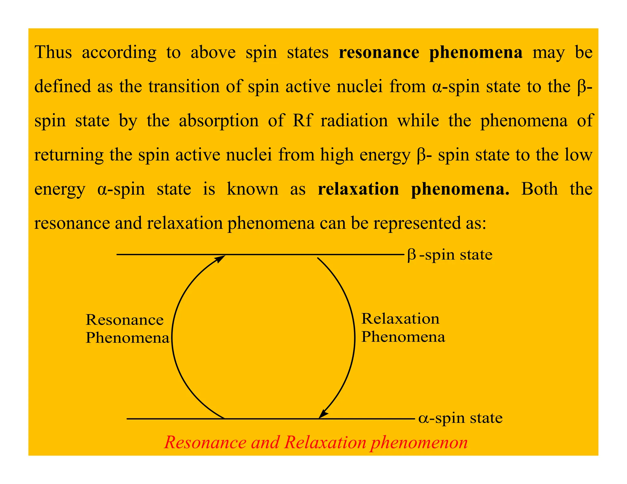
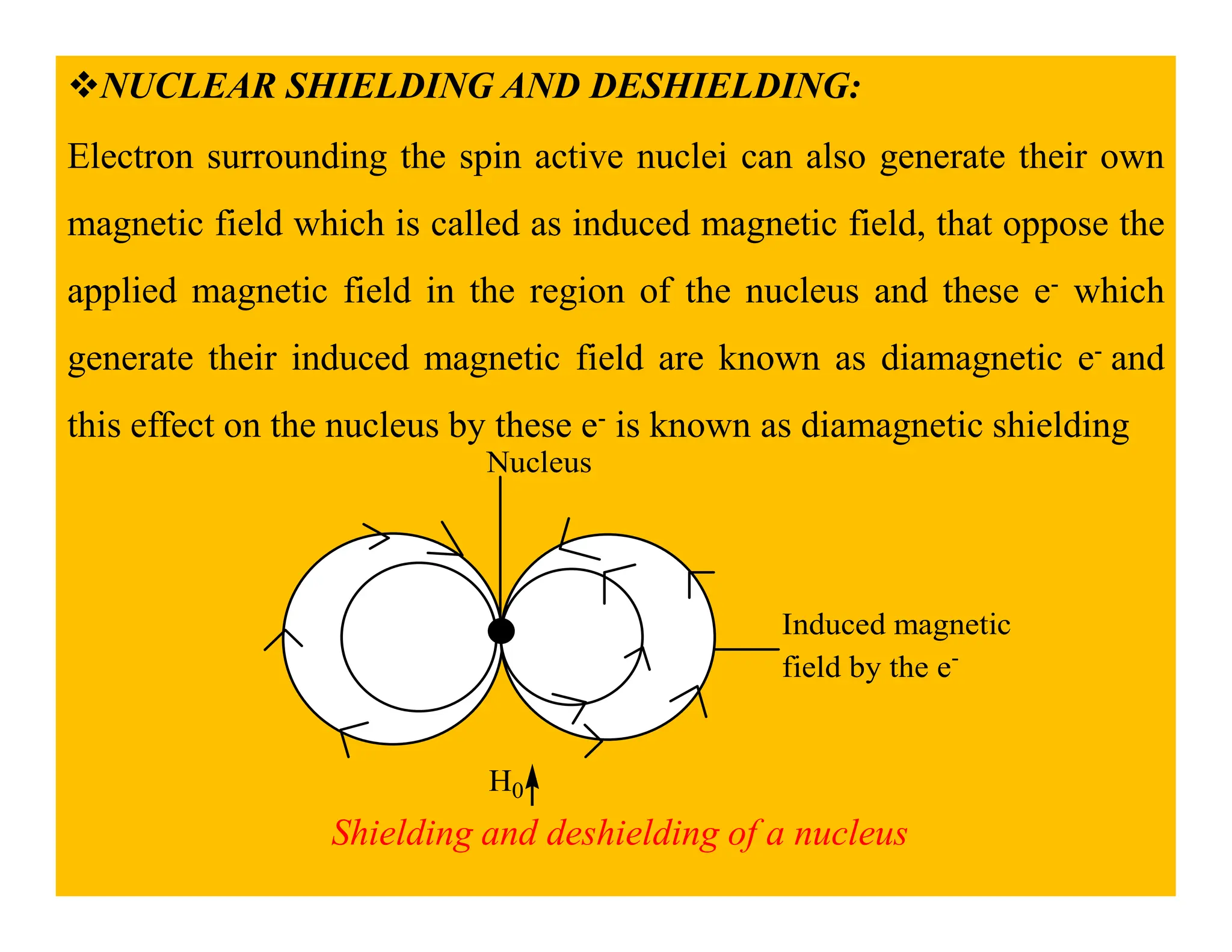
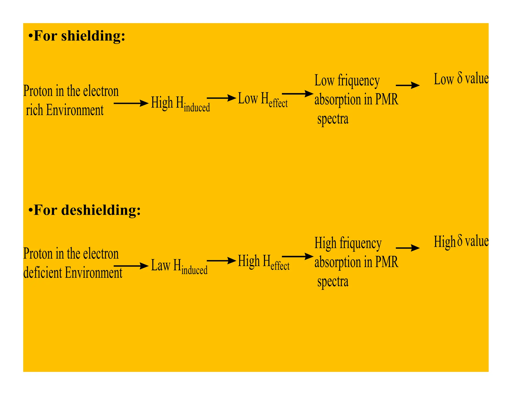
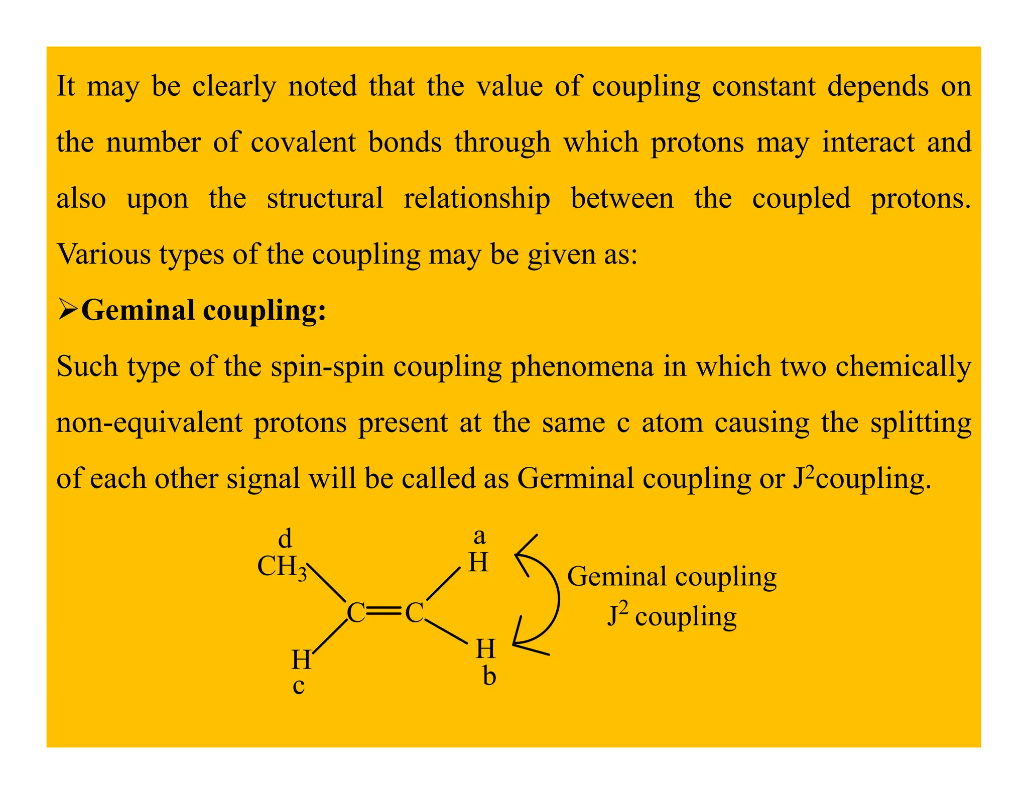
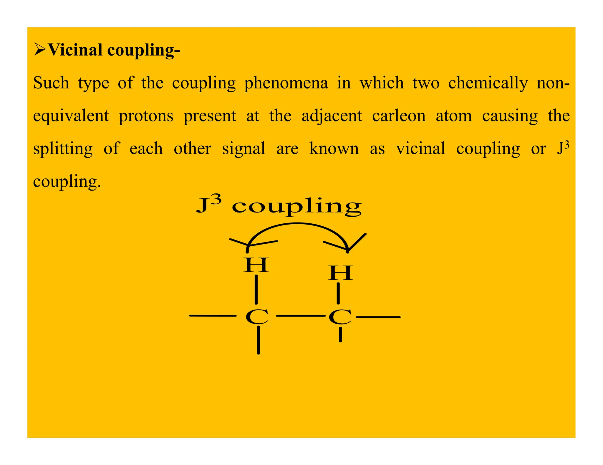
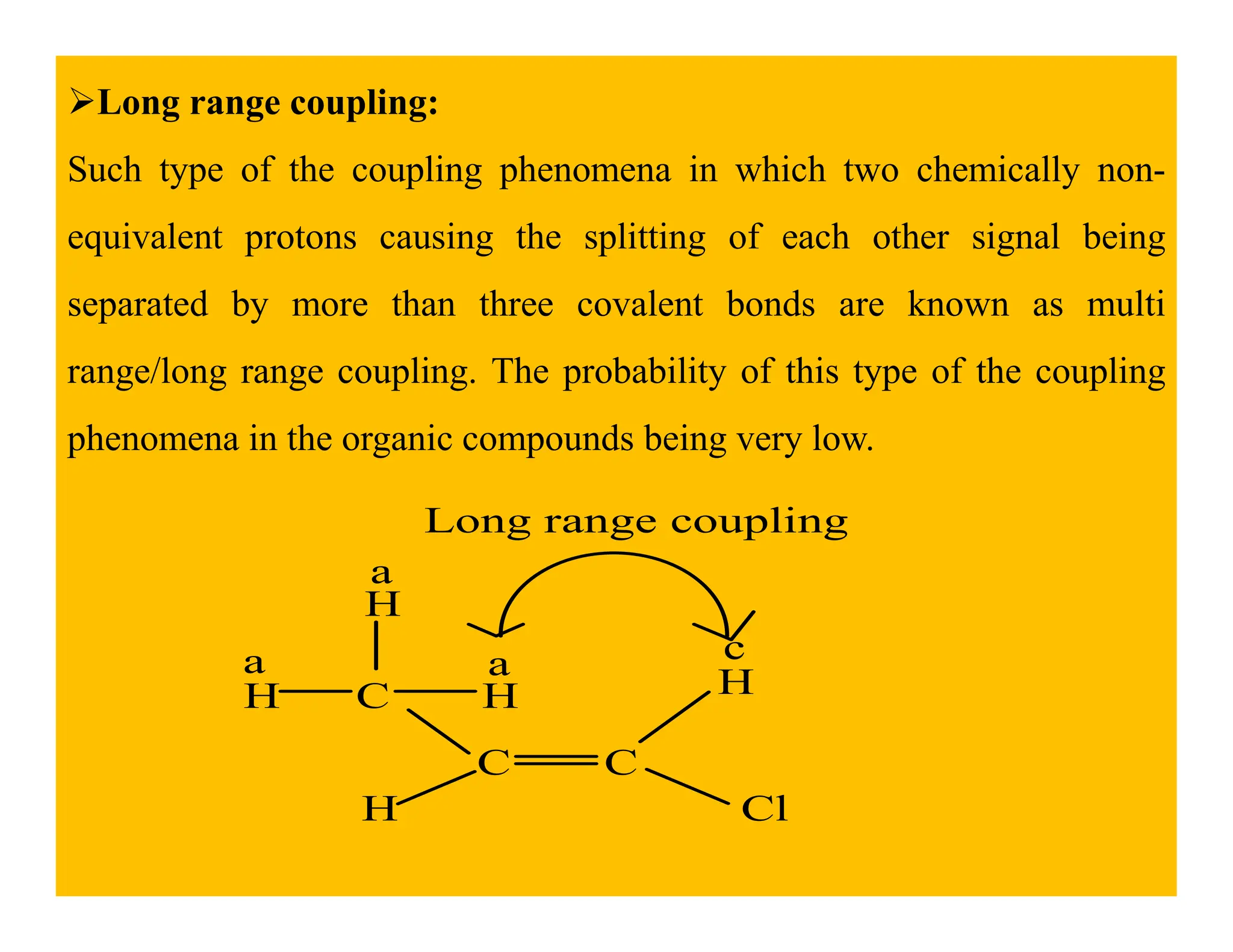
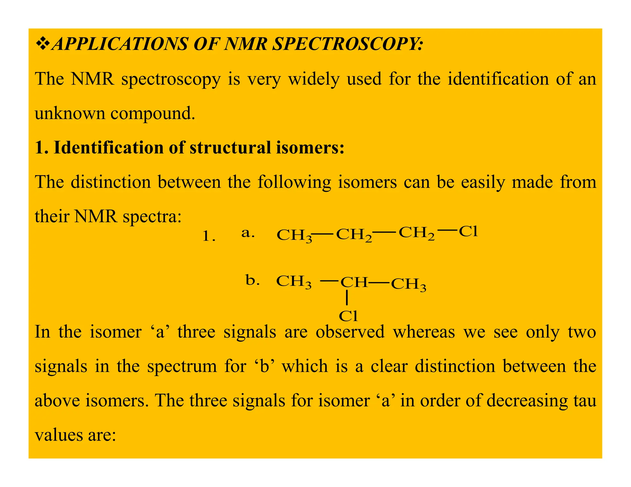
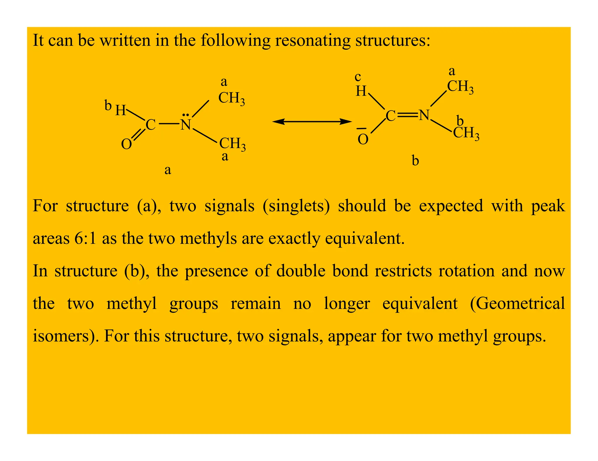
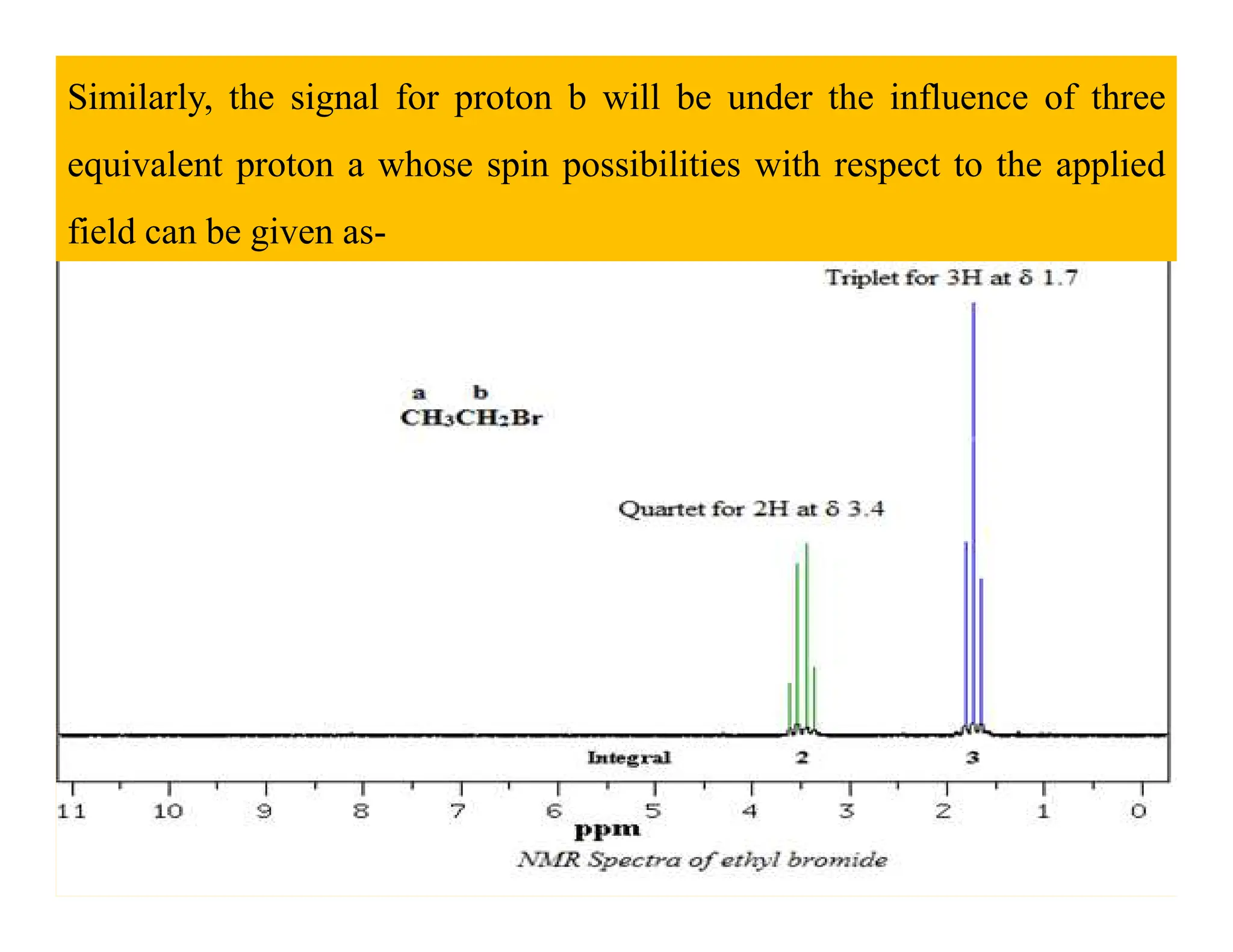
Programme Code- MSCCH-17/18/19
Course Code-CHE- 504
Block – 2 : Introduction to NMR Spectroscopy
Lecture-3
Presented by-
Dr. Charu C. Pant
Department of Chemistry
Uttarakhand Open University, Haldwani
LEARNING OUTCOMES
After studying this Chapter, you shall be able to:
To understand the basic concept of NMR.
To know about NMR active and inactive nuclei.
To know about the conditions of a nuclei to be called an
NMR active nuclei.
NMR active nuclei.
To know the types of relaxation processes a nuclei
undergoes.
To know when a nuclei is called shielded and deshielded.
INTRODUCTION:
The concept of NMR was represented at first in 1946 by two groups of
eminent physicist; Black Hensen and Packard at Stanford University
detected a signal from the Protons of water and Parcell, Torrey and
pound at Havard University observed a signal from the photons in
Paraffin wax. Black and Parcell were jointly awarded a Nobel Prize for
Physics in 1952 for this discovery.
NMR spectroscopy involvers transition of a nucleus from one spin state
to other with the resultant absorption of electromagnetic radiation by spin
active nuclei (having nuclei spin not equal to zero) when they are placed
in magnetic field. Nuclear magnetic resonance spectroscopy related to
the nuclei and only one type of nucleus at a particular timeline.
1H or C13, F19 when the frequency of the rotating magnetic field and that
of the processing nucleus (Lamar Frequency) become equal, they are
said to be in resonance absorption or emission of energy by the nucleus
can be obtained. Plot of the peak intensities versus the frequencies of
objection (represented by δ or τ) establish an NMR spectrum.
The 1H nucleus is most commonly studied by NMR spectroscopy
because of its high natural abundance (99.98%) and the fact that it is
present in the majority of organic compounds, the PMR or 1H NMR
spectrum provides information about the number of different types of
protons and also the chemical environment of each of them.
•A simple representation of NMR spectrum can be given as:
TMS
Intensity
δ
PROTON MAGNETIC RESONANCE (1H NMR) SPECTROSCOPY
1HNMR or PMR spectroscopy is the most widely applicable Nuclear
Magnetic Resonance spectroscopy for the structural determination of
various organic compounds but the other NMR spectroscopic methods
like C13 and P31 NMR spectroscopy; F19 spectroscopy can also be helpful
in the structural determination of the compounds.
Spin active nuclei:
All those nuclei which having the full integer or half integer nuclear spin
value are known as spin active nuclei. With the help of the number of the
electron/proton and neutron in the various nuclei, the spin active or
inactive nature for them can be defined as:
e-/p Neutron (n) Nuclear spin
(I)
Nuclei Examples
Even Even 0 Inactive 8O16
Odd Odd Full integer Active 7N14, 6C12
Even Odd Full integer Active 6C13
Odd Even Half integer Active 15P31, 1H1
Phenomena of energy absorption (Resonance & relaxation
phenomena):
In the absence of external magnetic field H0 the nuclear spin are
randomly oriented, However when the sample is placed in an external
magnetic field then the nuclei (proton) with the spin +half (1/2) are
aligned with the applied field in that lower energy α- spin stagehand the
nuclei with the spin -1/2 are aligned against to the external magnetic field
in the higher energy β- spin state that can be represented as:
β−-spin state
(I= -1/2)
H
∆E
α-spin state
(I= +1/2)
H0
Random arrangement of spin
active nuclei in the absance of H0
H0
Orientation of spinning nuclei in absence and presence of external
magnetic field
The value of energy difference b/w α and β spin state depends on the
strength of external magnetic field H0 according to the equation ∆E =
2µh0 during the PMR spectroscopy, and then it can be represented as:
E
∆
β -spin state
E
∆
α-spin state
E
H0
Energy states of nuclear spin
Thus according to above spin states resonance phenomena may be
defined as the transition of spin active nuclei from α-spin state to the β-
spin state by the absorption of Rf radiation while the phenomena of
returning the spin active nuclei from high energy β- spin state to the low
energy α-spin state is known as relaxation phenomena. Both the
resonance and relaxation phenomena can be represented as:
β
α
-spin state
-spin state
Resonance
Phenomena
Relaxation
Phenomena
Resonance and Relaxation phenomenon
NUCLEAR SHIELDING AND DESHIELDING:
Electron surrounding the spin active nuclei can also generate their own
magnetic field which is called as induced magnetic field, that oppose the
applied magnetic field in the region of the nucleus and these e- which
generate their induced magnetic field are known as diamagnetic e- and
this effect on the nucleus by these e- is known as diamagnetic shielding
Nucleus
H0
Nucleus
Induced magnetic
field by the e-
Shielding and deshielding of a nucleus
The external magnetic field is uniform over the entire molecule and
therefore cannot differentiate to the different types of the proton.
However the induced magnetic field generated by the e- around the
nucleus is not uniform, this situation makes the different spin active
nuclei 9proton) to be non-equivalent. Thus each proton in the different
electronic environment show slightly different magnetic field due to the
circulation of e- in the neighboring bond.
Thus the effective magnetic field for the different spin active nuclei can
be calculated through the following equations:
H effect = H0 – H induced
Form the above equation the shielded and deshielded proton concept can
be given as:
•For shielding:
Proton in the electron
rich Environment High Hinduced
Low Heffect
Low friquency
absorption in PMR
spectra
Low δvalue
•For deshielding:
Proton in the electron
deficient Environment Law Hinduced
High Heffect
High friquency
absorption in PMR
spectra
Highδvalue
CHEMICALLY EQUIVALENT & NON- EQUIVALENT
PROTONS:
Those protons present in the sample of spin active nuclei which having
same chemical environment are known as chemically equivalent protons.
The entire chemically equivalent protons appear as a single signal in the
PMR spectrum.
On the other hand those protons present in the sample of spin active
nuclei which having different chemical environment are known as
chemically non-equivalent proton. Chemically non-equivalent proton
represents the different signal in the PMR spectrum.
CH3
Br
H
H
C C
1.
a b
c
In a molecule reveals that thrre different types of protons, indicated by the letters a,b,c.
2.
CH3
CH3
H
H
C C
a b
There are two types of protons. Six methyl protons on the LHS are equivalent. The protons on RHS are
again equivalent
a
b
3. CH3-CHCl-C-Cl
H
H
a b
c
d
Protons marked c and d appear to be equivalent, but they are no actualy so.Thus it has four types of protons.
4.
CH2
CH2
CH2
CH2 CH2
CH2
a
a
a
a
a
a
In this case all the protons are equivalent giving rise to only one PMR signals
5. CH3
There are two types of protons, benzene ring protons and the methyl protons. Two signals will be observed.
6. CH3-C-CHO
H
H
a
b
b
c
There are three kind of protonsgiving rise tothree signals.
CHEMICAL SHIFT :
Chemical shift it expresses the difference in the resonance frequency of a
given proton compared to that of the methyl protons of TMS, under the
experimental conditions. In practice, this difference is divided by the
operating radio-frequency of the instrument and the chemical shift δ is
expressed, downfield from TMS, as per the given equation.
For example, if the observed shift from TMS is 200 Hz and the operating
frequency of the instrument id 100 MHz, then the chemical shift δ is
given by the following expression:
This frequency ratio (2.0 x 10⁻⁶) is multiplied by 10-6 in order to obtain
an easily handled number (2.0 x 10-6 x 106 ppm) and consequently the
chemical shift δ is expressed as part per million (106) of the operating
frequency.
frequency.
Thus,
Chemical shift =
νs - νTMS (Hz)
Operating friquency
x 106
TMS
Shielded region
Deshielded region
or
Up field region
Down field region
or
Chemical shift
δ ppm
Chemical shift region
Internal standard for NMR spectroscopy:
That compound which is used as a reference standard to represent the
NMR/PMR signal of the compounds is known as internal standard for
the NMR or PMR spectroscopy.
In case of PMR spectroscopy Tetramethylsilane (TMS) used as a internal
standard due to the following reasons:
1. Due to the more shielded nature of the proton of TMS in compare to
the protons of most of the organic compound.
2. It is chemically inert and miscible with large range of solvent.
3. It does not take part in intermolecular association with the sample.
4. Due to the volatile nature of TMS.
The internal standard for some other type of the NMR spectroscopic
methods can be given as:
F19 NMR – CFCl3
N15 NMR – NH4NO3
C13 NMR – TMS
SPIN-SPIN SPLITTING AND COUPLING CONSTANTS:
The coupling interaction between two or more protons, most often
through the bond, results in splitting of the spectral lines. This is called
spin-spin coupling. It is related to the possible combinations of the spin
orientations of the neighboring protons. The phenomena of splitting the
signal of any particular type of proton by the spin orientation of the non-
equivalent proton present adjacent to it is known as spin-spin coupling
phenomena.
Example:
CH3CHCl2
a
b
According to the NMR spectroscopy the signal of two different type of
the proton present in this compound can be give as:
TMS
O
δ
a
b
According to the spin-spin coupling phenomena the splitting of the
signal of Hb proton & Ha proton according to the spin orientation of non-
equivalent adjacent proton can be given as:
1. Spin orientation of Ha for the splitting of the signal of Hb :
TMS
b
Ho)
(
δ
2. Spin orientation of Hb for the splitting of the signal of Ha
TMS
Ho)
(
a
Coupling constant (J):
The distance between the centers of the two adjacent peaks in a multiplet
is usually constant and is called the coupling constant. The value of
coupling constant is independent of the external field. It is measured
Hertz (Hz) or in cps (cycle per second). It is denote by the letter J. In
other words, we can say the value of J remains the same whatever the
applied field. The value of J generally lies between 0 and 20 Hertz (Hz).
Always the value of coupling constant being same for the protons which
causing the splitting each other signal.
Now, let us consider a compound:
CH CH2
a
b
In this compound two signals are expected in the NMR spectrum. Under
the influence of two equivalent proton a, the signal for proton b will
appear as a triplet. The distance between any two adjacent peaks in a
multiplet will be exactly the same. The spin-spin coupling is given
below:
TMS
J
J
spin-spin coupling
It may be clearly noted that the value of coupling constant depends on
the number of covalent bonds through which protons may interact and
also upon the structural relationship between the coupled protons.
Various types of the coupling may be given as:
Geminal coupling:
Such type of the spin-spin coupling phenomena in which two chemically
non-equivalent protons present at the same c atom causing the splitting
of each other signal will be called as Germinal coupling or J2coupling.
C C
CH3
H
H
H
a
b
c
d
Geminal coupling
J2
coupling
Vicinal coupling-
Such type of the coupling phenomena in which two chemically non-
equivalent protons present at the adjacent carleon atom causing the
splitting of each other signal are known as vicinal coupling or J3
coupling.
J3
coupling
C C
H
H
Long range coupling:
Such type of the coupling phenomena in which two chemically non-
equivalent protons causing the splitting of each other signal being
separated by more than three covalent bonds are known as multi
range/long range coupling. The probability of this type of the coupling
phenomena in the organic compounds being very low.
C C
H
H Cl
C H
H
H
a
a
a
c
Long range coupling
APPLICATIONS OF NMR SPECTROSCOPY:
The NMR spectroscopy is very widely used for the identification of an
unknown compound.
1. Identification of structural isomers:
The distinction between the following isomers can be easily made from
their NMR spectra:
1. CH3 CH2 CH2 Cl
a.
1. 3 2
CH3 CH CH3
Cl
b.
In the isomer ‘a’ three signals are observed whereas we see only two
signals in the spectrum for ‘b’ which is a clear distinction between the
above isomers. The three signals for isomer ‘a’ in order of decreasing tau
values are:
1. A three proton triplet
2. A two proton sextet and
3. A two proton triplet For isomer (b), two signals have their
multiplicities as:
4. Doublet (6H)-up field and
5. Septet (1H)-downfield
2. Detection of hydrogen bonding:
Intermolecular hydrogen bonding shifts the absorption for a concerned
proton downfield. The extent of hydrogen bonding varies with the nature
of solvent, concentration of the solution and the temperature. The
intramolecular hydrogen bonding also shifts the absorption downfield.
The two types of hydrogen bonding can be distinguished as the
intramolecular hydrogen bonding is not concentration dependant.
3. Distinction between Cis-Trans Isomers and conformers:
The is and trans isomers of a compound can be easily distinguished as
the concerned protons have different values of the chemical shift as well
as the coupling constants. Similarly, the various conformation of a
compound, the axial and equatorial positions of the proton or group
carrying a proton can be distinguished from their different values of the
coupling.
4. Detection of some double bond character due to resonance:
In some compounds, the molecule acquires a little double bond character
due to resonance. Due to this, two signals can be expected for apparently
equivalent protons, It is due to the hindered rotation which changes the
geometry of the molecules. Consider N, N- dimethyl formamide.
It can be written in the following resonating structures:
C N
O
CH3
CH3
CH3
CH3
O
H
H
..
a
a
b
C N
a
b
c
a
b
For structure (a), two signals (singlets) should be expected with peak
areas 6:1 as the two methyls are exactly equivalent.
In structure (b), the presence of double bond restricts rotation and now
the two methyl groups remain no longer equivalent (Geometrical
isomers). For this structure, two signals, appear for two methyl groups.
INTERPRETATION OF PMR SPECTRA OF SIMPLE ORGANIC
MOLECULES:
NMR interpretation plays a important role in molecular identifications.
As interpreting NMR spectra, the structure of an unknown compound, as
well as known structures, can be assigned by several factors such as
chemical shift, spin multiplicity, coupling constants, and integration. For
the interpreting of the NMR spectra, the following points may be noted:
1. Molecular formula is determined by chemical analysis such as
elementary analysis.
2. Double-bond equivalent or Degree of Unsaturation: It is calculated
by a simple equation to estimate the number of the multiple bonds and
rings. It assumes that oxygen (O) and sulfur (S) are ignored and halogen
(Cl, Br) and nitrogen is replaced by CH. The resulting empirical formula
is CxHy
Double Bond Equivalent (DBE) =
(2x + 2) - y
2
3. Structure fragmentation is determined by chemical shift, spin
multiplicity, integral (peak area), and coupling constant(1J, 2J)
4. Molecular skeleton is built up using 2-dimensional NMR
spectroscopy.
5. Relative configuration is predicted by coupling constant (3J).
EXAMPLES:
1. Ethyl bromide (CH3CH2Br):
In this compound (Ethyl bromide) containing two set of equivalent
protons ‘a’ and ‘b’ types. The signal due to equivalent protons a will be
under the influence of neighboring proton b.
Similarly, the signal for proton b will be under the influence of three
equivalent proton a whose spin possibilities with respect to the applied
field can be given as-
The following peaks can be identified in the spectrum
(a) Triplet, δ 1.7, 3H (b) Quartet, δ 3.4, 2H
The proton at δ 1.7 is given by the three methyl protons which are
magnetically equivalent and are coupled with the two methylene protons
to give a shielding. While the quartet at δ 3.4 is from the two equivalent
methylene protons which are coupled with the three methylene protons
to produce a downfield quartet as a result of deshielding influence of
bromine.
2. Interpretation of PMR spectra of ethanol:
The 1H NMR spectrum of ethanol (Figure 1.9) shows the methyl peak
has been split into three peaks (triplet) and the methylene peak has been
split into four peaks (quartet). This occurs because there is a small
interaction between the two groups of protons. The space between the
peaks of the methyl triplet is equal to the space between the peaks of the
methylene quartet. This spacing is measured in Hertz and is called the
coupling constant, J.
PMR Spectrum of Ethyl alcohol
In the above PMR spectrum of ethyl alcohol the following observation
takes place:
•A triplet centered at 1.18 δ, equivalent to 3H, represents the CH3 (a)
proton.
•A singlet at 4.51 δ, equivalent to 1H, exhibit the OH (c) proton.
•A quartet centered at 3.63 δ, equivalent to 2H indicates the CH2 (b)
proton.
CHE-504 Lecture 3 Basics of NMR Spectroscopy by Dr. Charu C. Pant.pdf

CHE-504 Lecture 3 Basics of NMR Spectroscopy by Dr. Charu C. Pant.pdf

  • 1.
    Programme Code- MSCCH-17/18/19 CourseCode-CHE- 504 Block – 2 : Introduction to NMR Spectroscopy Block – 2 : Introduction to NMR Spectroscopy Lecture-3 Presented by- Dr. Charu C. Pant Department of Chemistry Uttarakhand Open University, Haldwani
  • 2.
    LEARNING OUTCOMES After studyingthis Chapter, you shall be able to: To understand the basic concept of NMR. To know about NMR active and inactive nuclei. To know about the conditions of a nuclei to be called an NMR active nuclei. NMR active nuclei. To know the types of relaxation processes a nuclei undergoes. To know when a nuclei is called shielded and deshielded.
  • 3.
    INTRODUCTION: The concept ofNMR was represented at first in 1946 by two groups of eminent physicist; Black Hensen and Packard at Stanford University detected a signal from the Protons of water and Parcell, Torrey and pound at Havard University observed a signal from the photons in Paraffin wax. Black and Parcell were jointly awarded a Nobel Prize for Physics in 1952 for this discovery. NMR spectroscopy involvers transition of a nucleus from one spin state to other with the resultant absorption of electromagnetic radiation by spin active nuclei (having nuclei spin not equal to zero) when they are placed in magnetic field. Nuclear magnetic resonance spectroscopy related to the nuclei and only one type of nucleus at a particular timeline.
  • 4.
    1H or C13,F19 when the frequency of the rotating magnetic field and that of the processing nucleus (Lamar Frequency) become equal, they are said to be in resonance absorption or emission of energy by the nucleus can be obtained. Plot of the peak intensities versus the frequencies of objection (represented by δ or τ) establish an NMR spectrum. The 1H nucleus is most commonly studied by NMR spectroscopy because of its high natural abundance (99.98%) and the fact that it is present in the majority of organic compounds, the PMR or 1H NMR spectrum provides information about the number of different types of protons and also the chemical environment of each of them.
  • 5.
    •A simple representationof NMR spectrum can be given as: TMS Intensity δ
  • 6.
    PROTON MAGNETIC RESONANCE(1H NMR) SPECTROSCOPY 1HNMR or PMR spectroscopy is the most widely applicable Nuclear Magnetic Resonance spectroscopy for the structural determination of various organic compounds but the other NMR spectroscopic methods like C13 and P31 NMR spectroscopy; F19 spectroscopy can also be helpful in the structural determination of the compounds. Spin active nuclei: All those nuclei which having the full integer or half integer nuclear spin value are known as spin active nuclei. With the help of the number of the electron/proton and neutron in the various nuclei, the spin active or inactive nature for them can be defined as:
  • 7.
    e-/p Neutron (n)Nuclear spin (I) Nuclei Examples Even Even 0 Inactive 8O16 Odd Odd Full integer Active 7N14, 6C12 Even Odd Full integer Active 6C13 Odd Even Half integer Active 15P31, 1H1 Phenomena of energy absorption (Resonance & relaxation phenomena): In the absence of external magnetic field H0 the nuclear spin are randomly oriented, However when the sample is placed in an external magnetic field then the nuclei (proton) with the spin +half (1/2) are aligned with the applied field in that lower energy α- spin stagehand the
  • 8.
    nuclei with thespin -1/2 are aligned against to the external magnetic field in the higher energy β- spin state that can be represented as: β−-spin state (I= -1/2) H ∆E α-spin state (I= +1/2) H0 Random arrangement of spin active nuclei in the absance of H0 H0 Orientation of spinning nuclei in absence and presence of external magnetic field
  • 9.
    The value ofenergy difference b/w α and β spin state depends on the strength of external magnetic field H0 according to the equation ∆E = 2µh0 during the PMR spectroscopy, and then it can be represented as: E ∆ β -spin state E ∆ α-spin state E H0 Energy states of nuclear spin
  • 10.
    Thus according toabove spin states resonance phenomena may be defined as the transition of spin active nuclei from α-spin state to the β- spin state by the absorption of Rf radiation while the phenomena of returning the spin active nuclei from high energy β- spin state to the low energy α-spin state is known as relaxation phenomena. Both the resonance and relaxation phenomena can be represented as: β α -spin state -spin state Resonance Phenomena Relaxation Phenomena Resonance and Relaxation phenomenon
  • 11.
    NUCLEAR SHIELDING ANDDESHIELDING: Electron surrounding the spin active nuclei can also generate their own magnetic field which is called as induced magnetic field, that oppose the applied magnetic field in the region of the nucleus and these e- which generate their induced magnetic field are known as diamagnetic e- and this effect on the nucleus by these e- is known as diamagnetic shielding Nucleus H0 Nucleus Induced magnetic field by the e- Shielding and deshielding of a nucleus
  • 12.
    The external magneticfield is uniform over the entire molecule and therefore cannot differentiate to the different types of the proton. However the induced magnetic field generated by the e- around the nucleus is not uniform, this situation makes the different spin active nuclei 9proton) to be non-equivalent. Thus each proton in the different electronic environment show slightly different magnetic field due to the circulation of e- in the neighboring bond. Thus the effective magnetic field for the different spin active nuclei can be calculated through the following equations: H effect = H0 – H induced Form the above equation the shielded and deshielded proton concept can be given as:
  • 13.
    •For shielding: Proton inthe electron rich Environment High Hinduced Low Heffect Low friquency absorption in PMR spectra Low δvalue •For deshielding: Proton in the electron deficient Environment Law Hinduced High Heffect High friquency absorption in PMR spectra Highδvalue
  • 14.
    CHEMICALLY EQUIVALENT &NON- EQUIVALENT PROTONS: Those protons present in the sample of spin active nuclei which having same chemical environment are known as chemically equivalent protons. The entire chemically equivalent protons appear as a single signal in the PMR spectrum. On the other hand those protons present in the sample of spin active nuclei which having different chemical environment are known as chemically non-equivalent proton. Chemically non-equivalent proton represents the different signal in the PMR spectrum.
  • 15.
    CH3 Br H H C C 1. a b c Ina molecule reveals that thrre different types of protons, indicated by the letters a,b,c. 2. CH3 CH3 H H C C a b There are two types of protons. Six methyl protons on the LHS are equivalent. The protons on RHS are again equivalent a b 3. CH3-CHCl-C-Cl H H a b c d Protons marked c and d appear to be equivalent, but they are no actualy so.Thus it has four types of protons. 4. CH2 CH2 CH2 CH2 CH2 CH2 a a a a a a In this case all the protons are equivalent giving rise to only one PMR signals 5. CH3 There are two types of protons, benzene ring protons and the methyl protons. Two signals will be observed. 6. CH3-C-CHO H H a b b c There are three kind of protonsgiving rise tothree signals.
  • 16.
    CHEMICAL SHIFT : Chemicalshift it expresses the difference in the resonance frequency of a given proton compared to that of the methyl protons of TMS, under the experimental conditions. In practice, this difference is divided by the operating radio-frequency of the instrument and the chemical shift δ is expressed, downfield from TMS, as per the given equation. For example, if the observed shift from TMS is 200 Hz and the operating frequency of the instrument id 100 MHz, then the chemical shift δ is given by the following expression:
  • 17.
    This frequency ratio(2.0 x 10⁻⁶) is multiplied by 10-6 in order to obtain an easily handled number (2.0 x 10-6 x 106 ppm) and consequently the chemical shift δ is expressed as part per million (106) of the operating frequency. frequency. Thus, Chemical shift = νs - νTMS (Hz) Operating friquency x 106
  • 18.
    TMS Shielded region Deshielded region or Upfield region Down field region or Chemical shift δ ppm Chemical shift region
  • 19.
    Internal standard forNMR spectroscopy: That compound which is used as a reference standard to represent the NMR/PMR signal of the compounds is known as internal standard for the NMR or PMR spectroscopy. In case of PMR spectroscopy Tetramethylsilane (TMS) used as a internal standard due to the following reasons: 1. Due to the more shielded nature of the proton of TMS in compare to the protons of most of the organic compound. 2. It is chemically inert and miscible with large range of solvent. 3. It does not take part in intermolecular association with the sample. 4. Due to the volatile nature of TMS.
  • 20.
    The internal standardfor some other type of the NMR spectroscopic methods can be given as: F19 NMR – CFCl3 N15 NMR – NH4NO3 C13 NMR – TMS SPIN-SPIN SPLITTING AND COUPLING CONSTANTS: The coupling interaction between two or more protons, most often The coupling interaction between two or more protons, most often through the bond, results in splitting of the spectral lines. This is called spin-spin coupling. It is related to the possible combinations of the spin orientations of the neighboring protons. The phenomena of splitting the signal of any particular type of proton by the spin orientation of the non- equivalent proton present adjacent to it is known as spin-spin coupling phenomena.
  • 21.
    Example: CH3CHCl2 a b According to theNMR spectroscopy the signal of two different type of the proton present in this compound can be give as: TMS O δ a b According to the spin-spin coupling phenomena the splitting of the signal of Hb proton & Ha proton according to the spin orientation of non- equivalent adjacent proton can be given as:
  • 22.
    1. Spin orientationof Ha for the splitting of the signal of Hb : TMS b Ho) ( δ
  • 23.
    2. Spin orientationof Hb for the splitting of the signal of Ha TMS Ho) ( a
  • 24.
    Coupling constant (J): Thedistance between the centers of the two adjacent peaks in a multiplet is usually constant and is called the coupling constant. The value of coupling constant is independent of the external field. It is measured Hertz (Hz) or in cps (cycle per second). It is denote by the letter J. In other words, we can say the value of J remains the same whatever the applied field. The value of J generally lies between 0 and 20 Hertz (Hz). Always the value of coupling constant being same for the protons which causing the splitting each other signal. Now, let us consider a compound: CH CH2 a b
  • 25.
    In this compoundtwo signals are expected in the NMR spectrum. Under the influence of two equivalent proton a, the signal for proton b will appear as a triplet. The distance between any two adjacent peaks in a multiplet will be exactly the same. The spin-spin coupling is given below: TMS J J spin-spin coupling
  • 26.
    It may beclearly noted that the value of coupling constant depends on the number of covalent bonds through which protons may interact and also upon the structural relationship between the coupled protons. Various types of the coupling may be given as: Geminal coupling: Such type of the spin-spin coupling phenomena in which two chemically non-equivalent protons present at the same c atom causing the splitting of each other signal will be called as Germinal coupling or J2coupling. C C CH3 H H H a b c d Geminal coupling J2 coupling
  • 27.
    Vicinal coupling- Such typeof the coupling phenomena in which two chemically non- equivalent protons present at the adjacent carleon atom causing the splitting of each other signal are known as vicinal coupling or J3 coupling. J3 coupling C C H H
  • 28.
    Long range coupling: Suchtype of the coupling phenomena in which two chemically non- equivalent protons causing the splitting of each other signal being separated by more than three covalent bonds are known as multi range/long range coupling. The probability of this type of the coupling phenomena in the organic compounds being very low. C C H H Cl C H H H a a a c Long range coupling
  • 29.
    APPLICATIONS OF NMRSPECTROSCOPY: The NMR spectroscopy is very widely used for the identification of an unknown compound. 1. Identification of structural isomers: The distinction between the following isomers can be easily made from their NMR spectra: 1. CH3 CH2 CH2 Cl a. 1. 3 2 CH3 CH CH3 Cl b. In the isomer ‘a’ three signals are observed whereas we see only two signals in the spectrum for ‘b’ which is a clear distinction between the above isomers. The three signals for isomer ‘a’ in order of decreasing tau values are:
  • 30.
    1. A threeproton triplet 2. A two proton sextet and 3. A two proton triplet For isomer (b), two signals have their multiplicities as: 4. Doublet (6H)-up field and 5. Septet (1H)-downfield 2. Detection of hydrogen bonding: Intermolecular hydrogen bonding shifts the absorption for a concerned proton downfield. The extent of hydrogen bonding varies with the nature of solvent, concentration of the solution and the temperature. The intramolecular hydrogen bonding also shifts the absorption downfield. The two types of hydrogen bonding can be distinguished as the intramolecular hydrogen bonding is not concentration dependant.
  • 31.
    3. Distinction betweenCis-Trans Isomers and conformers: The is and trans isomers of a compound can be easily distinguished as the concerned protons have different values of the chemical shift as well as the coupling constants. Similarly, the various conformation of a compound, the axial and equatorial positions of the proton or group carrying a proton can be distinguished from their different values of the coupling. 4. Detection of some double bond character due to resonance: In some compounds, the molecule acquires a little double bond character due to resonance. Due to this, two signals can be expected for apparently equivalent protons, It is due to the hindered rotation which changes the geometry of the molecules. Consider N, N- dimethyl formamide.
  • 32.
    It can bewritten in the following resonating structures: C N O CH3 CH3 CH3 CH3 O H H .. a a b C N a b c a b For structure (a), two signals (singlets) should be expected with peak areas 6:1 as the two methyls are exactly equivalent. areas 6:1 as the two methyls are exactly equivalent. In structure (b), the presence of double bond restricts rotation and now the two methyl groups remain no longer equivalent (Geometrical isomers). For this structure, two signals, appear for two methyl groups.
  • 33.
    INTERPRETATION OF PMRSPECTRA OF SIMPLE ORGANIC MOLECULES: NMR interpretation plays a important role in molecular identifications. As interpreting NMR spectra, the structure of an unknown compound, as well as known structures, can be assigned by several factors such as chemical shift, spin multiplicity, coupling constants, and integration. For the interpreting of the NMR spectra, the following points may be noted: 1. Molecular formula is determined by chemical analysis such as elementary analysis. 2. Double-bond equivalent or Degree of Unsaturation: It is calculated by a simple equation to estimate the number of the multiple bonds and rings. It assumes that oxygen (O) and sulfur (S) are ignored and halogen
  • 34.
    (Cl, Br) andnitrogen is replaced by CH. The resulting empirical formula is CxHy Double Bond Equivalent (DBE) = (2x + 2) - y 2 3. Structure fragmentation is determined by chemical shift, spin multiplicity, integral (peak area), and coupling constant(1J, 2J) 4. Molecular skeleton is built up using 2-dimensional NMR 4. Molecular skeleton is built up using 2-dimensional NMR spectroscopy. 5. Relative configuration is predicted by coupling constant (3J). EXAMPLES: 1. Ethyl bromide (CH3CH2Br): In this compound (Ethyl bromide) containing two set of equivalent protons ‘a’ and ‘b’ types. The signal due to equivalent protons a will be under the influence of neighboring proton b.
  • 35.
    Similarly, the signalfor proton b will be under the influence of three equivalent proton a whose spin possibilities with respect to the applied field can be given as-
  • 36.
    The following peakscan be identified in the spectrum (a) Triplet, δ 1.7, 3H (b) Quartet, δ 3.4, 2H The proton at δ 1.7 is given by the three methyl protons which are magnetically equivalent and are coupled with the two methylene protons to give a shielding. While the quartet at δ 3.4 is from the two equivalent methylene protons which are coupled with the three methylene protons to produce a downfield quartet as a result of deshielding influence of bromine.
  • 37.
    2. Interpretation ofPMR spectra of ethanol: The 1H NMR spectrum of ethanol (Figure 1.9) shows the methyl peak has been split into three peaks (triplet) and the methylene peak has been split into four peaks (quartet). This occurs because there is a small interaction between the two groups of protons. The space between the peaks of the methyl triplet is equal to the space between the peaks of the methylene quartet. This spacing is measured in Hertz and is called the coupling constant, J.
  • 38.
    PMR Spectrum ofEthyl alcohol In the above PMR spectrum of ethyl alcohol the following observation takes place: •A triplet centered at 1.18 δ, equivalent to 3H, represents the CH3 (a) proton. •A singlet at 4.51 δ, equivalent to 1H, exhibit the OH (c) proton. •A quartet centered at 3.63 δ, equivalent to 2H indicates the CH2 (b) proton.