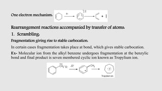
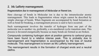
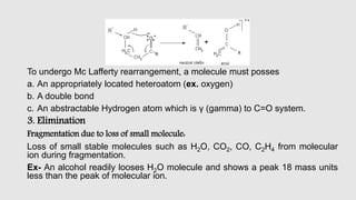
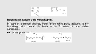
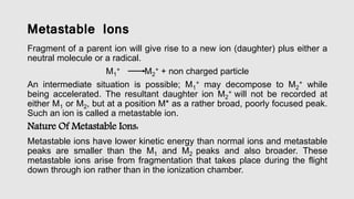
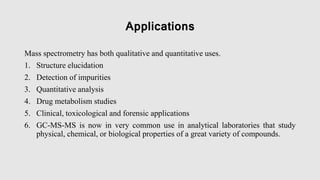
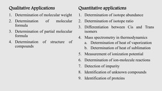
Introduction
working principle
fragmentation process
general rules for fragmentation
general modes of fragmentation
metastable ions
isotopic peaks
applications
1. Mass Fragmentation & Its Rules
Advanced Instrumental Analysis
M. Pharmacy II-Sem
Mehul H Jain
Pharmaceutical Analysis
2. Introduction
• Mass spectroscopy is one of the most generally applicable tools providing both
qualitative and quantitative information about the atomic and molecular materials.
• Here the compound under the investigation is bombarded with a beam of electrons
which produce an ionic molecule or ionic fragments of the original species. The
resulting charging particle is then separated according their masses.
Definition
It is instrumental technique in which sample is converted to rapidly moving positive
ions by which electron bombardment and charged particles are separated according
to their masses.
Mass spectra
It is a plot of relative abundance against the ratio of mass/charge (m/e)
3. Working Principle
• In this technique, molecules are bombarded with a beam of energetic electrons.
• The molecules are ionized and broken up into many fragments, some of which are
positive ions.
• Each kind of ion has a particular ratio of mass to charge, i.e., m/e ratio.
• For most ions, the charge is one and thus, m/e ratio is simply the molecular mass
of the ion.
• The ion pass through magnetic and electric fields to reach detector where they are
detected and signals are recorded to give a mass spectra.
4.
5. Fragmentation Process
• Bombardment of molecules by an electron beam with energy between 10-
15ev usually results in the ionization of molecules by removal of one
electron (Molecular ion formation).
• When the energy of electron beam is increased between 50-70ev, these
molecular ions acquire a high excitation resulting in their break down into
various fragments.
• This process is called “ Fragmentation process”.
6. General Rules For Fragmentation
1. The relative height of the molecular ion peak is greatest for the straight
chain compound and decreases as the degree of branching increases.
2. The relative height of the Molecular ion peak usually decreases with
increasing molecular weight in a homologous series.
3. Cleavage is favoured at alkyl substituted carbon atoms; the more
substituted, the more likely is cleavage. This is a consequence of the
increased stability of a tertiary carbon atom over a secondary, which in
turn is more stable than a primary.
CH3
+ < RCH2
+ < R2CH+ < R3C+
7. Stevensons Rule:
1. When an ion fragments, the positive charge will remain on the fragment of lowest
ionization potential.
2. Generally the largest substituent at a branch is eliminated most readily as a radical
because a long chain radical can achieve some stability by delocalization of the
lone electron.
Ex- cleavage of 1-methyl pentane
1-methyl pentane largest fragment
3. In this fragmentation, positive charge remains on the more high substituted
fragments, i.e. the one with lower ionization potential.
4. Double bonds, cyclic structures and especially aromatic or hetero aromatic rings
stabilize the Molecular ion and thus increase the probability of its appearance.
8. 5. Double bonds favour allylic cleavage and give the resonance stabilized allylic
carbonium.
Ex: Mass spectrum of 1-butene
6. Saturated rings tend to lose alkyl side chains at the a carbon atom. This positive
charge tends to stay with the ring fragment.
Ex: Mass spectrum of n-propyl cyclohexene
9. 7. In alkyl substituted aromatic compounds, cleavage is very probable at the bond β
to the ring, giving the resonance stabilized benzyl ion or more likely, the
tropylium ion.
Ex: mass spectra of n-butyl benzene.
8. Cleavage is often associated with elimination of small, stable, neutral molecules
such as carbon monoxide, olefins, water, ammonia, hydrogen sulphide, hydrogen
cyanide, mercaptans, ketone, or alcohols, often with rearrangement.
10. General Modes Of Fragmentation
Fragmentation of the molecular ion takes place in following modes:
Simple cleavage
1. Homolytic cleavage
2. Heterolytic cleavage
3. Retro Diels-Alder reaction
Rearrangement reactions accompanied by transfer of atoms.
1. Scrambling
2. Mc Lafferty rearrangement
3. Elimination
12. 2. Heterolytic cleavage:
Fragmentation by movement of two electrons:
• In this type of cleavage both the electrons of the bond are taken over by one of the
atoms; the fragments are an even electron cation and a radical with the positive
charge residing on the alkyl group. It is designated by a conventional arrow (↶ or
↷) to signify the transfer of a pair of electrons in the direction of the charged site.
3. Retro Diels-Alder reaction:
Elimination by multiple bond rupture:
Cyclohexene is broken down to Diene and Dienophile. It can be explained by one
electron mechanism.
13. One electron mechanism:
Rearrangement reactions accompanied by transfer of atoms:
1. Scrambling:
Fragmentation giving rise to stable carbocation:
In certain cases fragmentation takes place at bond, which gives stable carbocation.
Ex- Molecular ion from the alkyl benzene undergoes fragmentation at the benzylic
bond and final product is seven membered cyclic ion known as Tropylium ion.
14. 2. Mc Lafferty rearrangement:
Fragmentation due to rearrangement of Molecular or Parent ion:
Here cleavage of bonds in Molecular ion is due to the intramolecular atomic
rearrangement. This leads to fragmentation whose origin cannot be described by
simple cleavage of bonds. When fragments are accompanied by bond formation as
well as bond for breaking, a rearrangement process is said to have occurred.
Such rearrangement involves the transfer of hydrogen from one part of the
molecular ion to another via, preferably, a six-membered cyclic transition state. This
process is favoured energetically because as many bonds are formed as are broken.
Compounds containing hydrogen atom at position gamma to carbonyl group
have been found to a relative intense peak. This is probably due to
rearrangement and fragmentation is accompanied by the loss of neutral
molecule. This rearrangement is known as Mc Lafferty rearrangement.
The rearrangement results in the formation of charged enols and a neutral
olefins.
15. To undergo Mc Lafferty rearrangement, a molecule must posses
a. An appropriately located heteroatom (ex. oxygen)
b. A double bond
c. An abstractable Hydrogen atom which is γ (gamma) to C=O system.
3. Elimination
Fragmentation due to loss of small molecule:
Loss of small stable molecules such as H2O, CO2, CO, C2H4 from molecular
ion during fragmentation.
Ex- An alcohol readily looses H2O molecule and shows a peak 18 mass units
less than the peak of molecular ion.
16. Fragmentation adjacent to the branching point:
In case of branched alkanes, bond fission takes place adjacent to the
branching point. Hence this leads to the formation of more stable
carbocation
Ex: 3-methyl pentane
17. Metastable Ions
Fragment of a parent ion will give rise to a new ion (daughter) plus either a
neutral molecule or a radical.
M1
+ M2
+ + non charged particle
An intermediate situation is possible; M1
+ may decompose to M2
+ while
being accelerated. The resultant daughter ion M2
+ will not be recorded at
either M1 or M2, but at a position M* as a rather broad, poorly focused peak.
Such an ion is called a metastable ion.
Nature Of Metastable Ions:
Metastable ions have lower kinetic energy than normal ions and metastable
peaks are smaller than the M1 and M2 peaks and also broader. These
metastable ions arise from fragmentation that takes place during the flight
down through ion rather than in the ionization chamber.
18. Molecular ions formed in the ionization chamber do one of the following things:
1. Either they decompose completely and very rapidly in the ion source and never
reach the collector (as in case of highly branched molecular ions with life times
less than 10-5 seconds).
2. Or else they survive long enough to reach the collector and be recorded there
(life times longer than 10-5).
Significance of Metastable ions:
• Metastable ions are useful in helping to establish fragments routes.
• Metastable ion peak can also be used to distinguish between fragmentation
Processes, which occur in few microseconds.
19. Isotopic Peaks
• Mass spectrum of certain compounds show peaks that occur at one or two m/e
units greater than the parent ion.
• These peaks are attributable to those ions which have same chemical formula but
different isotopic compositions. The size of the various peaks depends on the
relative natural abundance of the isotopes.
• In organic compounds, there is generally a small peak appearing at one mass unit
higher than the parent peak (M+1) due to small but observable abundance of C, H,
O, N, S isotopes. If the same sample contains two heavy isotopes like Cl, Br, then
an additional smaller peak occurs at M+2. Isotope peak provides a useful means
for determining the molecular formula of a compound.
20. Applications
Mass spectrometry has both qualitative and quantitative uses.
1. Structure elucidation
2. Detection of impurities
3. Quantitative analysis
4. Drug metabolism studies
5. Clinical, toxicological and forensic applications
6. GC-MS-MS is now in very common use in analytical laboratories that study
physical, chemical, or biological properties of a great variety of compounds.
21. Qualitative Applications
1. Determination of molecular weight
2. Determination of molecular
formula
3. Determination of partial molecular
formula
4. Determination of structure of
compounds
Quantitative applications
1. Determination of isotope abundance
2. Determination of isotope ratio
3. Differentiation between Cis and Trans
isomers
4. Mass spectrometry in thermodynamics
a. Determination of heat of vaporization
b. Determination of heat of sublimation
5. Measurement of ionization potential
6. Determination of ion-molecule reactions
7. Detection of impurity
8. Identification of unknown compounds
9. Identification of proteins
22. References
1. Sharma Y.R. Elementary organic spectroscopy principles and chemical
applications. 1st ed. S. Chand and Company ltd; New Delhi: 2008.
2. Chatwal G.R, Anand S.K. Instrumental methods of chemical analysis. 1st
ed. Himalaya Publishing house; Mumbai: 2004.
3. S. Ravi Shankar. Text book of pharmaceutical analysis. 3rd ed. Rx
publication; Tirunelveli: 2006.
4. Skoog DA, West DM. principle of instrumental analysis. 2nd edition.