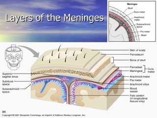
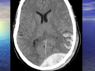
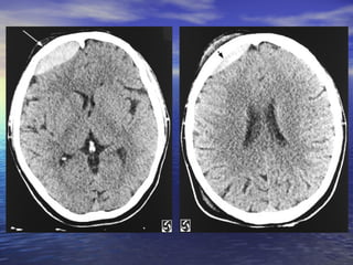
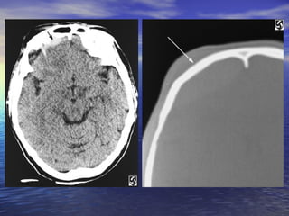
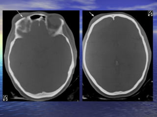
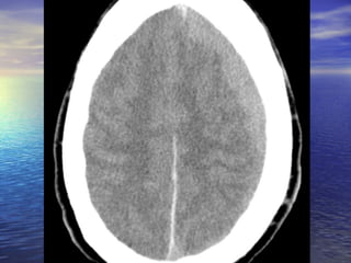
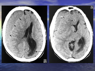
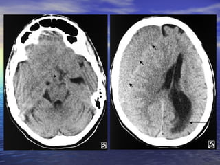
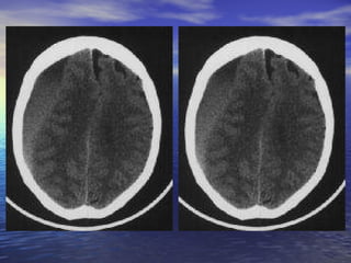
Neuroradiology Traumatic Hemorrhage By: Luke Aldo, MSIV LECOM Erie, Pennsylvania
Layers of the Meninges
Epidural Hematoma Accumulation of blood in the potential space between dura mater and bone EDH is considered to be the most serious complication of head injury, requiring immediate diagnosis and surgical intervention (mortality rate associated with epidural hematoma has been estimated to be 5-50%)
Pathophysiology Usually results from a brief linear contact force to the calvaria that causes separation of the periosteal dura from bone and disruption of interposed vessels due to shearing stress  Skull fractures occur in 85-95% of adult cases  Extension of the hematoma usually is limited by suture lines owing to the tight attachment of the dura at these locations. The  temporoparietal  region and the  middle meningeal artery  are involved most commonly (66%)
Frequency Epidural hematoma complicates 2% of cases of head trauma (approximately 40,000 cases per year) Alcohol and other forms of intoxication have been associated with a higher incidence of epidural hematoma Sex more frequent in men, with a male-to-female ratio of 4:1 Age   rare in individuals younger than 2 years rare in individuals older than 60 years because the dura is tightly adherent to the calvaria
History Head trauma Lucid interval  between the initial loss of consciousness at the time of impact and a delayed decline in mental status (10-33% of cases) Headache Nausea/vomiting Seizures Focal neurological deficits (eg, visual field cuts, aphasia, weakness, numbness)
Diagnostic Imaging Noncontrast CT scanning  of the head (imaging study of choice for intracranial EDH) not only visualizes skull fractures, but also directly images an epidural hematoma It appears as a  hyperdense   biconvex or lenticular-shaped  mass situated between the brain and the skull, though regions of hypodensity may be seen with serum or fresh blood MRI  also demonstrates the evolution of an epidural hematoma, though this imaging modality may not be appropriate for patients in unstable condition


Subdural Hematoma Rapidly clotting blood collection below the inner layer of the dura but external to the brain and arachnoid membrane Typically, low-pressure venous bleeding of  bridging veins  (between the cortex and venous sinuses) dissects the arachnoid away from the dura and layers out along the cerebral convexity It conforms to the shape of the brain and the cranial vault, exhibiting  concave inner margins and convex outer margins (crescent shape) Frequency is related directly to the incidence of blunt head trauma  It’s the most common type of intracranial mass lesion, occurring in about a third of those with severe head injuries
Mortality/Age Mortality Simple SDH (no parenchymal injury) is associated with a mortality rate of about 20% Complicated SDH (parenchymal injury) is associated with a mortality rate of about 50%  Age It’s associated with age factors related to the risk of blunt head trauma More common in people older than 60 years (bridging veins are more easily damaged/falls are more common) Bilateral SDHs are more common in infants since adhesions existing in the subdural space are absent at birth Interhemispheric SDHs are often associate with child abuse
History Usually involves moderately severe to severe blunt  head trauma   Acute deceleration injury from a fall or motor vehicle accident, but rarely associated with skull fracture Generally  loss of consciousness Any degree or type of coagulopathy should heighten suspicion of SDH Commonly seen in alcoholics because they’re prone to thrombocytopenia, prolonged bleeding times, and blunt head trauma Patients on anticoagulants can develop SDH with minimal trauma and warrant a lowered threshold for obtaining a head CT scan
Diagnostic Imaging MRI is superior for demonstrating the size of an acute SDH and its effect on the brain, however noncontrast head CT is the primary means of making a diagnosis and suffice for immediate management purposes Noncontrast head CT scan  (imaging study of choice for acute SDH)  The SDH appears as a  hyperdense  (white)  crescentic mass  along the inner table of the skull, most commonly over the cerebral convexity in the parietal region. The second most common area is above the tentorium cerebelli Contrast-enhanced CT or MRI is widely recommended for imaging 48-72 hours after head injury because the lesion becomes isodense in the subacute phase In the chronic phase, the lesion becomes hypodense and is easy to appreciate on a noncontrast head CT scan


Summary Epidural Hematoma Potential space between the dura in the inner table of the skull Can’t cross sutures Skull fractures in temporoparietal region Middle meningeal artery Lenticular or biconvex shape Lucid interval Common in alcoholics Medical emergency CT without contrast Evacuate via burr holes Subdural Hematoma Between the dura mater and the arachnoid mater Can cross sutures Cortical bridging veins Crescent shape Loss of consciousness Common in elderly Common in alcoholics Medical emergency CT without contrast Evacuate via burr holes
Bibliography Abramson, Nina, MD.  Subdural Hematoma.  Brigham Radiology: 1994 Nov. Azmoun, Leyla, MD.  Epidural Hematoma. Brigham Radiology: 1995 Nov. Liebeskine, David, MD.  Epidural Hematoma. Emedicine.com: 2006 Apr; 1-10. Scaletta, Tom, MD.  Subdural Hematoma.  Emedicine.com: 2006 May; 1-10.

Neuroradiology Head Trauma

  • 1.
    Neuroradiology Traumatic HemorrhageBy: Luke Aldo, MSIV LECOM Erie, Pennsylvania
  • 2.
    Layers of theMeninges
  • 3.
    Epidural Hematoma Accumulationof blood in the potential space between dura mater and bone EDH is considered to be the most serious complication of head injury, requiring immediate diagnosis and surgical intervention (mortality rate associated with epidural hematoma has been estimated to be 5-50%)
  • 4.
    Pathophysiology Usually resultsfrom a brief linear contact force to the calvaria that causes separation of the periosteal dura from bone and disruption of interposed vessels due to shearing stress Skull fractures occur in 85-95% of adult cases Extension of the hematoma usually is limited by suture lines owing to the tight attachment of the dura at these locations. The temporoparietal region and the middle meningeal artery are involved most commonly (66%)
  • 5.
    Frequency Epidural hematomacomplicates 2% of cases of head trauma (approximately 40,000 cases per year) Alcohol and other forms of intoxication have been associated with a higher incidence of epidural hematoma Sex more frequent in men, with a male-to-female ratio of 4:1 Age rare in individuals younger than 2 years rare in individuals older than 60 years because the dura is tightly adherent to the calvaria
  • 6.
    History Head traumaLucid interval between the initial loss of consciousness at the time of impact and a delayed decline in mental status (10-33% of cases) Headache Nausea/vomiting Seizures Focal neurological deficits (eg, visual field cuts, aphasia, weakness, numbness)
  • 7.
    Diagnostic Imaging NoncontrastCT scanning of the head (imaging study of choice for intracranial EDH) not only visualizes skull fractures, but also directly images an epidural hematoma It appears as a hyperdense biconvex or lenticular-shaped mass situated between the brain and the skull, though regions of hypodensity may be seen with serum or fresh blood MRI also demonstrates the evolution of an epidural hematoma, though this imaging modality may not be appropriate for patients in unstable condition
  • 8.
  • 9.
  • 10.
  • 11.
  • 12.
    Subdural Hematoma Rapidlyclotting blood collection below the inner layer of the dura but external to the brain and arachnoid membrane Typically, low-pressure venous bleeding of bridging veins (between the cortex and venous sinuses) dissects the arachnoid away from the dura and layers out along the cerebral convexity It conforms to the shape of the brain and the cranial vault, exhibiting concave inner margins and convex outer margins (crescent shape) Frequency is related directly to the incidence of blunt head trauma It’s the most common type of intracranial mass lesion, occurring in about a third of those with severe head injuries
  • 13.
    Mortality/Age Mortality SimpleSDH (no parenchymal injury) is associated with a mortality rate of about 20% Complicated SDH (parenchymal injury) is associated with a mortality rate of about 50% Age It’s associated with age factors related to the risk of blunt head trauma More common in people older than 60 years (bridging veins are more easily damaged/falls are more common) Bilateral SDHs are more common in infants since adhesions existing in the subdural space are absent at birth Interhemispheric SDHs are often associate with child abuse
  • 14.
    History Usually involvesmoderately severe to severe blunt head trauma Acute deceleration injury from a fall or motor vehicle accident, but rarely associated with skull fracture Generally loss of consciousness Any degree or type of coagulopathy should heighten suspicion of SDH Commonly seen in alcoholics because they’re prone to thrombocytopenia, prolonged bleeding times, and blunt head trauma Patients on anticoagulants can develop SDH with minimal trauma and warrant a lowered threshold for obtaining a head CT scan
  • 15.
    Diagnostic Imaging MRIis superior for demonstrating the size of an acute SDH and its effect on the brain, however noncontrast head CT is the primary means of making a diagnosis and suffice for immediate management purposes Noncontrast head CT scan (imaging study of choice for acute SDH) The SDH appears as a hyperdense (white) crescentic mass along the inner table of the skull, most commonly over the cerebral convexity in the parietal region. The second most common area is above the tentorium cerebelli Contrast-enhanced CT or MRI is widely recommended for imaging 48-72 hours after head injury because the lesion becomes isodense in the subacute phase In the chronic phase, the lesion becomes hypodense and is easy to appreciate on a noncontrast head CT scan
  • 16.
  • 17.
  • 18.
  • 19.
  • 20.
    Summary Epidural HematomaPotential space between the dura in the inner table of the skull Can’t cross sutures Skull fractures in temporoparietal region Middle meningeal artery Lenticular or biconvex shape Lucid interval Common in alcoholics Medical emergency CT without contrast Evacuate via burr holes Subdural Hematoma Between the dura mater and the arachnoid mater Can cross sutures Cortical bridging veins Crescent shape Loss of consciousness Common in elderly Common in alcoholics Medical emergency CT without contrast Evacuate via burr holes
  • 21.
    Bibliography Abramson, Nina,MD. Subdural Hematoma. Brigham Radiology: 1994 Nov. Azmoun, Leyla, MD. Epidural Hematoma. Brigham Radiology: 1995 Nov. Liebeskine, David, MD. Epidural Hematoma. Emedicine.com: 2006 Apr; 1-10. Scaletta, Tom, MD. Subdural Hematoma. Emedicine.com: 2006 May; 1-10.

Editor's Notes

  • #9 Slide #8 Epidural Hematoma Case A 32 y/o man presented to the ER after slipping on ice in his driveway. The patient fell backwards and hit his head on the ground. The patient got up and returned to his house and told his wife what had happened. After about 20 minutes of sitting the patient began complaining of a headache. Approximately one hour after the fall the patient became disoriented and obtunded. The patient's wife immediately called an ambulance, which brought him in to the ER. On arrival the patient was obtunded to the point that he could not answer or respond to questions. The CT on the left was taken shortly after arrival. Diagnosis: Epidural Hematoma On CT, epidural hematomas appear as well-defined, high attenuation lenticular or biconvex extra-axial collections. Associated mass effect with sulcal effacement and midline shift is frequently present. Overlying linear skull fractures can often be visualized on bone windows. If an epidural hematoma appears heterogeneous, containing irregular areas of lower attenuation, this can indicate active extravasation of fresh unclotted blood, requiring immediate surgical attention. Alternatively, areas of low attenuation can represent serum extruded from the clot.
  • #10 Slide #9 CT of the head obtained without intravenous contrast enhancement shows a biconvex high-attenuation epidural hematoma adjacent to the right frontal lobe ( arrows ). The lesion extends superiorly to the level of the body of the lateral ventricle ( arrow )
  • #11 Slide #10 and inferiorly to the roof of the right orbit (arrow). Mild mass effect is exerted on the subjacent brain parenchyma. A fracture is visible extending through the right side of frontal bone (arrow)
  • #12 Slide #11 to the roof of the right orbit (arrow) with associated extracranial soft tissue swelling (arrow).
  • #17 Slide #16 Subdural Hematoma Case: A 26 yo female presents to her family practice physician complaining of a headache that has persisted for over one month and sporadic blurry vision that has worsened over the last week. The headache is only slightly relieved with Ibuprofen and is worse when doing strenuous activity. The blurry vision comes and goes and can last minutes to hours when it is present. The neurologic and physical exams are normal except for some slight papilledema. The patient was then sent for a CT exam, the results of which are shown here. Diagnosis: Subdural Hematoma
  • #18 Slide #17 Axial CT images of the brain show a large isodense right-sided subdural hematoma ( short arrows ) extending from the high convexities to the low frontal lobe. It is producing extensive right to left midline shift with subfalcine ( arrow )
  • #19 Slide #18 and right uncal (arrow) herniation. There is trapping of the ventricles and left temporal horn with acute ependymal cerebrospinal fluid seepage, predominantly in the left periatrial and occipital regions (long arrow).
  • #20 Slide #19 Subdural Hematoma