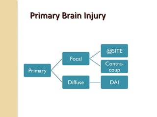
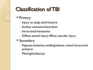
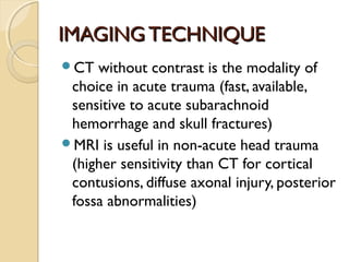
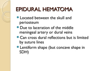
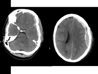
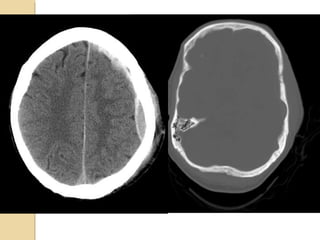
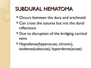
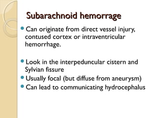
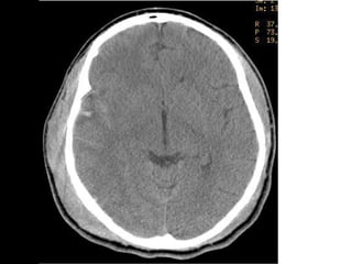
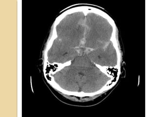
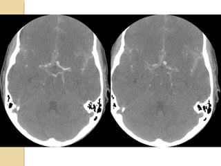
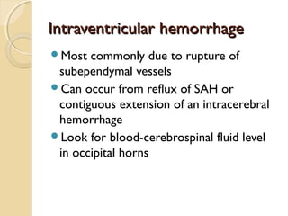
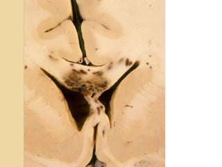
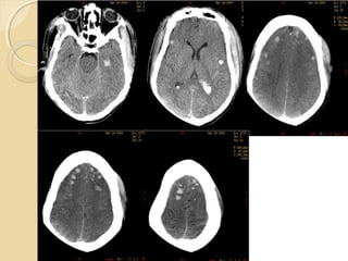
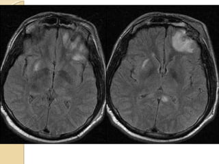
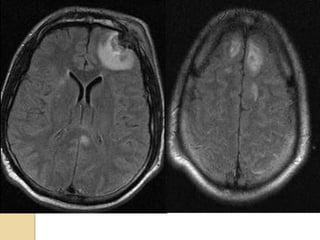
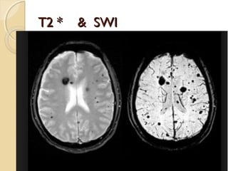
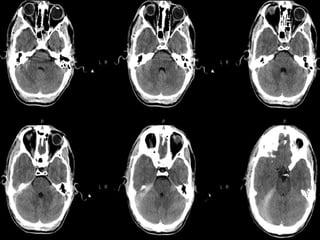
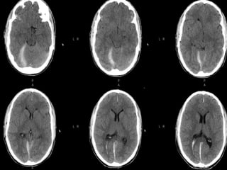
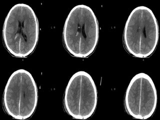
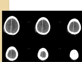
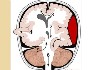
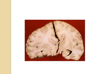
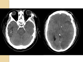
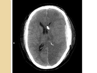
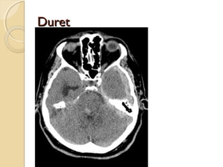
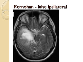
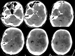
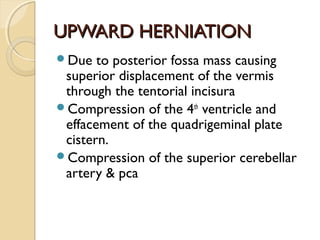
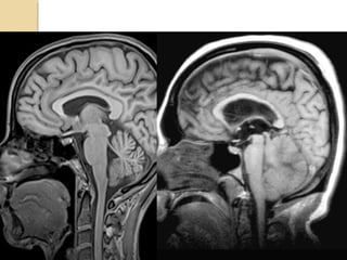
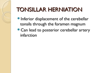
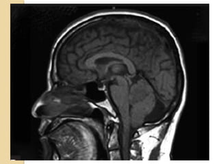
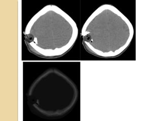
Brain imaging is important in trauma to identify injuries from primary impact and secondary complications. CT is best for acute trauma to detect fractures and hemorrhages while MRI is more sensitive for diffuse injuries. Common primary injuries seen include fractures, contusions, hematomas, shearing injuries and hemorrhages in various locations. Secondary complications can include swelling, infection and herniations putting pressure on vessels.
5. IMAGINGTECHNIQUEIMAGINGTECHNIQUE
CT without contrast is the modality of
choice in acute trauma (fast, available,
sensitive to acute subarachnoid
hemorrhage and skull fractures)
MRI is useful in non-acute head trauma
(higher sensitivity than CT for cortical
contusions, diffuse axonal injury, posterior
fossa abnormalities)
8. EPIDURAL HEMATOMAEPIDURAL HEMATOMA
Located between the skull and
periosteum
Due to laceration of the middle
meningeal artery or dural veins
Can cross dural reflections but is limited
by suture lines
Lentiform shape (but concave shape in
SDH)
9.
10.
11. SUBDURAL HEMATOMASUBDURAL HEMATOMA
Occurs between the dura and arachnoid
Can cross the sutures but not the dural
reflections
Due to disruption of the bridging cortical
veins
Hypodense(hyperacute, chronic),
isodense(subacute), hyperdense(acute)
15. Subarachnoid hemorrageSubarachnoid hemorrage
Can originate from direct vessel injury,
contused cortex or intraventricular
hemorrhage.
Look in the interpeduncular cistern and
Sylvian fissure
Usually focal (but diffuse from aneurysm)
Can lead to communicating hydrocephalus
16.
17.
18.
19. Intraventricular hemorrhageIntraventricular hemorrhage
Most commonly due to rupture of
subependymal vessels
Can occur from reflux of SAH or
contiguous extension of an intracerebral
hemorrhage
Look for blood-cerebrospinal fluid level
in occipital horns
22. CONTUSION/LACERATIONSCONTUSION/LACERATIONS
Most common source of traumatic SAH
Contusion: must involve the superficial gray
matter
Laceration: contusion + tear of pia-arachnoid
Affects the crests of gyri
Hemorrhage present ½ cases and occur at right
angles to the cortical surface
Located near the irregular bony contours: poles
of frontal lobes, temporal lobes, inferior
cerebellar hemispheres
23.
24. Intraparenchymal hematomaIntraparenchymal hematoma
Focal collections of blood that most
commonly arise from shear-strain injury
to intraparenchymal vessels.
Usually located in the frontotemporal
white matter or basal ganglia
Hematoma within normal brain
DDx: DAI, hemorrhagic contusion
25.
26. DIFFUSE AXONAL INJURYDIFFUSE AXONAL INJURY
Rarely detected on CT ( 20% of DAI
lesions are hemorrhagic)
MRI:T1,T2,T2 GRE, SWI
27. DAIDAI
Due to acceleration/deceleration to
whtie matter + hypoxia
Patients have severe LOC at impact
Grade 1: axonal damage in WM only -67%
Grade 2:WM + corpus callosum
(posterior > anterior) – 21%
Grade 3:WM + CC + brainstem
28. DAIDAI
Hours:
◦ hemorrhages and tissue tears
◦ Axonal swellings
◦ Axonal bulbs
Days/weeks: clusters of microglia and
macrophages, astrocytosis
Months/years:Wallerian degeneration
34. BRAINSTEM INJURYBRAINSTEM INJURY
By direct or indirect forces
Most commonly associated with DAI
Involves the dorsolateral midbrain and upper
pons and is usually hemorrhagic
Duret hemorrhage is an example of indirect
damage: tearing of the pontine perforators
leading to hemorrhage in the setting
transtentorial herniation
<20% of brainstem lesions are seen on CT
44. UNCAL HERNIATIONUNCAL HERNIATION
Displacement of the medial temporal lobe
through the tentorial notch
Displacement of the midbrain
Effacement of the suprasellar cistern
Displacement of the contralateral cerebral
peduncle against the tentorium
Widening of the ipsilateral cerebello pontine
angle
Compression of the posterior cerebral artery
49. UPWARD HERNIATIONUPWARD HERNIATION
Due to posterior fossa mass causing
superior displacement of the vermis
through the tentorial incisura
Compression of the 4th
ventricle and
effacement of the quadrigeminal plate
cistern.
Compression of the superior cerebellar
artery & pca
53. Skull FracturesSkull Fractures
Thin skull #’s common place.
Risk of # associated intracranial injuries?
CT to R/o
1. Open
2. Closed
3. Comminuted
4. Diastatic
5. Depressed
54. SIGNIFICANT SKULL FRACTURESSIGNIFICANT SKULL FRACTURES
“Depressed”: inner table is depressed by
the thickness of the skull.
Overlie major venous sinus, motor
cortex, middle meningeal artery
Pass through sinuses
Look for sutural diastasis (lambdoid)
55.
56.
57. TEMPORAL BONE FRACTURESTEMPORAL BONE FRACTURES
Look for opacification of the mastoid
Longitudinal: 70%, parallel to long axis of
petrous bone, conductive hearing loss
(from ossicular dislocation), facial nerve
paralysis (20%)
Transverse: 20%, sensorineural hearing loss,
facial nerve paralysis (50%)
Complex
Complications: meningitis, abscess
60. SCALP INJURYSCALP INJURY
Cephalohematoma: blood between the bone
and periosteum. Cannot cross the suture lines.
Subgaleal hematoma: blood between the
periosteum and aponeurosis. Can cross the
suture lines.
Caput Succ: swelling across the midline with
scalp moulding. Resolves spontaneously.