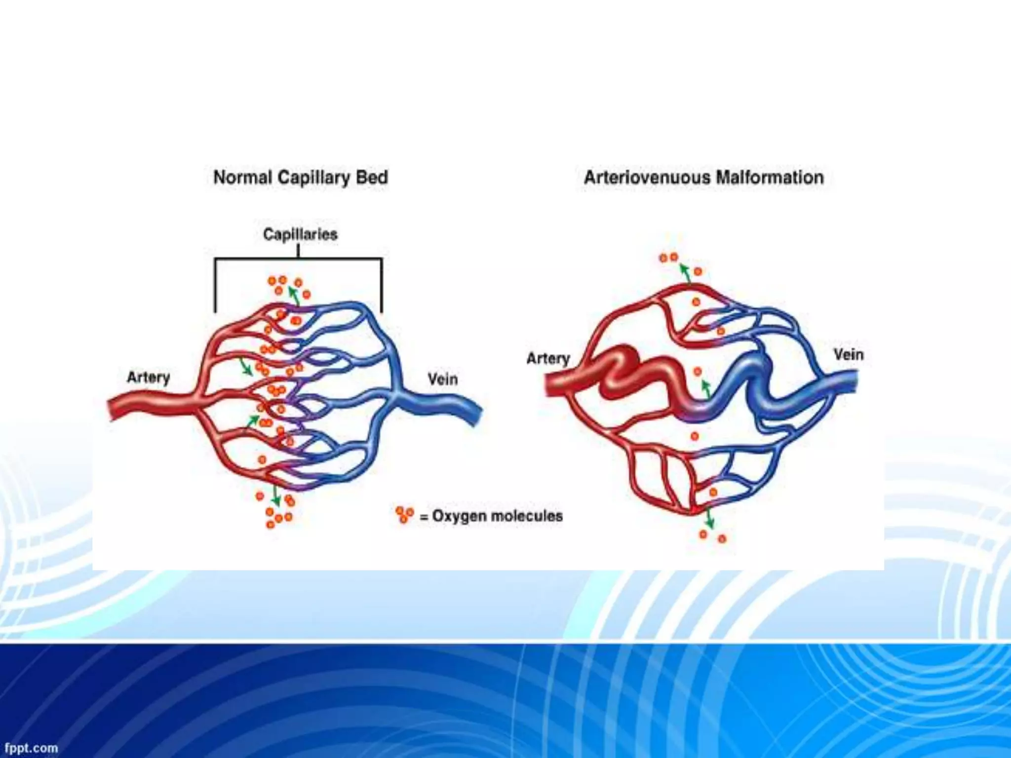
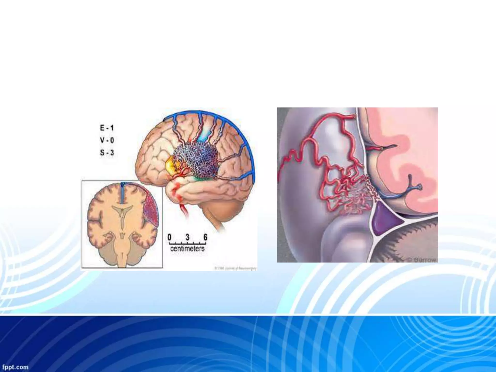
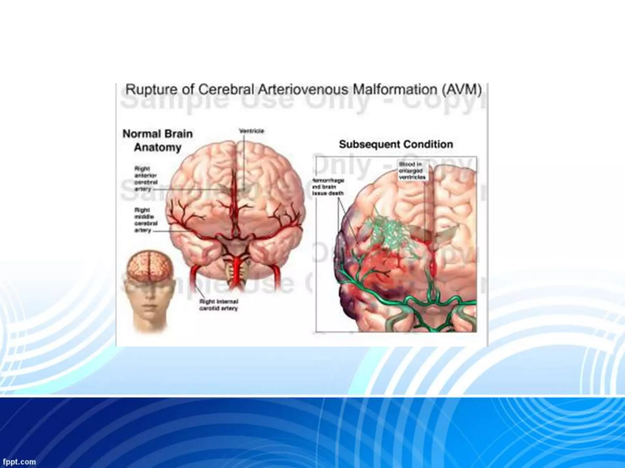
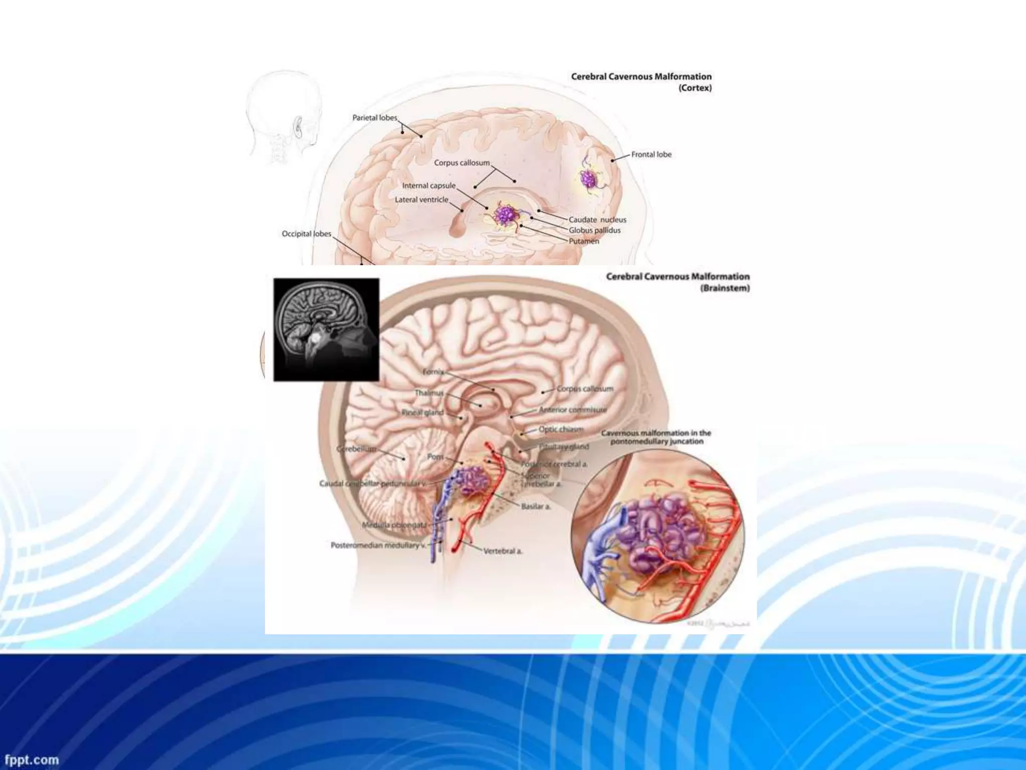
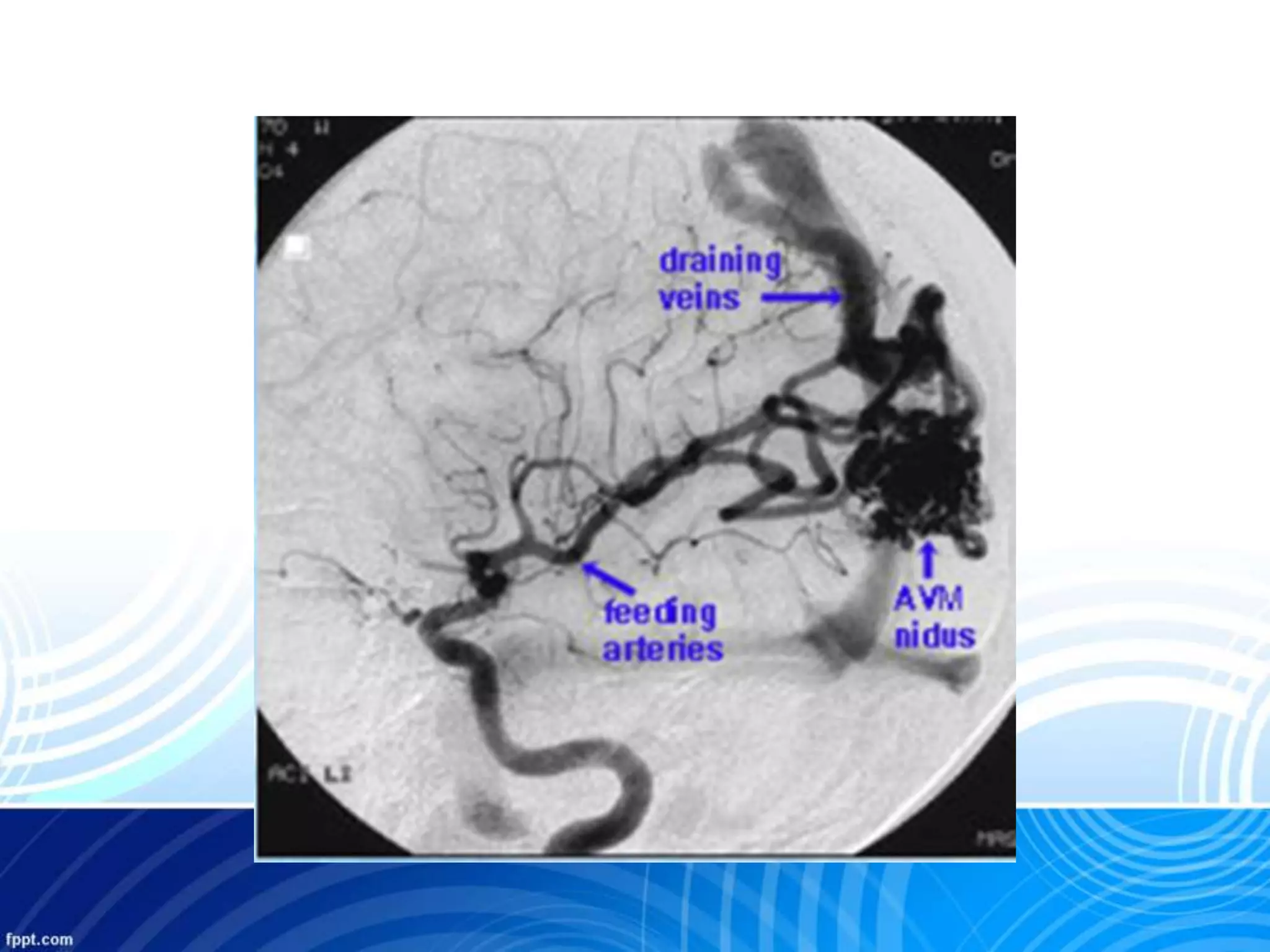
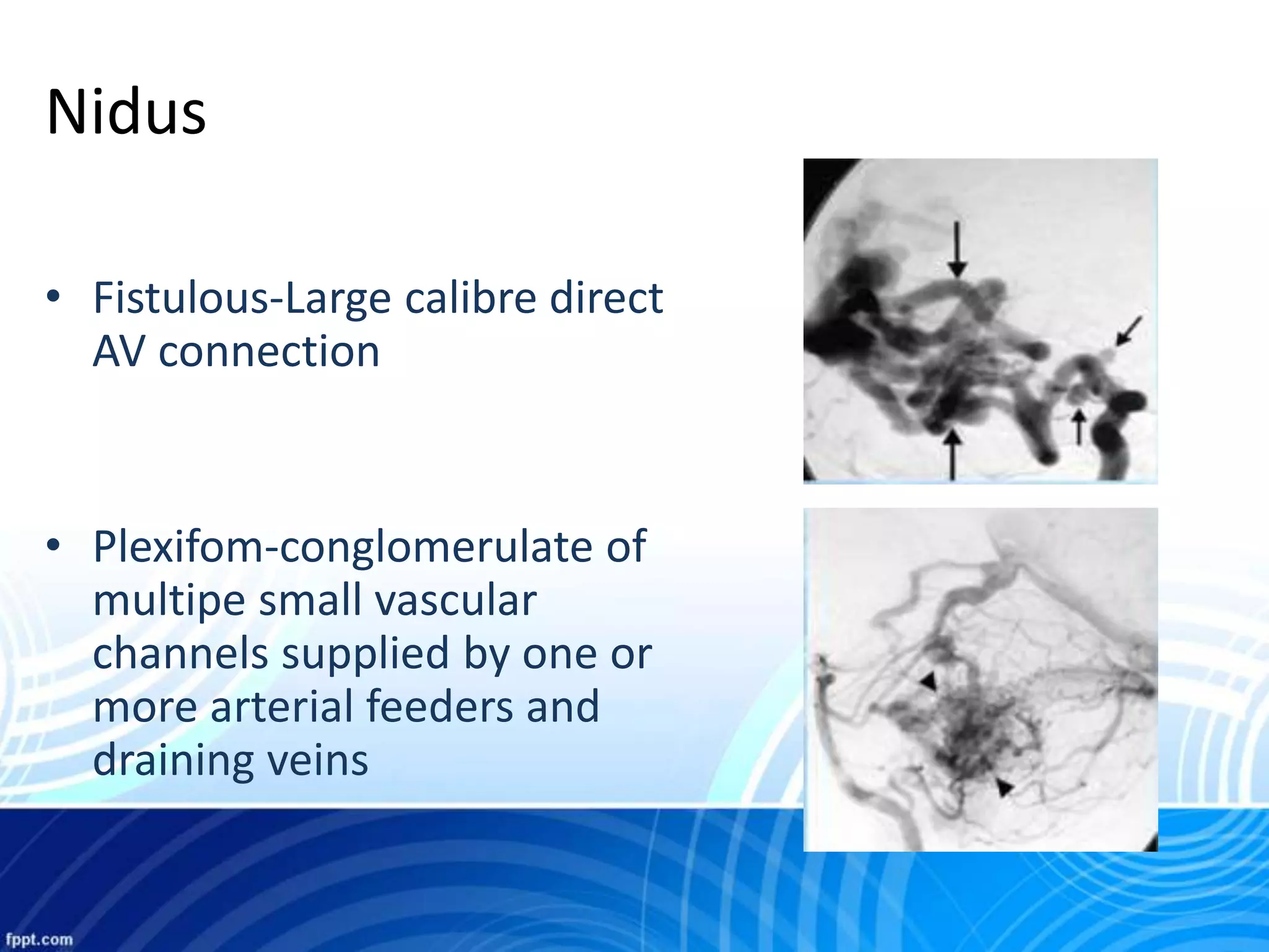
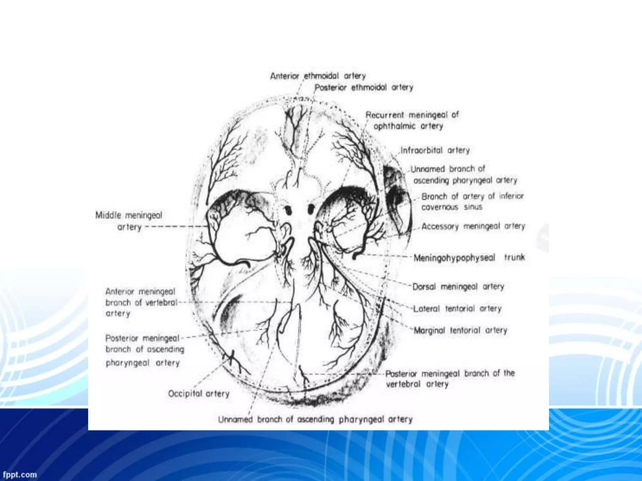
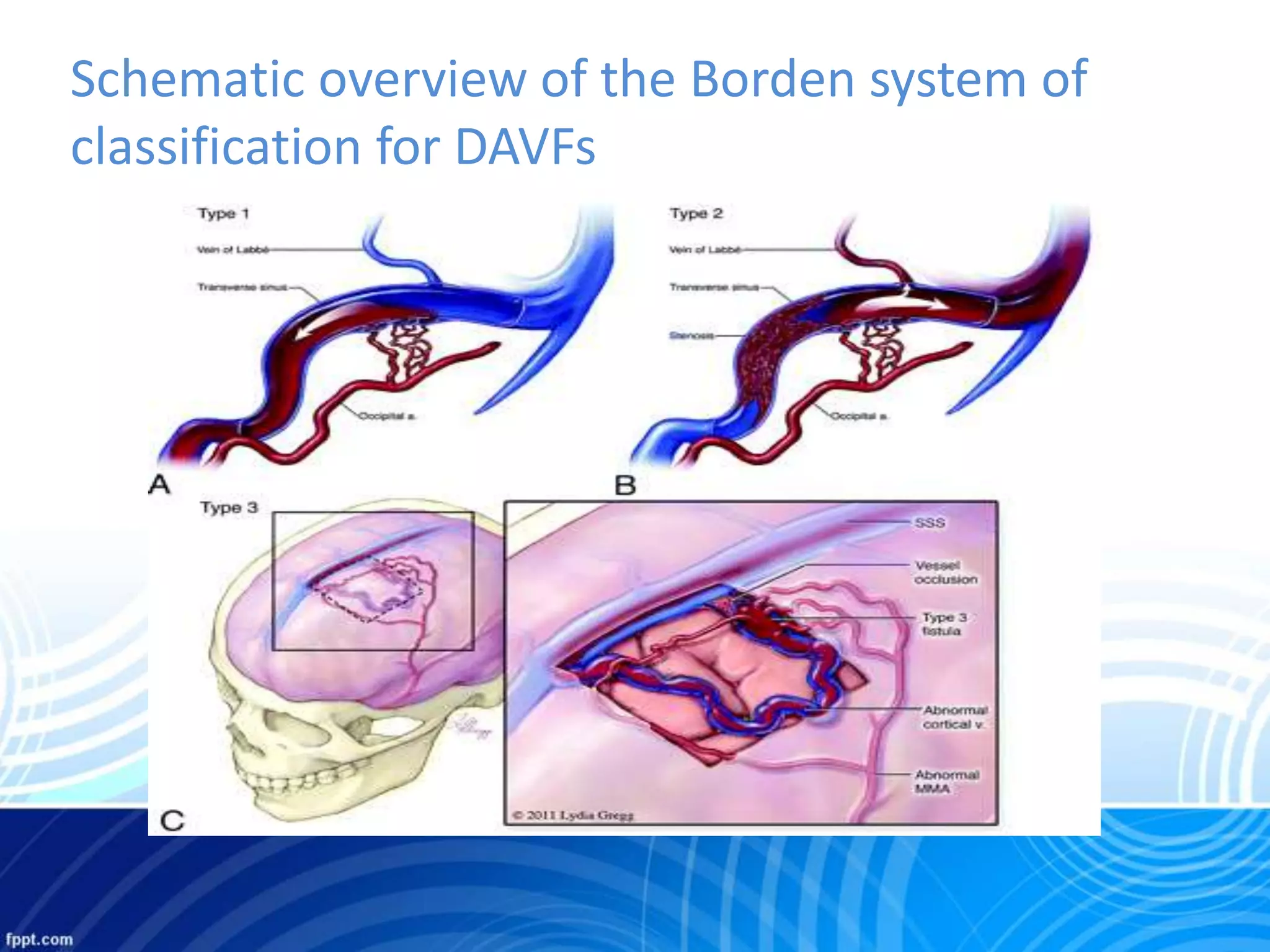
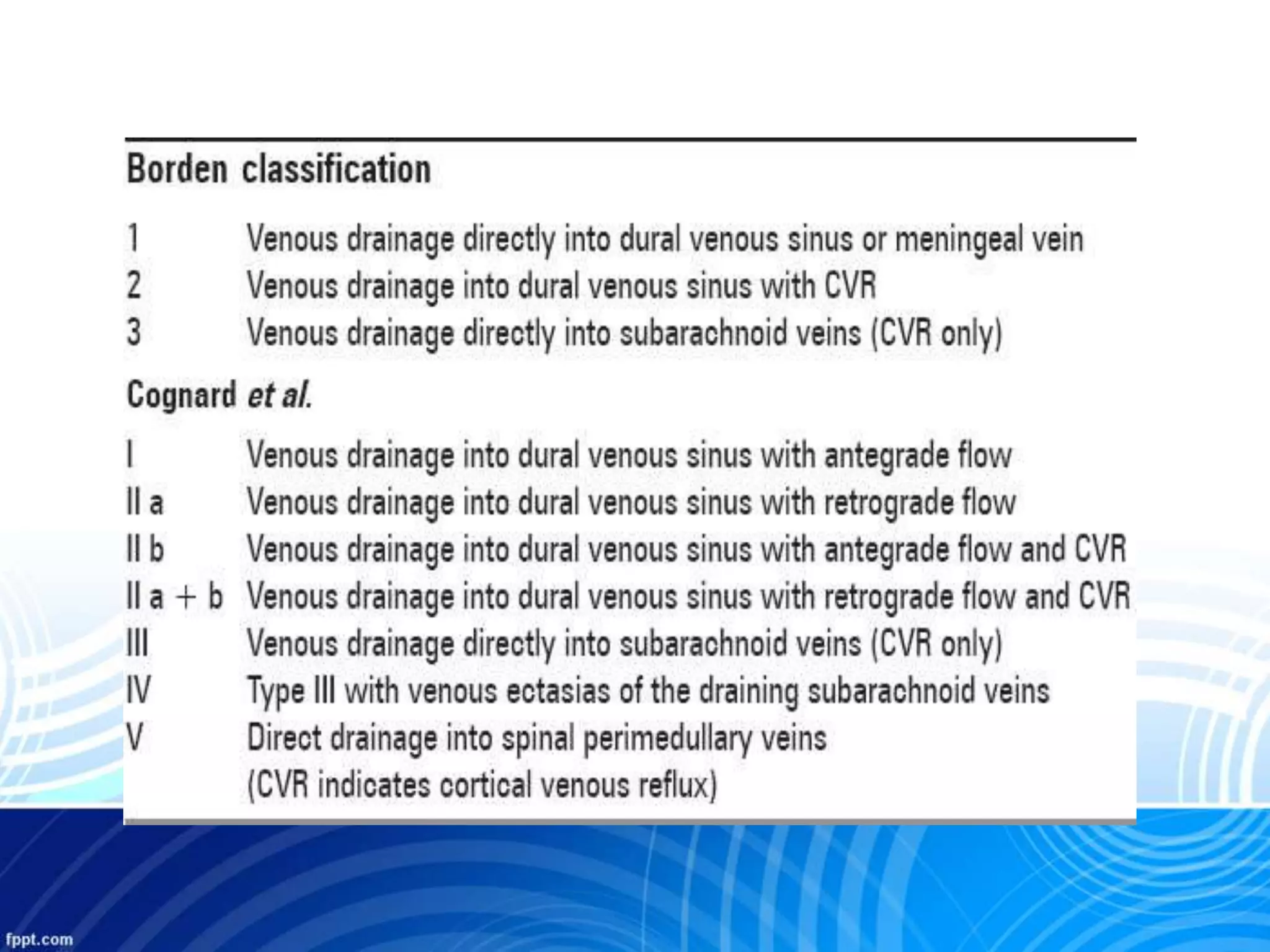
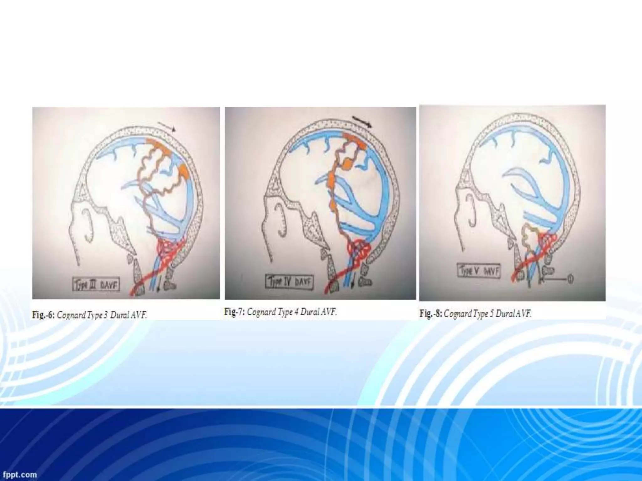
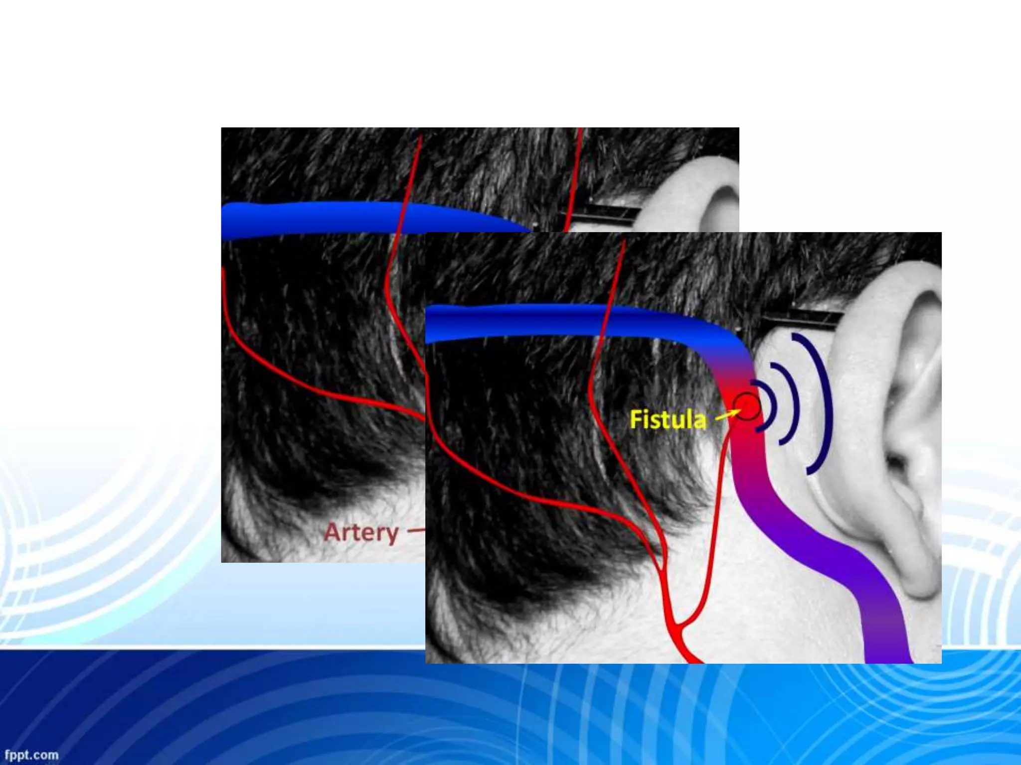
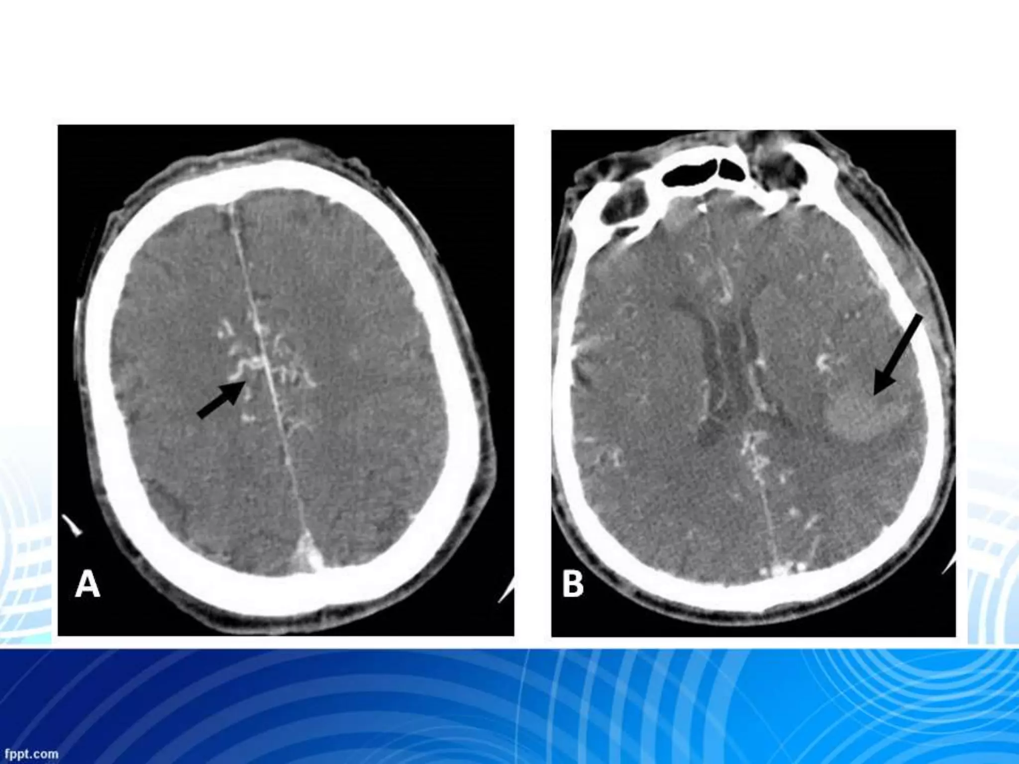
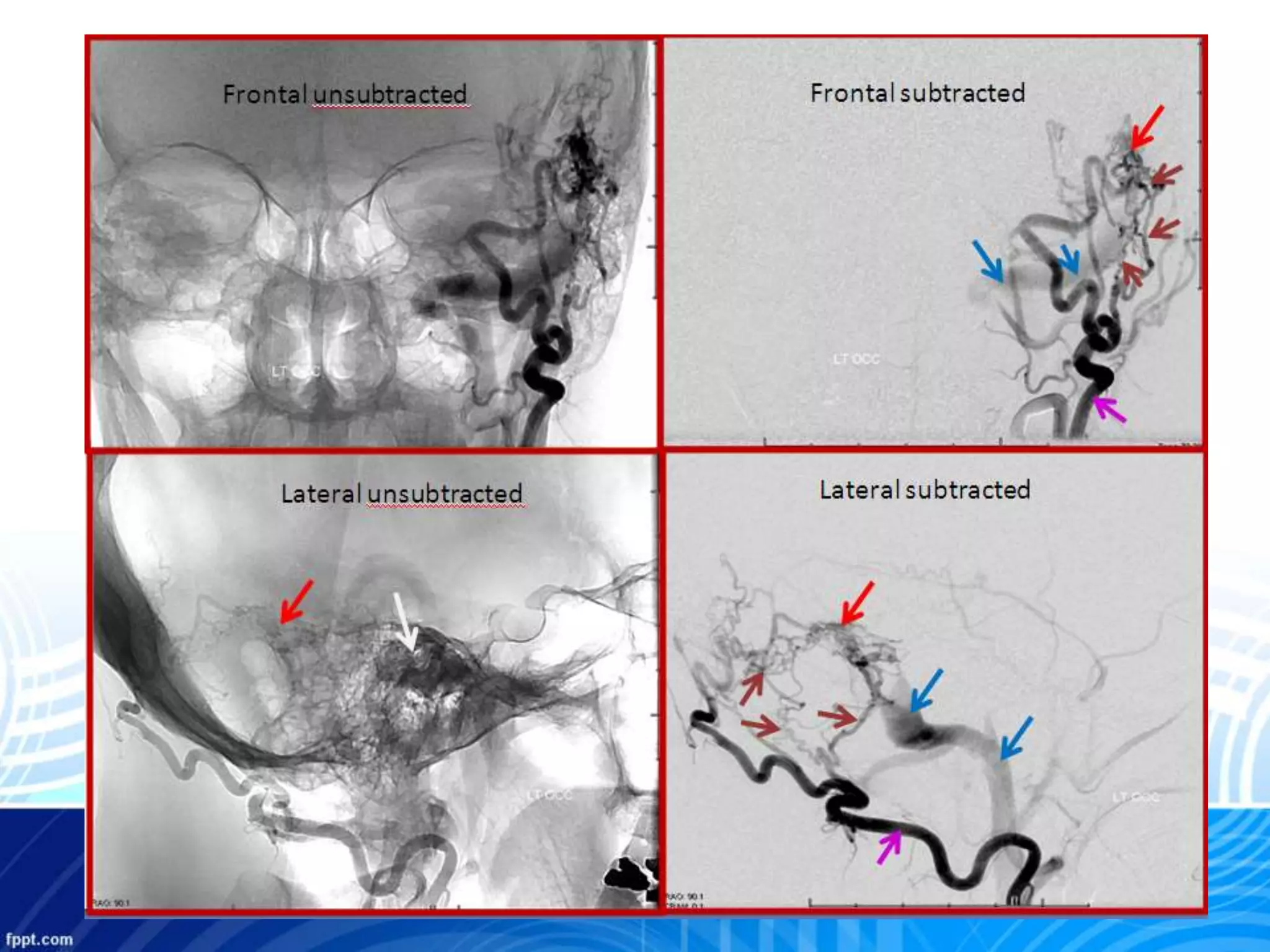
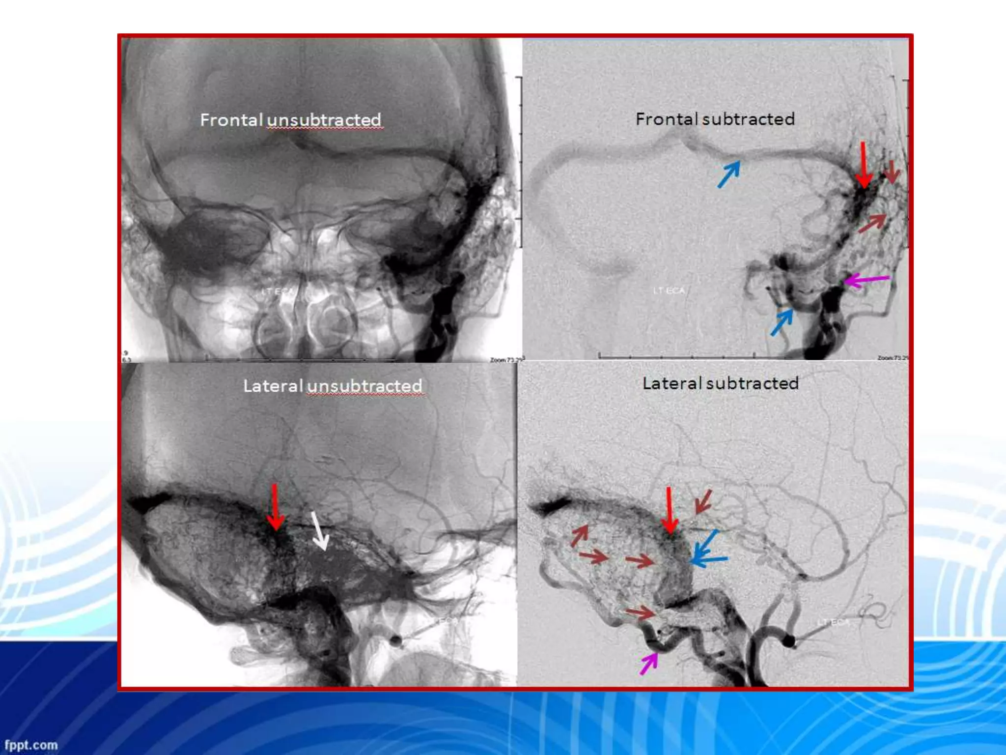
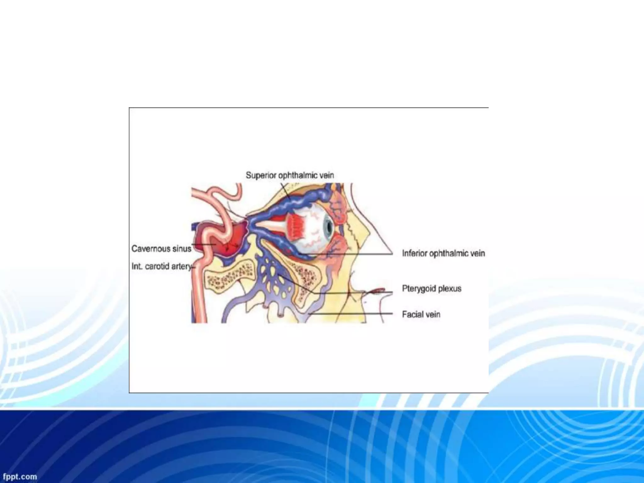
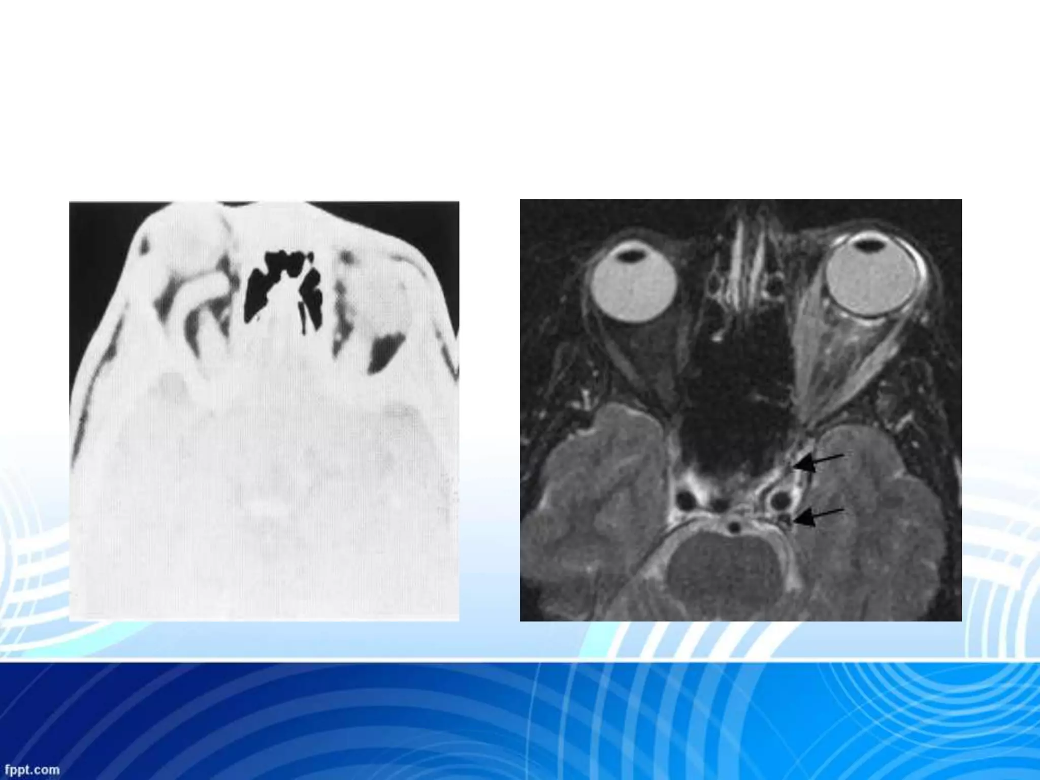
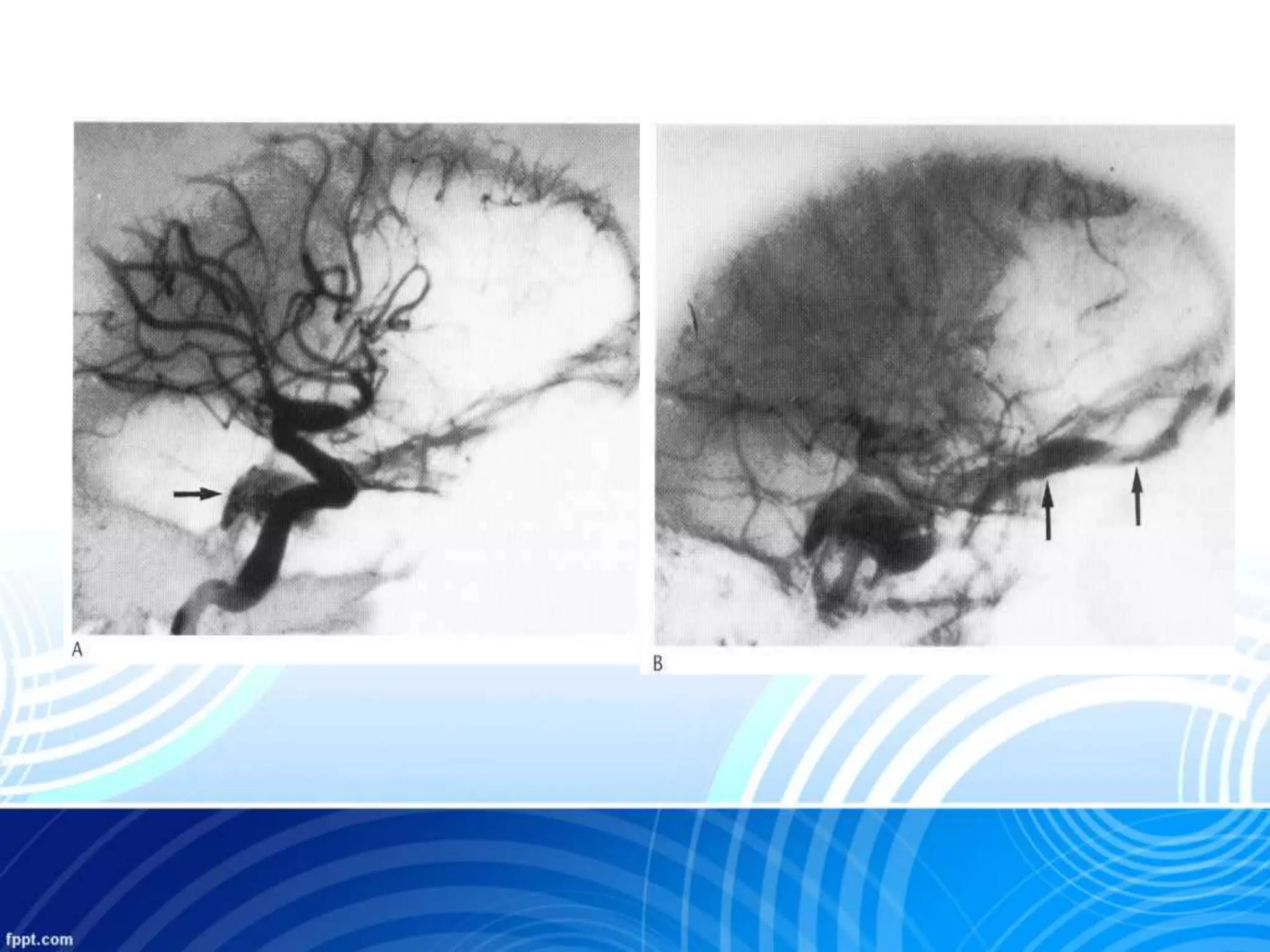
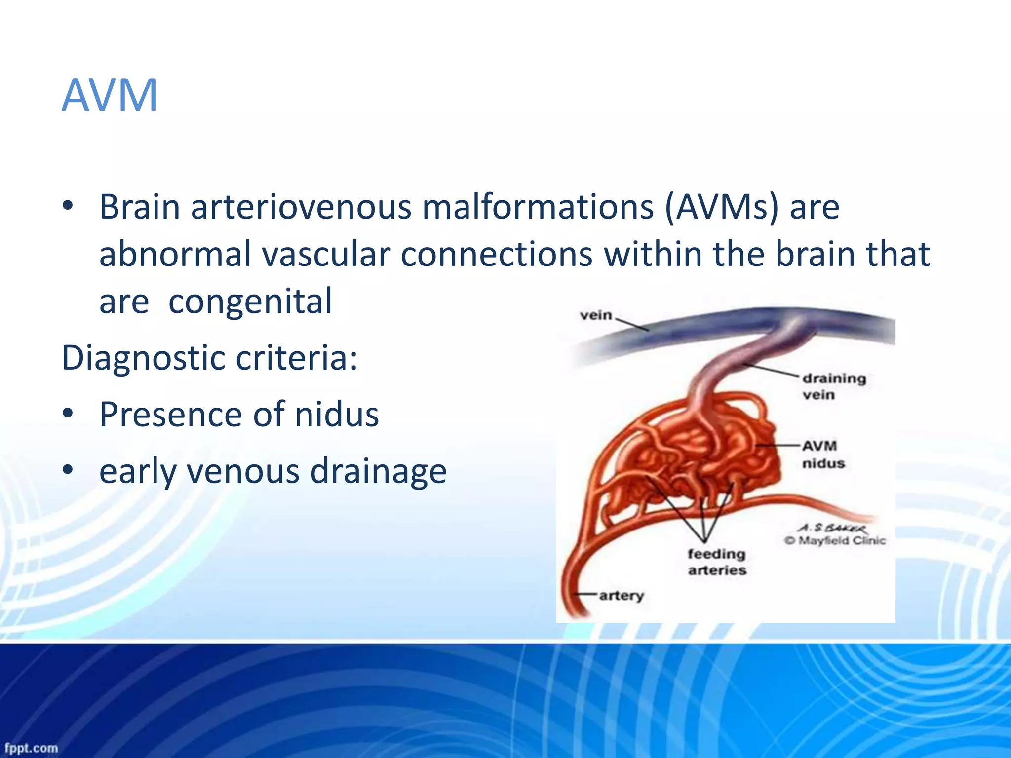
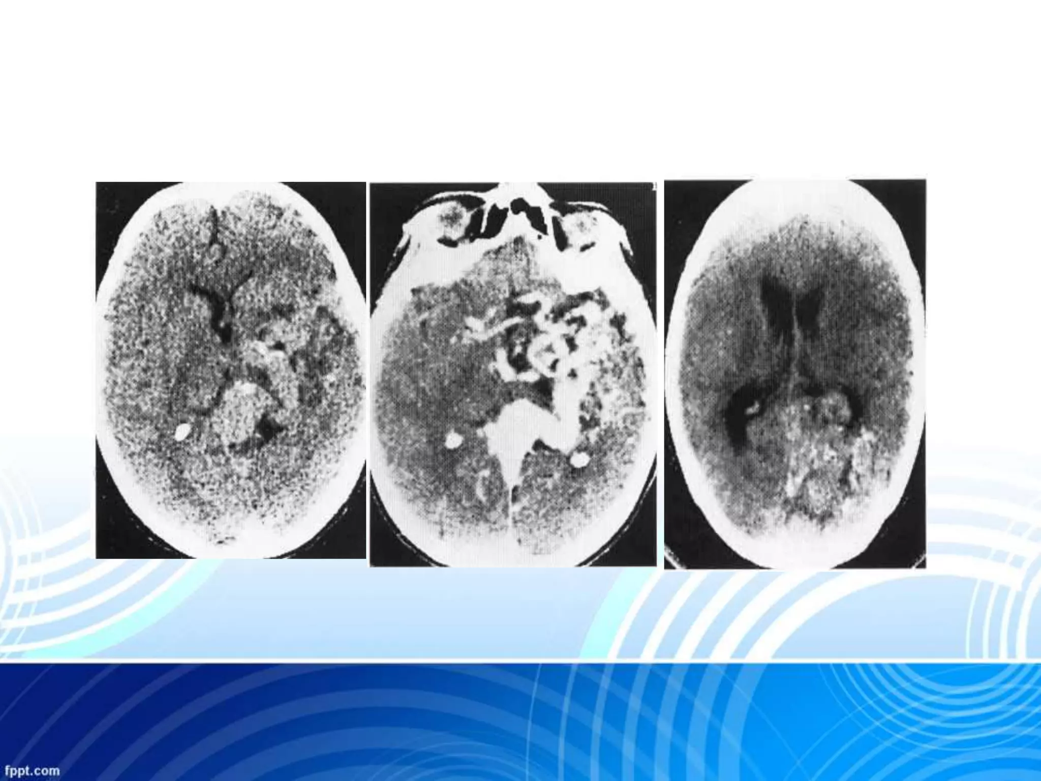
Dural arteriovenous fistula
& AV malformation
Dr.Aftab Qadir
Vascular malformations involving the brain are divided
into
• arteriovenous malformations (AVM)
• developmental venous anomalies (DVA)
• cavernous malformations
• capillary telangiectasia.
Angiography
• Selective : evaluation of AVM and entire cerebral
circulation
• Super selective : Microcatheters advanced into distal
aspect of arterial feeders to study anatomical
structure
Angiography
• Demonstrating the nidus
• Multiple arterial feeders
• Thrombosed dural sinus
• Flow reversal in sinus and cortical vein
• Arterial territories supplying the AVM
• Feeding pedicles
• High flow arteriopathy(stenose,ectasia,aneyrysm)
• Venous drainage(territories,deep,superficial)
• Individual draining veins
• High flow venous angiopathy (dural sinuses,venous
stenoses,occlusions )
• Venous drainage of normal brain parenchyma
Goals selective angiography
Nidus
• Fistulous-Large calibre direct
AV connection
• Plexifom-conglomerulate of
multipe small vascular
channels supplied by one or
more arterial feeders and
draining veins
Dural arteriovenous fistula
• Dural arteriovenous fistulas (AVF) are pathologic
abnormal vascular connections between dural
arteries with the venous sinuses or cortical veins
Schematic overview of the Borden system of
classification for DAVFs
Location
• transverse / sigmoid sinus most common
• cavernous sinus (indirect caroticocavernous fistula)
• superior sagittal sinus
• straight sinus
• other venous sinuses
• anterior cranial fossa
• tentorium
Clinical presentation
• Pulsatile tinnitus
• cranial nerve palsies
• orbital symptoms
• raised intracranial pressure
• focal neurological deficits
Complications
• haemorrhage
• subdural haemorrhage (SDH)
• intracranial haemorrhage (ICH)
• subarachnoid haemorrhage (SAH)
• intracranial hypertension
• venous congestion and oedema
Imaging
a) CT scan
b) MRI
c) Cerebral angiogram
CT
• NECT: usually normal
• CECT: Normal /small tortous dural feeders
Enlarged dural sinuses
Enlarged superior opthalmic vein in CCF
Enlarged cortical draining veins
Curvilinear subcortical calcifications
Curvilinear subcortical calcifications can be seen at CT
in patients with long-standing cortical venous reflux,
possibly due to chronic venous congestion
MRI
• TIWI and T2WI- Flow voids and thrombosed sinus
• T2 & Flair-Hyperintensities due to venous
congestion/ischemia
• T1C+ diffuse dural enhancement
• MRV- Occluded sinus and collateral flow
• 3D PC MRA with low velocity encoding- identify
feeding arteries,fistula and flow reversal in draining
veins
Angiography
• Subtraction selective angiography of internal and
external cartotid arteries or both
-dAVF second most common site
-Abnormal communication between carotid artery &
cavernous sinus
-Shunt between the meningeal branches of internal or
external carotid and dural veins near the cavernous
sinus
Carotid Cavernous Fistula (CCF)
CT & MRI
CT:
• – Marked dilation & enhancement of cavernous sinus
• – May see prominent SOV
MRI:
• – Abnormal flow voids in cavernous sinus
• – Enlargement of cavernous sinus
AVM
• Brain arteriovenous malformations (AVMs) are
abnormal vascular connections within the brain that
are congenital
Diagnostic criteria:
• Presence of nidus
• early venous drainage
High and Low flow malformations
High flow
• arteriovenous malformation (AVM)
• parenchymal AVM
• dural arteriovenous fistula (DAVF)
• mixed AVM
Low flow
• capillary telangiectasia
• cavernous haemangioma
• venous malformations
• developmental venous anomaly (DVA) (venous angiomas)
• vein of Galen malformation (can be high flow as well)
CT
• NECT:Iso/Hyperdense serpentine vessels
• calcificationin 25-30%
• Variable hemorrahage
• CECT: Strong enhancement
MRI
• Variable
• Punctate appearance on T2 due to single void
• Large arteries and veins are characteristic
• Intracerebral hematoma with high signal on T1
• Surrounding halo –hyposignal on T2 indicating
hemosidrren
• Post contrast not facilitate detactability like CECT
VEIN OF GALEN MALFORMATION
• Arteriovenous fistula involving a neurysmal dilatation
of median prosencephalic vein (MPV)
• Most common extracardiac cause of high-output
congestive heart failure in newborns
• < 1% of cerebral vascular malformations
Venous angiomas
• aberrant venous drainage of one or both cerebral
hemispheres.
• They are shown on MRI as a slightly curvilinear
structure traversing cerebral substance, representing
a single draining vein into which numerous cerebral
veins converge.
• They are often incidental findings, but may be
associated with (parenchyma) haemorrhage or focal
brain damage
Telangiectasia
• Shown on MRI as small lesion
• Hypointense on T1
• T1 C+ (GAD) : may demonstrate ill defined focal
enhancement
• Hyperintense on T2
cavernous hemangioma
• Slow flow lesions
• Accounts for 10-15%
• No intervening brain
• No mass effect on imaging
• CT usually normal if small
lesion or in absence of
calciication and hemorrgage
• Characteristic feature is
hypointense rim because of
hemosiderin
Few case
15-year-old boy who presented
with sudden onset of headaches followed by seizures
Thank You

Dural arteriovenous fistula & AVM

  • 1.
    Dural arteriovenous fistula &AV malformation Dr.Aftab Qadir
  • 2.
    Vascular malformations involvingthe brain are divided into • arteriovenous malformations (AVM) • developmental venous anomalies (DVA) • cavernous malformations • capillary telangiectasia.
  • 8.
    Angiography • Selective :evaluation of AVM and entire cerebral circulation • Super selective : Microcatheters advanced into distal aspect of arterial feeders to study anatomical structure
  • 9.
    Angiography • Demonstrating thenidus • Multiple arterial feeders • Thrombosed dural sinus • Flow reversal in sinus and cortical vein
  • 12.
    • Arterial territoriessupplying the AVM • Feeding pedicles • High flow arteriopathy(stenose,ectasia,aneyrysm) • Venous drainage(territories,deep,superficial) • Individual draining veins • High flow venous angiopathy (dural sinuses,venous stenoses,occlusions ) • Venous drainage of normal brain parenchyma Goals selective angiography
  • 13.
    Nidus • Fistulous-Large calibredirect AV connection • Plexifom-conglomerulate of multipe small vascular channels supplied by one or more arterial feeders and draining veins
  • 14.
    Dural arteriovenous fistula •Dural arteriovenous fistulas (AVF) are pathologic abnormal vascular connections between dural arteries with the venous sinuses or cortical veins
  • 16.
    Schematic overview ofthe Borden system of classification for DAVFs
  • 22.
    Location • transverse /sigmoid sinus most common • cavernous sinus (indirect caroticocavernous fistula) • superior sagittal sinus • straight sinus • other venous sinuses • anterior cranial fossa • tentorium
  • 23.
    Clinical presentation • Pulsatiletinnitus • cranial nerve palsies • orbital symptoms • raised intracranial pressure • focal neurological deficits
  • 24.
    Complications • haemorrhage • subduralhaemorrhage (SDH) • intracranial haemorrhage (ICH) • subarachnoid haemorrhage (SAH) • intracranial hypertension • venous congestion and oedema
  • 26.
    Imaging a) CT scan b)MRI c) Cerebral angiogram
  • 27.
    CT • NECT: usuallynormal • CECT: Normal /small tortous dural feeders Enlarged dural sinuses Enlarged superior opthalmic vein in CCF Enlarged cortical draining veins Curvilinear subcortical calcifications
  • 30.
    Curvilinear subcortical calcificationscan be seen at CT in patients with long-standing cortical venous reflux, possibly due to chronic venous congestion
  • 31.
    MRI • TIWI andT2WI- Flow voids and thrombosed sinus • T2 & Flair-Hyperintensities due to venous congestion/ischemia • T1C+ diffuse dural enhancement • MRV- Occluded sinus and collateral flow • 3D PC MRA with low velocity encoding- identify feeding arteries,fistula and flow reversal in draining veins
  • 35.
    Angiography • Subtraction selectiveangiography of internal and external cartotid arteries or both
  • 39.
    -dAVF second mostcommon site -Abnormal communication between carotid artery & cavernous sinus -Shunt between the meningeal branches of internal or external carotid and dural veins near the cavernous sinus Carotid Cavernous Fistula (CCF)
  • 41.
    CT & MRI CT: •– Marked dilation & enhancement of cavernous sinus • – May see prominent SOV MRI: • – Abnormal flow voids in cavernous sinus • – Enlargement of cavernous sinus
  • 44.
    AVM • Brain arteriovenousmalformations (AVMs) are abnormal vascular connections within the brain that are congenital Diagnostic criteria: • Presence of nidus • early venous drainage
  • 45.
    High and Lowflow malformations High flow • arteriovenous malformation (AVM) • parenchymal AVM • dural arteriovenous fistula (DAVF) • mixed AVM Low flow • capillary telangiectasia • cavernous haemangioma • venous malformations • developmental venous anomaly (DVA) (venous angiomas) • vein of Galen malformation (can be high flow as well)
  • 47.
    CT • NECT:Iso/Hyperdense serpentinevessels • calcificationin 25-30% • Variable hemorrahage • CECT: Strong enhancement
  • 49.
    MRI • Variable • Punctateappearance on T2 due to single void • Large arteries and veins are characteristic • Intracerebral hematoma with high signal on T1 • Surrounding halo –hyposignal on T2 indicating hemosidrren • Post contrast not facilitate detactability like CECT
  • 52.
    VEIN OF GALENMALFORMATION • Arteriovenous fistula involving a neurysmal dilatation of median prosencephalic vein (MPV) • Most common extracardiac cause of high-output congestive heart failure in newborns • < 1% of cerebral vascular malformations
  • 57.
    Venous angiomas • aberrantvenous drainage of one or both cerebral hemispheres. • They are shown on MRI as a slightly curvilinear structure traversing cerebral substance, representing a single draining vein into which numerous cerebral veins converge. • They are often incidental findings, but may be associated with (parenchyma) haemorrhage or focal brain damage
  • 61.
    Telangiectasia • Shown onMRI as small lesion • Hypointense on T1 • T1 C+ (GAD) : may demonstrate ill defined focal enhancement • Hyperintense on T2
  • 63.
    cavernous hemangioma • Slowflow lesions • Accounts for 10-15% • No intervening brain • No mass effect on imaging • CT usually normal if small lesion or in absence of calciication and hemorrgage • Characteristic feature is hypointense rim because of hemosiderin
  • 64.
  • 70.
    15-year-old boy whopresented with sudden onset of headaches followed by seizures
  • 72.

Editor's Notes

  • #71 Spetzler-Martin grade 1 temporal brain AVM in a 15-year-old boy who presented with sudden onset of headaches followed by seizures. Axial CT scan reveals A small hyperattenuating lesion in the right temporal lobe, compatible with a small intraparenchymal hematoma.(b) Lateral right internal carotid angiogram Demonstrates a small(<3-cm) brain AVM supplied mainly by the temporal branches of the right MCA, with superficial drainage into the right vein of Labbe.