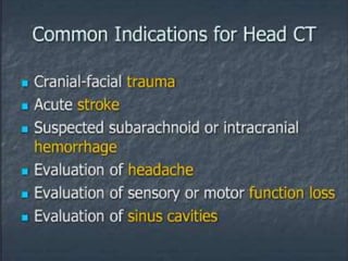
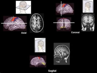
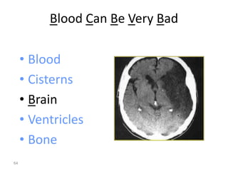
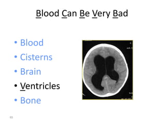
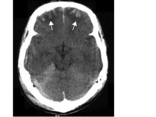
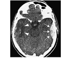
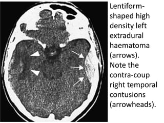
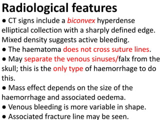
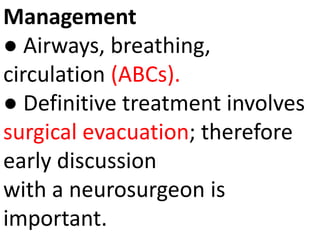
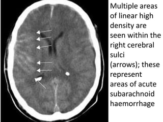
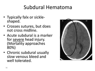
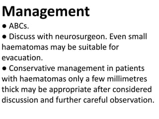
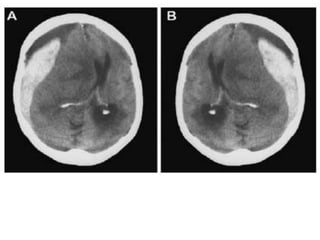
How to Read a Head CT
Dr: Muhammad Faisal. FCPS1(R4)
1
Sphenoid
sinus
Medulla
oblongata
cerebellum
4 Fourth ventricle
5 Middle cerebellar
peduncle
6 Sigmoid sinus
7 Petrous temporal
bone and mastoid
air cells
8 Cerebellopontine
angle
9 Pons
10 Pituitary fossa
11 Cerebellar
vermis
12 Basilar artery
13 Prepontine
cistern
14 Dorsum sellae
15 Temporal horn
of lateral
ventricle
16 Ambient
cistern
17
Interpeduncular
cistern
18 Cerebral
peduncle
19 Sylvian
fissure
20 Supra
vermian
cistern
21 Frontal
horn of
lateral
ventricle
21a Third
ventricle
22 Head of
caudate
nucleus
23 Insular
cortex
24 External
capsule
25 Lentiform
nucleus
26 Thalamus
27 Interhemispheric
fissure
28 Anterior limb of
internal capsule
29 Genu of internal
capsule
30 Posterior limb of
internal capsule
31 Trigone of lateral
ventricle and calcified
choroid plexus
32 Occipital horn of
lateral ventricle
CSF Production
• Produced in choroid plexus in the lateral
ventricles  Foramen of Monroe  IIIrd
Ventricle  Acqueduct of Sylvius  IVth
Ventricle  Lushka/Magendie
• 0.5-1 cc/min
• Adult CSF volume is approx. 150 cc’s.
• Adult CSF production is approx. 500-700
cc’s per day.
51
53
2nd Key Level Sagittal View
2nd Key Level
Circummesencephalic
Cistern
Cisterns at Cerebral Peduncles Level
54
Right and left sylvian fissures (black arrows)
meeting at the suprasellar cistern.
A = Left
frontal horn
B = Left
sylvian fissure
C = Third
ventricle
D = Ambient
cistern
Head CT
“Blood Can Be Very Bad”
61
Blood Can Be Very Bad
• Blood
• Cisterns
• Brain
• Ventricles
• Bone
62
Blood Can Be Very Bad
• Blood
• Cisterns
• Brain
• Ventricles
• Bone
63
Blood Can Be Very Bad
• Blood
• Cisterns
• Brain
• Ventricles
• Bone
64
Blood Can Be Very Bad
• Blood
• Cisterns
• Brain
• Ventricles
• Bone
65
Blood Can Be Very Bad
• Blood
• Cisterns
• Brain
• Ventricles
• Bone
66
B is for Blood
• Blood becomes hypodense
at approximately 2 weeks.
67
•Blood becomes
isodense at
approximately 1 week.
• Acute blood is bright white
on CT (once it clots).
B is for Blood
• Blood becomes
hypodense at
approximately 2 weeks.
68
• Blood becomes
isodense at
approximately 1 week.
• Acute blood is bright white
on CT (once it clots).
B is for Blood
• Blood becomes
hypodense at
approximately 2 weeks.
69
• Blood becomes
isodense at
approximately 1 week.
• Acute blood is bright white
on CT (once it clots).
THE 5Ss OF ANY HAEMATOMA!
• Clots are defined by:
• Size
• Symptoms and signs
• Shifts
• Side
• site
Cerebral
contusions in
both frontal
lobes
(arrows). The
adjacent low
density
represents
local
oedema
Cerebral contusion
Characteristics
● Commonest form of traumatic intra-axial injury.
● Contusions occur at the inferior and polar surfaces
of the frontal and
temporal lobes.
● Injuries may be coup or contra-coup.
● Cerebral contusions are also produced secondary
to depressed skull fractures and are associated with
other intracranial injuries.
Clinical features
● Usually associated with a
brief loss of consciousness.
● Beware of the elderly
patients, alcoholics and patients
taking anticoagulants as they
are at increased risk of
haemorrhage.
Radiological features
● Non-contrast computed tomography (CT)
useful in the early posttraumatic period.
● Contusions are seen as multiple focal
areas of low or mixed attenuation
intermixed with tiny areas of increased
density representing petechial
haemorrhage.
● Magnetic resonance imaging (MRI) is the
best modality for demonstration of
oedema and contusion distribution.
Management
● Secure airway whilst the cervical spine is protected.
Supplemental oxygen.
Assess and stabilise breathing and circulation.
● If Glasgow coma scale (GCS) 8 discuss with
anaesthetist as a definitive (secured) airway is
required.
● Early discussion with radiologist and neurosurgeon.
● Titrate opioid analgesia.Cleanse and close scalp
injuries.Discuss with a neurosurgeon
regarding intravenous (i.v.) antibiotics, steroid and
mannitol use.
Lentiform-
shaped high
density left
extradural
haematoma
(arrows).
Note the
contra-coup
right temporal
contusions
(arrowheads).
Extradural haematoma
Characteristics EPIDURAL
● The majority of these are arterial (middle meningeal
artery) with a small proportion being of venous origin.
● Commonly unilateral and associated with a fracture in
adults. Skull fractures are often absent in children due to
skull elasticity.
● Haematoma forms between the inner table of skull and
the dura.
● May have associated injuries, such as a subdural
haematoma (SDH) or contusions.
● Arterial bleeding usually develops and presents rapidly
within 1 hour of injury whereas venous haematomas may
present after several days.
Clinical features
● Classically present following a head injury
with initial loss of consciousness
followed by a lucid interval, prior to a second
decrease in the level of
consciousness.
● Beware as only about 30% of patients present
in this way.
● The symptomatology depends on how quickly
the haematoma expands.
Progressive sleepiness, headache, nausea and
vomiting are suspicious
Radiological features
● CT signs include a biconvex hyperdense
elliptical collection with a sharply defined edge.
Mixed density suggests active bleeding.
● The haematoma does not cross suture lines.
● May separate the venous sinuses/falx from the
skull; this is the only type of haemorrhage to do
this.
● Mass effect depends on the size of the
haemorrhage and associated oedema.
● Venous bleeding is more variable in shape.
● Associated fracture line may be seen.
Management
● Airways, breathing,
circulation (ABCs).
● Definitive treatment involves
surgical evacuation; therefore
early discussion
with a neurosurgeon is
important.
85
Multiple areas
of linear high
density are
seen within the
right cerebral
sulci
(arrows); these
represent
areas of acute
subarachnoid
haemorrhage
Subarachnoid Hemorrhage
88
Subarachnoid haemorrhage
Characteristics
● Spontaneous subarachnoid haemorrhage
(SAH) usually occurs secondary to a ruptured
aneurysm or arteriovenous malformation.
● Acquired aneurysms are commonest in the
circle ofWillis; at bifurcations with turbulent
flow.
● Commonest before 50 years of age, but may
occur at any age.
● Free blood causes irritation of the meninges.
Clinical features
● Acute severe headache often described as the worst
ever, although a mild headache does not exclude a
SAH.
● Vomiting, pallor and profuse sweating may occur.
● Neck stiffness and focal neurological signs seizures.
● coma.
● Complications include hydrocephalus (acute
obstructive and delayed communicating), cerebral
vasospasm leading to infarction and transtentorial
herniation secondary to raised ICP.
● Mimics many other conditions including
encephalitis, meningitis, acute
glaucoma and migraine amongst others
Radiological features
● Non-contrast CT is sensitive within 4–5
hours of onset.
● Look for acute haemorrhage (increased
density) in the cortical sulci, basal
cisterns, Sylvian fissures, superior
cerebellar cisterns and in the ventricles.
● MRI is relatively insensitive within the
first 48 hours but is useful after this
time and in recurrent bleeds to pick up
subtle haemosiderin deposition
Complications: The H.I.G.H
of SAH
HIGH stands for
Hydrocephalus,
Infarction,
Giant aneurysms
and Haematoma.
Management
● ABCs. Beware as patients may rapidly progress to
coma and require intubation.
● Supplemental oxygen, i.v. access and cardiac
monitoring are essential.
● Consider other causes for a decreased level of
consciousness.
● Check routine bloods and refer suspicious patients
for further investigation,
e.g. lumbar puncture or CT scan.
● A normal CT does not exclude a SAH.This group of
patients require a lumbar puncture.
(a) Acute SDH;
(b) Subacute SDH
(c) Chronic SDH
Subdural Hematoma
• Typically falx or sickle-
shaped.
• Crosses sutures, but does
not cross midline.
• Acute subdural is a marker
for severe head injury.
(Mortality approaches
80%)
• Chronic subdural usually
slow venous bleed and
well tolerated.
96
Subdural haematoma
Characteristics
● SDH commonly occurs in the elderly and in children
.
● Occur in the subdural space, i.e. the potential space
between pia arachnoid membrane and dura.
● Caused by traumatic tearing of bridging veins in the
subdural space.
● Often secondary to deceleration injuries, or direct
trauma in which there is movement of the brain in
relation to the skull. Beware forceful coughing/
sneezing or vomiting in the elderly.
● No consistent relationship to skull fractures.
Clinical features
● Often insidious due to the slow
build up of pressure.The resultant
mass effect over time can lead to
significant ischaemic damage.
● Clinical presentation depends on
the amount of trauma sustained and
the
speed of haematoma accumulation.
Radiological features
● CT shows a crescentic fluid collection between
the brain and inner skull.
Concave inner margin with minimal brain
substance displacement.
● Crosses suture lines but not dural reflections.
● In the acute phase the fluid collections appear
to be of high density; subacute phase (2–4
weeks post-injury) the collection is isodense to
brain
and in the chronic phase (4 weeks post-injury)
the collection is of low density.
Management
● ABCs.
● Discuss with neurosurgeon. Even small
haematomas may be suitable for
evacuation.
● Conservative management in patients
with haematomas only a few millimetres
thick may be appropriate after considered
discussion and further careful observation.
Intraventricular/
Intraparenchymal Hemorrhage
Schematic illustration showing the difference
between acute epidural haematoma (A) and acute
subdural haematoma (B). Can you confidently
distinguish the AEDH from the ASDH?
A and B. On which side of the brain is this clot? It is so easy to
inadvertently flip the CT films or the convention adopted may
be different from what you are used to; so ALWAYS CHECK
THE LABELLING OFLEFT AND RIGHT! See
How to Read a Head CT.pptx

How to Read a Head CT.pptx

  • 1.
    How to Reada Head CT Dr: Muhammad Faisal. FCPS1(R4) 1
  • 41.
  • 42.
    4 Fourth ventricle 5Middle cerebellar peduncle 6 Sigmoid sinus 7 Petrous temporal bone and mastoid air cells 8 Cerebellopontine angle 9 Pons 10 Pituitary fossa
  • 43.
    11 Cerebellar vermis 12 Basilarartery 13 Prepontine cistern 14 Dorsum sellae 15 Temporal horn of lateral ventricle
  • 44.
  • 45.
    20 Supra vermian cistern 21 Frontal hornof lateral ventricle 21a Third ventricle
  • 46.
    22 Head of caudate nucleus 23Insular cortex 24 External capsule 25 Lentiform nucleus 26 Thalamus
  • 47.
    27 Interhemispheric fissure 28 Anteriorlimb of internal capsule 29 Genu of internal capsule 30 Posterior limb of internal capsule 31 Trigone of lateral ventricle and calcified choroid plexus 32 Occipital horn of lateral ventricle
  • 51.
    CSF Production • Producedin choroid plexus in the lateral ventricles  Foramen of Monroe  IIIrd Ventricle  Acqueduct of Sylvius  IVth Ventricle  Lushka/Magendie • 0.5-1 cc/min • Adult CSF volume is approx. 150 cc’s. • Adult CSF production is approx. 500-700 cc’s per day. 51
  • 53.
    53 2nd Key LevelSagittal View 2nd Key Level Circummesencephalic Cistern
  • 54.
    Cisterns at CerebralPeduncles Level 54
  • 55.
    Right and leftsylvian fissures (black arrows) meeting at the suprasellar cistern.
  • 57.
    A = Left frontalhorn B = Left sylvian fissure C = Third ventricle D = Ambient cistern
  • 61.
    Head CT “Blood CanBe Very Bad” 61
  • 62.
    Blood Can BeVery Bad • Blood • Cisterns • Brain • Ventricles • Bone 62
  • 63.
    Blood Can BeVery Bad • Blood • Cisterns • Brain • Ventricles • Bone 63
  • 64.
    Blood Can BeVery Bad • Blood • Cisterns • Brain • Ventricles • Bone 64
  • 65.
    Blood Can BeVery Bad • Blood • Cisterns • Brain • Ventricles • Bone 65
  • 66.
    Blood Can BeVery Bad • Blood • Cisterns • Brain • Ventricles • Bone 66
  • 67.
    B is forBlood • Blood becomes hypodense at approximately 2 weeks. 67 •Blood becomes isodense at approximately 1 week. • Acute blood is bright white on CT (once it clots).
  • 68.
    B is forBlood • Blood becomes hypodense at approximately 2 weeks. 68 • Blood becomes isodense at approximately 1 week. • Acute blood is bright white on CT (once it clots).
  • 69.
    B is forBlood • Blood becomes hypodense at approximately 2 weeks. 69 • Blood becomes isodense at approximately 1 week. • Acute blood is bright white on CT (once it clots).
  • 70.
    THE 5Ss OFANY HAEMATOMA! • Clots are defined by: • Size • Symptoms and signs • Shifts • Side • site
  • 72.
    Cerebral contusions in both frontal lobes (arrows).The adjacent low density represents local oedema
  • 73.
    Cerebral contusion Characteristics ● Commonestform of traumatic intra-axial injury. ● Contusions occur at the inferior and polar surfaces of the frontal and temporal lobes. ● Injuries may be coup or contra-coup. ● Cerebral contusions are also produced secondary to depressed skull fractures and are associated with other intracranial injuries.
  • 74.
    Clinical features ● Usuallyassociated with a brief loss of consciousness. ● Beware of the elderly patients, alcoholics and patients taking anticoagulants as they are at increased risk of haemorrhage.
  • 76.
    Radiological features ● Non-contrastcomputed tomography (CT) useful in the early posttraumatic period. ● Contusions are seen as multiple focal areas of low or mixed attenuation intermixed with tiny areas of increased density representing petechial haemorrhage. ● Magnetic resonance imaging (MRI) is the best modality for demonstration of oedema and contusion distribution.
  • 77.
    Management ● Secure airwaywhilst the cervical spine is protected. Supplemental oxygen. Assess and stabilise breathing and circulation. ● If Glasgow coma scale (GCS) 8 discuss with anaesthetist as a definitive (secured) airway is required. ● Early discussion with radiologist and neurosurgeon. ● Titrate opioid analgesia.Cleanse and close scalp injuries.Discuss with a neurosurgeon regarding intravenous (i.v.) antibiotics, steroid and mannitol use.
  • 79.
    Lentiform- shaped high density left extradural haematoma (arrows). Notethe contra-coup right temporal contusions (arrowheads).
  • 81.
    Extradural haematoma Characteristics EPIDURAL ●The majority of these are arterial (middle meningeal artery) with a small proportion being of venous origin. ● Commonly unilateral and associated with a fracture in adults. Skull fractures are often absent in children due to skull elasticity. ● Haematoma forms between the inner table of skull and the dura. ● May have associated injuries, such as a subdural haematoma (SDH) or contusions. ● Arterial bleeding usually develops and presents rapidly within 1 hour of injury whereas venous haematomas may present after several days.
  • 82.
    Clinical features ● Classicallypresent following a head injury with initial loss of consciousness followed by a lucid interval, prior to a second decrease in the level of consciousness. ● Beware as only about 30% of patients present in this way. ● The symptomatology depends on how quickly the haematoma expands. Progressive sleepiness, headache, nausea and vomiting are suspicious
  • 83.
    Radiological features ● CTsigns include a biconvex hyperdense elliptical collection with a sharply defined edge. Mixed density suggests active bleeding. ● The haematoma does not cross suture lines. ● May separate the venous sinuses/falx from the skull; this is the only type of haemorrhage to do this. ● Mass effect depends on the size of the haemorrhage and associated oedema. ● Venous bleeding is more variable in shape. ● Associated fracture line may be seen.
  • 84.
    Management ● Airways, breathing, circulation(ABCs). ● Definitive treatment involves surgical evacuation; therefore early discussion with a neurosurgeon is important.
  • 85.
  • 87.
    Multiple areas of linearhigh density are seen within the right cerebral sulci (arrows); these represent areas of acute subarachnoid haemorrhage
  • 88.
  • 89.
    Subarachnoid haemorrhage Characteristics ● Spontaneoussubarachnoid haemorrhage (SAH) usually occurs secondary to a ruptured aneurysm or arteriovenous malformation. ● Acquired aneurysms are commonest in the circle ofWillis; at bifurcations with turbulent flow. ● Commonest before 50 years of age, but may occur at any age. ● Free blood causes irritation of the meninges.
  • 90.
    Clinical features ● Acutesevere headache often described as the worst ever, although a mild headache does not exclude a SAH. ● Vomiting, pallor and profuse sweating may occur. ● Neck stiffness and focal neurological signs seizures. ● coma. ● Complications include hydrocephalus (acute obstructive and delayed communicating), cerebral vasospasm leading to infarction and transtentorial herniation secondary to raised ICP. ● Mimics many other conditions including encephalitis, meningitis, acute glaucoma and migraine amongst others
  • 91.
    Radiological features ● Non-contrastCT is sensitive within 4–5 hours of onset. ● Look for acute haemorrhage (increased density) in the cortical sulci, basal cisterns, Sylvian fissures, superior cerebellar cisterns and in the ventricles. ● MRI is relatively insensitive within the first 48 hours but is useful after this time and in recurrent bleeds to pick up subtle haemosiderin deposition
  • 92.
    Complications: The H.I.G.H ofSAH HIGH stands for Hydrocephalus, Infarction, Giant aneurysms and Haematoma.
  • 93.
    Management ● ABCs. Bewareas patients may rapidly progress to coma and require intubation. ● Supplemental oxygen, i.v. access and cardiac monitoring are essential. ● Consider other causes for a decreased level of consciousness. ● Check routine bloods and refer suspicious patients for further investigation, e.g. lumbar puncture or CT scan. ● A normal CT does not exclude a SAH.This group of patients require a lumbar puncture.
  • 95.
    (a) Acute SDH; (b)Subacute SDH (c) Chronic SDH
  • 96.
    Subdural Hematoma • Typicallyfalx or sickle- shaped. • Crosses sutures, but does not cross midline. • Acute subdural is a marker for severe head injury. (Mortality approaches 80%) • Chronic subdural usually slow venous bleed and well tolerated. 96
  • 97.
    Subdural haematoma Characteristics ● SDHcommonly occurs in the elderly and in children . ● Occur in the subdural space, i.e. the potential space between pia arachnoid membrane and dura. ● Caused by traumatic tearing of bridging veins in the subdural space. ● Often secondary to deceleration injuries, or direct trauma in which there is movement of the brain in relation to the skull. Beware forceful coughing/ sneezing or vomiting in the elderly. ● No consistent relationship to skull fractures.
  • 98.
    Clinical features ● Ofteninsidious due to the slow build up of pressure.The resultant mass effect over time can lead to significant ischaemic damage. ● Clinical presentation depends on the amount of trauma sustained and the speed of haematoma accumulation.
  • 99.
    Radiological features ● CTshows a crescentic fluid collection between the brain and inner skull. Concave inner margin with minimal brain substance displacement. ● Crosses suture lines but not dural reflections. ● In the acute phase the fluid collections appear to be of high density; subacute phase (2–4 weeks post-injury) the collection is isodense to brain and in the chronic phase (4 weeks post-injury) the collection is of low density.
  • 100.
    Management ● ABCs. ● Discusswith neurosurgeon. Even small haematomas may be suitable for evacuation. ● Conservative management in patients with haematomas only a few millimetres thick may be appropriate after considered discussion and further careful observation.
  • 101.
  • 102.
    Schematic illustration showingthe difference between acute epidural haematoma (A) and acute subdural haematoma (B). Can you confidently distinguish the AEDH from the ASDH?
  • 106.
    A and B.On which side of the brain is this clot? It is so easy to inadvertently flip the CT films or the convention adopted may be different from what you are used to; so ALWAYS CHECK THE LABELLING OFLEFT AND RIGHT! See