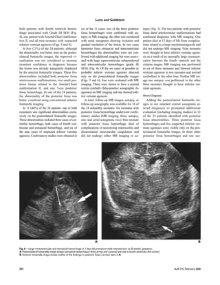
Sonography of the neonatal brain is an important diagnostic tool, but the posterior fossa is often poorly visualized due to its location. This study evaluated adding images through the posterolateral fontanelle to the standard anterior fontanelle approach. Of 1292 neonatal sonograms reviewed, 24 showed posterior fossa abnormalities. Posterolateral fontanelle images better displayed abnormalities in 23 cases and increased diagnostic confidence in 18 cases. In 11 cases, it was the only view revealing abnormalities. Follow-up confirmed most findings, though 4 cases of suspected vermian defects were false positives. Adding posterolateral fontanelle imaging significantly improved posterior fossa evaluation.
![Sonographic Visualization of
Neonatal Posterior Fossa
Abnormalities Through the
Posterolateral Fontanelle
Joseph A. Luna 1 OBJECTIVE. This study was performed to determine whether imaging through the poste-
Ruth B. Goldstein 2 rolateral fontanelle in addition to the anterior fontanelle during neonatal cranial sonography
improves diagnostic accuracy or examiner confidence in the diagnosis of neonatal posterior
fossa abnormalities.
MATERIALS AND METHODS. In 1995 we changed our protocol of neonatal cranial
sonography to include imaging through the posterolateral fontanelle in all patients. The re-
ports of all sonography performed in the first 15 months of this protocol were reviewed, and
two radiologists reviewed the images of all patients in whom a posterior fossa abnormality
was diagnosed with posterolateral fontanelle images masked and then with posterolateral fon-
tanelle images available.
RESULTS. In total, 1292 sonograms were obtained in 462 patients. In 200 patients, the
sonographic findings were abnormal; of these 200 patients, 24 (12%) had posterior fossa ab-
normalities (nine posterior fossa hemorrhages, four Arnold-Chiari malformations (type II),
two posterior fossa arteriovenous malformations, and nine partial vermian defects). The pos-
terolateral fontanelle images showed the posterior fossa abnormality better than the anterior
fontanelle images did in 23 (96%) of the 24 patients, increased confidence in the diagnosis of
18 (75%) of the 24 patients, and was the only technique to reveal the posterior fossa abnor-
mality in 11 (46%) of the 24 patients. Nearly all pathologic correlations with imaging con-
firmed the posterolateral fontanelle findings except for the diagnosis of inferior vermian
agenesis, which was presumed to be a false-positive diagnosis in four patients in whom MR
imaging showed no abnormalities.
CONCLUSION. Additional imaging through the posterolateral fontanelle during routine
neonatal cranial sonography added considerable benefit. False-positive diagnosis of vermian
defects is a troubling problem but may be avoided with careful attention to the midline sagittal
sonographic images of the vermis and fourth ventricle.
S
onography of the neonatal brain is an conventional anterior fontanelle images obtained
important tool in the assessment of in all cranial sonography performed at our insti-
neonates, particularly premature ne- tution. The purpose of this review of our first 15
onates at significant risk for intracranial hemor- months’ experience is to determine the benefit of
rhage. The procedure has shown good sensitivity adding posterolateral fontanelle imaging to the
and specificity in the detection of neonatal intra- conventional anterior fontanelle examination.
cranial abnormalities, particularly in the suprat-
entorial region [1–3]. When scanning through the Materials and Methods
anterior fontanelle, the most poorly evaluated re- The studies were performed in an academic refer-
Received May 14, 1999; accepted after revision gion is the posterior fossa. This is largely because ral center with a 42-bed regional intensive care unit
July 22, 1999. approved for extracorporeal membrane oxygenation
the posterior fossa is farthest from the transducer
1
Kaiser Permanente, 4647 Zion Ave., San Diego, CA 92120. (ECMO) and pediatric cardiothoracic surgery.
and because many of its structures are parallel to
2 We reviewed the reports of all of the cranial sono-
Department of Radiology, University of California, the insonating beam [4–7]. Several articles have
505 Parnassus Ave., M-396, San Francisco, CA 94143-0628. grams obtained during the first 15 months of using
recommended adjunctive imaging through poste- routine posterolateral fontanelle sonography in addi-
Address correspondence to R. B. Goldstein.
rior and posterolateral fontanelles to improve vi- tion to our standard cranial sonography (February
AJR 2000;174:561–567
sualization of the posterior fossa [4–8]. 1995 through April 1996). Each patient whose sono-
0361–803X/00/1742–561 In 1995 we added posterolateral fontanelle graphic report indicated a posterior fossa abnormal-
© American Roentgen Ray Society angled axial images of the cerebellum to the ity was identified.
AJR:174, February 2000 561](https://image.slidesharecdn.com/ustransfontanelarhemorragia-090930142403-phpapp01/85/Us-Transfontanelar-Hemorragia-1-320.jpg)


![Luna and Goldstein
pected inferior vermian agenesis the conven- abnormalities. In 23 (96%) of the 24 sono- a small apparent communication between the
tional images showed an equivocal area that graphically diagnosed posterior fossa ab- fourth ventricle and the cisterna magna that
was interpreted as definitely abnormal on the normalities, two examiners independently was mistakenly thought to represent an infe-
posterolateral fontanelle images. concluded that the abnormality was better seen rior vermian defect. The erroneous diagnoses
using the posterolateral fontanelle images. were all made early in our study. This imag-
Interobserver Agreement Even if the posterior fossa abnormality could ing pitfall has been described in the fetal
Good interobserver agreement was seen. The be identified on the anterior fontanelle images, sonography literature (angled axial and coro-
examiners agreed with the diagnosis and “im- the examiners in this study concluded that add- nal images of the posterior fossa similar to
provement” or “no improvement” of the postero- ing the posterolateral fontanelle images re- our posterolateral fontanelle images are eas-
lateral fontanelle image for the diagnosis in 23 of sulted in increased confidence in diagnosis in ily and commonly obtained) [15]. Angled ax-
the 24 neonates. The examiners differed in the 18 (75%) of the 24 cases. ial images of the cerebellum can make a
confidence of the findings on the anterior fon- In 11 (46%) of the 24 patients, the postero- prominent vallecula appear similar to a par-
tanelle images in only one patient. One examiner lateral fontanelle approach allowed detection tial (inferior) vermian defect. Barkovich et
interpreted the images as having normal findings; of significant findings that were not clearly al. [16], who used MR imaging to investigate
the other interpreted the image as showing a seen on anterior fontanelle imaging, especially Dandy-Walker syndrome, found that the nor-
“possible abnormal area.” Both examiners inter- in the seven neonates with posterior fossa mally formed vermis may tilt forward from
preted this patient’s posterolateral fontanelle im- hemorrhage. One hemorrhage was well seen its usual position and create the impression
ages as showing posterior fossa hemorrhage. with anterior fontanelle imaging. The other six of an inferior vermian defect on angled axial
were poorly seen or not detected at all using and coronal images. We believe that scrutiny
anterior fontanelle imaging, yet confidently of midline sagittal images of the vermis cere-
Discussion identified using posterolateral fontanelle imag- belli obtained through the anterior fontanelle
Cranial sonography of the neonate is a ing. Six of these hemorrhages were confirmed may serve to arbitrate in equivocal cases
widely accepted technique for evaluating the by autopsy (n = 1), MR imaging (n = 3), or fol- (Figs. 5B and 6B). A midline sagittal image
neonatal brain. Initial reports describing the low-up sonography ( n = 2). There were no allows display of the precise cerebellar anat-
technique stressed imaging through the anterior false-positive diagnoses of posterior fossa hem- omy because the nodulus of the vermis cov-
fontanelle [9, 10]. Imaging through the anterior orrhage. Only three of the nine neonates with ering the inferior roof of the fourth ventricle
fontanelle allows excellent evaluation of the posterior fossa hemorrhage had died at the time can be better seen, indicating whether the
common sites of germinal matrix hemorrhage of this writing. Whereas large and catastrophic inferior vermis is intact or deficient. This ex-
and the cerebral ventricles, but a weakness of hemorrhages have generally been described in perience emphasizes the caution with
anterior fontanelle imaging is its evaluation of the literature, posterolateral fontanelle imaging which the diagnosis of inferior vermian
the posterior fossa [4, 5, 7]. Taylor et al. [2] and allowed the detection of some relatively small agenesis should be made.
Babcock et al. [3] described neonates in whom cerebellar hemorrhages in this study. Two large In conclusion, ours is a retrospective study,
significant posterior fossa hemorrhage was autopsy studies of low-birth-weight premature and the overall sensitivity of the posterolateral
missed on sonography performed exclusively neonates have reported an incidence of posterior fontanelle images for posterior fossa abnor-
through the anterior fontanelle. fossa hemorrhage between 16% and 21% even malities is not evaluated. However, in our re-
It has recently been hypothesized that bring- though this hemorrhage is less common than view of 15 months of experience with these
ing the high-frequency transducer closer and supraventricular and intraventricular hemor- images, the posterolateral fontanelle images
more perpendicular to many of the posterior rhage [12, 13]. In one of these studies [13] the revealed three posterior fossa hemorrhages not
fossa structures should improve image clarity. findings of cerebellar hemorrhage fell into two detected with standard anterior fontanelle im-
Several recent studies have confirmed im- groups: large hemorrhages destroying one third aging and confirmed three more that were only
proved visualization of the normal posterior or more of the cerebellar parenchyma and small suspected on the standard images. Nearly ev-
fossa using imaging through a variety of poste- hemorrhages no larger than 5 mm. ery abnormality observed was considered bet-
rior acoustic windows, including the foramen The observation of small cerebellar hem- ter displayed on the posterolateral fontanelle
magnum [4], the posterior fontanelle [5, 7], orrhages using the posterolateral fontanelle images, and adding the posterolateral fonta-
and the posterolateral fontanelle [8, 11]. approach in premature neonates has recently nelle images allowed increased confidence in
The technique for obtaining images been described [14]. The clinical signifi- diagnosis in 75% of the posterior fossa
through the posterolateral fontanelle is cance of these otherwise undiagnosed hem- abnormalities. Only 1 or 2 min of additional
learned easily and quickly. After only a few orrhages is currently under investigation. scan time is needed to obtain these images.
“learning cases” the images may be obtained Unfortunately, better visualization of The potential pitfall of overdiagnosing inferior
in less than 5 min, and our sonographers con- structures not well visualized in the past can vermian agenesis should be anticipated and
sistently obtain the images in less than 2 min, lead to false-positive diagnoses when one easily avoided.
usually in less than 1 min. embarks on using this new technique. In this
On the basis of these early reports and our study at least four false-positive diagnoses
own experience, we added routine posterolat- occurred as a result of adding the posterolat- References
eral fontanelle imaging to our standard cranial eral fontanelle images; all were misdiag-
1. Taylor GA, Fitz CR, Kapur S, Short BL. Cere-
sonography. Posterior fossa anatomy is un- noses of possible inferior vermian agenesis brovascular accidents in neonates treated with
equivocally better displayed using the postero- (Fig. 5) that deserve mention. These errors extracorporeal membrane oxygenation: sonographic–
lateral fontanelle, as are posterior fossa were made as a result of misinterpretation of pathologic correlation. AJR 1989;153:355–361
566 AJR:174, February 2000](https://image.slidesharecdn.com/ustransfontanelarhemorragia-090930142403-phpapp01/85/Us-Transfontanelar-Hemorragia-6-320.jpg)
![Sonography of Neonatal Posterior Fossa Abnormalities
2. Taylor GA, Fiktz CR, Glass P, Short, BL. CT of sound: anatomic and sonographic correlation. 12. Grunnet ML, Shields WD. Cerebellar hemor-
cerebrovascular injury after neonatal extracorporeal Early Hum Dev 1995;42:141–152 rhage in the premature infant. J Pediatr 1976;88:
membrane oxygenation: implications for neurode- 8. Buckley KM, Taylor GA, Estroff JA, Barnewolt 605–608
velopmental outcome. AJR 1989;153:121–126 CE, Share JC, Paltiel HJ. Use of the mastoid fon- 13. Martin R, Roessmann U, Fanaroff A. Massive in-
3. Babcock DS, Han BK, Weiss RG, Ryckman RC. tanelle for improved sonographic visualization of tracerebellar hemorrhage in low-birth-weight in-
Brain abnormalities in infants on extracorporeal the neonatal midbrain and posterior fossa. AJR fants. J Pediatr 1976;89:290–293
membrane oxygenation: sonographic and CT 1997;168:1021–1025 14. Merrill J, Piecuch RF, Fell SC, Barkovich AJ, Gold-
findings. AJR 1989;153:571–576 9. Shuman WP, Rogers JV, Mack LA, Alvord EC, stein RB. A new pattern of cerebellar hemorrhages
4. Sudakoff G, Montazemi M, Rifkin M. The fora- Christie DP. Real-time sonographic sector scan- in preterm infants. Pediatrics 1998;102:62–66
men magnum: the underutilized acoustic window ning of the neonatal cranium: technique and nor- 15. Laing FC, Frates MC, Brown DL, Benson CB, Di
to the posterior fossa. J Ultrasound Med 1993;4: mal anatomy. AJR 1981;137:821–828 Salvo DN, Doubilet PM. Sonography of the fetal
205–210 10. Grant EG, Borts FT, Schellinger D, McCullough posterior fossa: false appearance of mega-cisterna
5. Maertens P. Imaging through the posterior fon- DC, Sivasubramanian KN, Smith Y. Real-time ul- magna and Dandy-Walker variant. Radiology
tanelle. J Child Neurol 1989;4 [suppl]:S62–S67 trasonography of neonatal intraventricular 1994;192:247–251
6. Anderson N, Fulton J. Sonography through the hemorrhage and comparison with computed to- 16. Barkovich AJ, Kjos BO, Norman D, Edwards
posterior fontanelle in diagnosing neonatal intra- mography. Radiology 1981;139:687–691 MS. Revised classification of posterior fossa
ventricular hemorrhage. AJNR 1991;12:368–370 11. Yousefzadeh D, Naidich T. US anatomy of the cysts and cystlike malformations based on the re-
7. Anderson NG, Hay R, Hutchings M, Whitehead posterior fossa in children: correlation with brain sults of multiplanar MR imaging. AJR 1989;153:
M, Darlow B. Posterior fontanelle cranial ultra- sections. Radiology 1985;156:353–361 1289–1300
AJR:174, February 2000 567](https://image.slidesharecdn.com/ustransfontanelarhemorragia-090930142403-phpapp01/85/Us-Transfontanelar-Hemorragia-7-320.jpg)


![NSG AND HIE [Autosaved].............pptx](https://cdn.slidesharecdn.com/ss_thumbnails/nsgandhieautosaved-250908133023-52fc5444-thumbnail.jpg?width=640&height=640&fit=bounds)


