SlideShare a Scribd company logo
INTRACRANIAL BLEEDING
Authors
1. Nelson Ssewante MBChB V
2. Ronald Mujuni MBChB V
Tutor
Mr. Muhindo A.
Department of Surgery, Makerere University.
Outline
1. Introduction with relevant anatomy
2. Neurologic assessment
3. Epidural hemorrhage
4. Subdural hemorrhage
5. Subarachnoid hemorrhage
6. Intracerebral hemorrhage
7. Intraventricular hemorrhage
Introduction
• Intracranial bleeding is the pathological accumulation of blood
within the cranial vault.
• This can be with in the meningeal spaces or the brain parenchyma
• It can be classified as intra-axial (parenchymal or intraventricular)
or extra-axial hemorrhage(Epidural or subdural or subarachnoid)
Relevant anatomy
• The brain parenchyma is protected by the bony shield called the cranium.
• The brain is divided into;
i. Cerebral hemispheres
ii. The midbrain
iii. The brain stem(pons and medulla)
iv. The cerebellum
• It is covered by 3 protective membranes, from inside out; the pia matter,
arachnoid and dura matter.
Blood supply
• Arterial supply • Venous drainage
Ventricular system Coverings of the brain
Neurologic Assessment
• History
• Examination Findings
• Mental status examination(GCS)
• Cranial nerve examination
• Motor examination
• Deep tendon reflexes
• Sensation
• Cerebellar function
Epidural Hemorrhage
• EDH is collection of blood in the extradural space between the dura and
skull
• It commonly results from rupture of Middle meningeal artery(85%)
• Other vessels may include; middle meningeal vein, Dural sinus or
diploic veins
Epidemiology of EDH
• EDH is one of the most serious complications of head injury, requiring
immediate diagnosis and surgical intervention.
• Occurs in ~2% of all patients with head injuries and 5−15% of patients with
fatal head injuries
• EDH may be acute (58%), subacute (31%) or chronic (11%).
• It is more common in young adults. Rare in children <2 years and after 60
years
Pathophysiology
Direct blow on the head(due to RTA, Falls Coup, Counter-coup
injuries)
Fracture of the scalp(Commonly the temporal
bone)
Disruption of the integrity between the dura and
the bone
Tearing of the vessels(Commonly middle meningeal
artery)
Bleeding with resultant hematoma
formation
Gradual stripping of the dura from the skull & collection
of blood
Hematoma
expansion
Clinical presentation
• Classic sequence (seen in <30%):
 Post-traumatic reduced LOC, a lucid interval of several hours, then obtundation, hemiparesis, ipsilateral
pupillary dilatation, and coma.
• Signs and symptoms depend on severity;
 Headache
 Nausea & Vomiting
 Amnesia
 Altered LOC
 Aphasia
 Seizures, HTN, and respiratory distress may also occur
• Deterioration can take hours to days
Diagnosis
• Laboratory
 CBC, grouping and crossmatch
 Prothrombin time(PT)/activated partial thromboplastin time (aPTT)
 Serum chemistries, including electrolytes, blood urea nitrogen (BUN), creatinine, and glucose
 Toxicology screen and serum alcohol level
• The diagnosis of EDH must be considered when the plain skull X-rays show a
fracture
• CT scan is modality of choice
 a hyperdense, biconvex (lenticular) mass adjacent to the skull
Big Arrow: Fracture Small arrows: Epidural
hematoma
Management
• Candidates for non-surgical management include;
I. Small EDH (≤1 cm maximal thickness)
II. Subacute or chronic EDH
III. Minimal midline shift(<5mm)
IV. No focal deficit
• These should be admitted and close neurological observation with serial CT done.
Continued…
Indication for surgery
 Any symptomatic EDH.
 An acute asymptomatic EDH >1 cm
 EDH in pediatric patients
 Failure of non-surgical (conservative) management.
Objectives of surgery;
 To remove the clot (to reduce ICP & eliminate focal deficit)
 Achieve absolute hemostasis.
 Prevent reaccumulation by placing dural hitch sutures (tack-up sutures).
Continued…
Surgery
• Craniotomy and hematoma evacuation is the mainstay surgical treatment
• This involves identification and ligation of the bleeding vessels
• Appropriate antibiotics should be initiated preoperatively
• Appropriate analgesia
• Mannitol pre-operatively if elevated ICP or signs of brain herniation
Prognosis
• Good with prompt management,
• Worse prognosis if bilateral Babinski or
decerebration pre-operative
• Death is usually due to respiratory arrest from
uncal herniation (injury to the midbrain)
Subdural hemorrhage
• Collection of blood between the dura and the arachnoid membranes
• It is due to injury to the cortical veins and/or laceration of cortex of
brain
• It can be acute or chronic
Epidemiology
• Acute SDH complicates approximately 11% of mild to severe head
injuries and 20% of severe traumatic brain injuries.
• The elderly and chronic alcoholics are at a high risk due to brain
atrophy.
Pathophysiology
• SDH usually results from tearing of a bridging veins running from the
cerebral cortex to the dural sinuses.
• Arterial rupture can also result in SDH in 20-30% of cases.
• This commonly follows acceleration injuries.
• Brain atrophy can create traction on arachnoid leading to tearing of veins
since the dura is firmly adherent to the skull
• Intracranial hypotension(Low CSF pressure) is another mechanism that can
result in SDH. This is due to a reduction in brain buoyancy with resultant
traction on vessels.
Continued…
Chronic SDH
• It occurs approximately 2 weeks after acute SDH.
• There's increased collagen synthesis and deposition of fibroblasts on the outer
surface of the dura
• This creates a thin inner membrane that completely encapsulates the clot.
• Chronic SDH may liquefy to form a hygroma, and the membranes may calcify.
• In some cases, hematoma expansion can occur as a result of recurrent bleeding
(Acute on Chronic) or osmotic draw of water.
Clinical presentation
• Common presentation include;
 Altered level of consciousness
 Headache
 Focal deficits
 Features of raised intracranial pressure (such as blurred vision, N/V, worsening headache)
 Seizure activity
• Clinical features of an acute SDH occur quickly, whilst those of a chronic SDH have a
latent period of weeks (or even months) before symptoms appear.
• In children, it is important to survey for other injuries with suspected SDH as there
might be signs of non-accidental injury.
Diagnosis
• Patients should have initial routine bloods, including FBC, CRP, U&Es, LFTs, and a
clotting, which will also aid in assessing for differential diagnosis.
• Non-contrast CT scan is the gold standard
 a crescent-shaped hematoma over one hemisphere, with or without associated midline shift
• Others include; MRI, Angiography
Acute on chronic SDH. Acute component is denser than
chronic. Arrow heads are defining the outline of the
hematoma.
A: Chronic SDH B: Acute SDH
Management
The decision between surgical or medical management of SDH is
based upon;
• Glasgow Coma Scale score
• CT findings; mainly SDH clot thickness, degree of midline shift, and
presence of associated brain lesion
• Neurologic examination, including pupillary signs
• Clinical stability or deterioration over time
• Acuity of SDH
• Presence and severity of comorbidities and associated trauma
• Age
Continued…
Medical SDH care focuses on;
(a) Management of intracranial hypertension (ICP < 22 mmHg)
(b) Maintenance of adequate cerebral perfusion (CPP > 60 mmHg)
(c) Seizure prevention and treatment,
(d) Maintenance of normothermia, eucarbia, euglycemia, and euvolemia,
(e) Early initiation of enteral feeding, mobilization, and physical therapy
Continued…
Surgery
• Craniotomy is the recommended surgical technique for patients with
acute SDH and coma
• Decompressive craniectomy
• Burr hole trepanation/trephination
Subarachnoid Hemorrhage
• The extravasation of blood into the subarachnoid space
between the pial and arachnoid membrane.
• It occurs in various clinical contexts, the most common being
head trauma.
• Nontraumatic (or spontaneous) cases occurs in the setting of a
ruptured cerebral aneurysm or arteriovenous malformation
(AVM).
Etiology
Risk factors
• HTN
• Pregnancy/parturition in patients with
pre-existing AVMs, eclampsia
• Oral contraceptive pill
• Substance abuse (cigarette smoking,
cocaine, alcohol)
• Conditions associated with high
incidence of aneurysms
Clinical features
• Sudden onset of severe headache with vomiting.
• Features of raised intracranial pressure.
• Photophobia.
• Neck stiffness.
• Focal neurological deficits: hemiplegia, dysphasia.
• Eye changes: ptosis, dilated pupil, changes in the eyeball movements.
• Sudden loss of consciousness.
• Features of brain oedema and cerebral ischemia.
• In 40% of recovered patients, rebleeding occurs in 6–8 weeks which is commonly fatal.
Grade Hunt & Hess system Fischer system WFNS system
1 Asymptomatic or minimal headache and slight
neck stiffness
70% survival
No subarachnoid blood seen
on CT scan
GCS 15, no motor deficit.
2 Severe headache and neck stiffness, No
neurologic deficit except cranial nerve palsy
60% survival
Diffuse or vertical layers of
SAH less than 1 mm thick
GCS 13-14 without deficit
3 Drowsy; minimal neurologic deficit
50% survival
Diffuse clot and/or vertical
layer greater than 1 mm
thick
GCS 13-14 with focal
neurological deficit
4 Stuporous; moderate to severe hemiparesis;
possibly early decerebrate rigidity and
vegetative disturbances
20% survival
Intracerebral or
intraventricular clot
with diffuse or no
subarachnoid blood
GCS 7-12, with or without
deficit.
5 Deep coma; decerebrate rigidity; moribund
10% survival
N/A GCS <7 , with or without
deficit
Grading systems for SAH
Diagnostic evaluation
• Routine Labs
• Lumbar puncture.
 In subarachnoid hemorrhage, blood stained CSF is collected.
• CT scan.
• Carotid and vertebral angiogram.
Treatment
• Clipping, or wrapping the aneurysm.
• Craniotomy and proceed.
• Ligation of common carotid artery-there is risk of hemiplegia.
• Therapeutic embolization.
• Excision of vascular malformations.
• Coiling of aneurysm with instruments.
Intracerebral hemorrhage
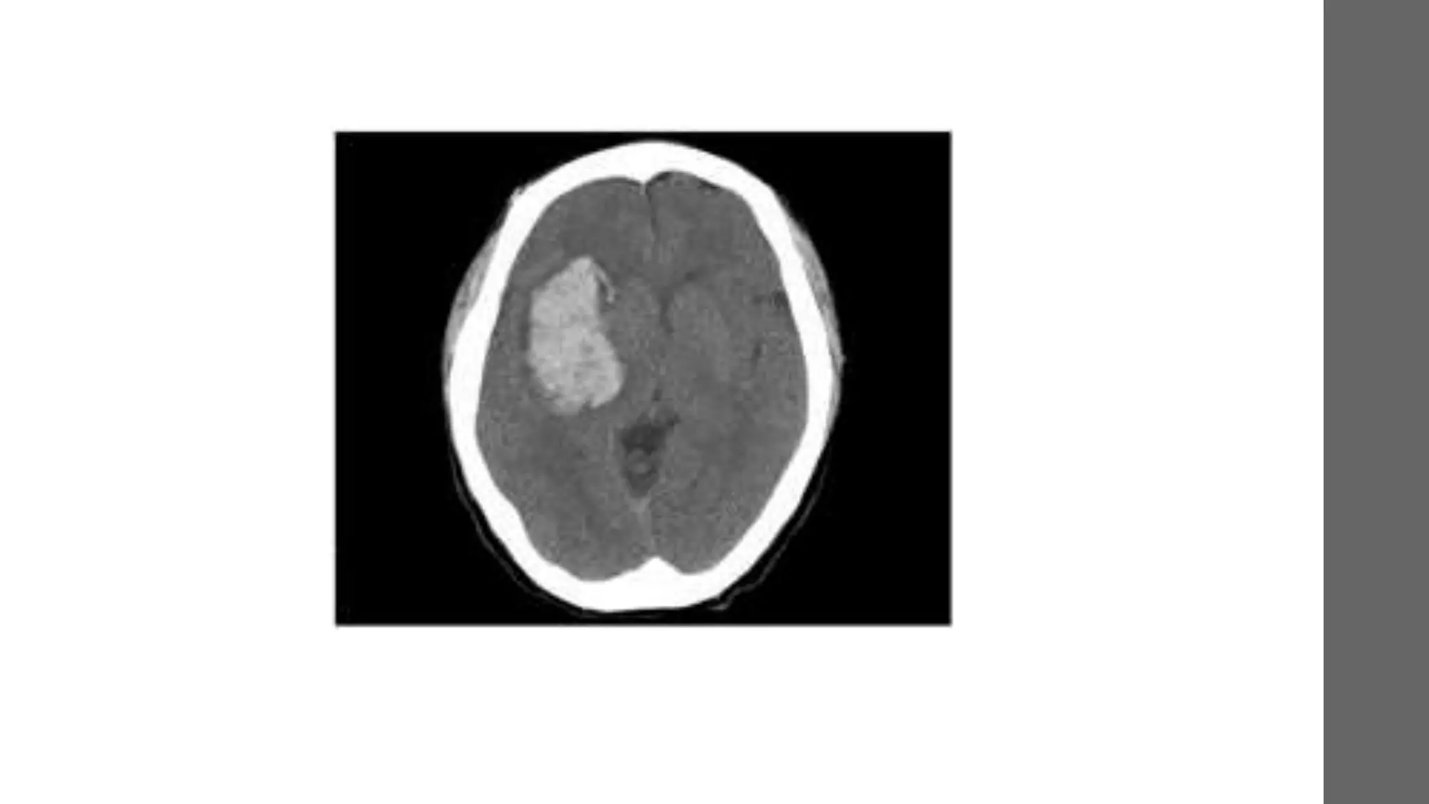
• Bleeding within brain parenchyma. It is the second most common
cause of stroke.
• Common sites of involvement
 Basal Ganglia/Internal Capsule (50%)
 Thalamus (15%)
 Cerebral White Matter (15%)
 Cerebellum/Brainstem – usually pons (15%)
 Other (5%)
Etiology
• Traumatic
• Hypertensive vasculopathy
 Commonly occur at the branching points between small penetrating arteries and their
parental vessels- area of maximal pressure
 usually causes bleeds at putamen, thalamus, pons, and cerebellum
• Cerebral amyloid angiopathy(CAA)
 Usually asymptomatic, CAA is an important cause of primary lobar ICH in older adults
 Due to deposition of amyloid in small- to medium-sized blood vessels of the brain and
leptomeninges.
Etiology continued…
Others
• Hemorrhagic transformation (reperfusion post stroke, surgery, strenuous exercise)
• Vascular anomalies
 Aneurysms, AVMs, and venous sinus thrombosis
• Tumors (1%): often malignant (e.g. GBM, lymphoma, metastases)
• Drugs (amphetamines, cocaine, alcohol, anticoagulants)
• Coagulopathy (iatrogenic, leukemia, TTP, aplastic anemia)
• CNS infections (fungal, granulomas, herpes simplex encephalitis)
Risk factors
• Increasing age (mainly >55 years)
• Male gender
• HTN
• Black/Asian > Caucasian
• Previous CVA of any type (23x risk)
• Both acute and chronic heavy alcohol use; cocaine, amphetamines
• Liver disease
• Anticoagulants
Clinical features
• TIA-like symptoms often precede ICH, can localize to site of impending
hemorrhage
• Gradual onset of symptoms over minutes-hours, usually during activity
• Headache, Nausea and Vomiting, and decreased LOC are common
• Focal deficits depending on location of ICH
Continued…
• Putaminal hemorrhage
 Hemiplegia, hemisensory loss, homonymous hemianopsia, gaze palsy, stupor, and coma.
• Internal capsule hemorrhage
 Mild dysarthria, contralateral hemiparesis and sensory deficit
• Thalamic hemorrhage
 Hemiparesis, hemisensory loss, and occasionally transient homonymous hemianopsia.
• Lobar hemorrhage
 Vary in their neurologic signs depending upon location(common in Parietal or occipital)
 These bleeds are associated with a higher incidence of seizures
• Pontine hemorrhage
 Characterized by deep coma, total paralysis, pinpointed pupil that is reactive to light or ocular
bobbing, facial palsy, deafness, and dysarthria when the patient is awake
Investigations
Labs
• CBC, PT, aPTT, electrolytes, blood urea nitrogen, creatinine, and glucose
Neuroimaging
• CT or MRI is mandatory to confirm the diagnosis
• CT Angiography
 spot sign (contrast in the hematoma) demonstrate high likelihood of clot growth
A small focus of enhancement is seen peripherally
on CTA source images, consistent with the spot
sign (arrow)
Medical management
• Initial Resuscitation
• ICH is managed as an emergence.
• Control raised ICP
 Avoid corticosteroid use
• Seizure prophylaxis (phenytoin or levetiracetam)
• Correct any electrolyte abnormalities
• Assess for coagulation disorders and correct if any
Cyrus K Dastur, and Wengui Yu Stroke Vasc Neurol 2017;2:21-29
Surgical management
Indications of craniotomy
• Symptoms of raised ICP or mass effect
• Rapid deterioration (especially if signs of brainstem compression)
• Favourable location (e.g. cerebellar, non-dominant hemisphere)
• Young patient (<50 yr)
Interventricular Hemorrhage
• Also known as intraventricular bleeding, is a bleeding into the brain's ventricular
system
• The ventricular system is contains freely flowing CSF.
• These include paired lateral ventricles, the 3rd ventricle, and 4th ventricle.
Etiology
• Primary IVH(30% of cases)
 HTN
 Intraventricular trauma
 Aneurysm
 Vascular malformations
 Tumors
• Secondary(70% of cases)
 These results as an expansion of existing parenchymal or subarachnoid
hemorrhage
Clinical presentation
• Sudden onset of headache
• Nausea and vomiting.
• Alteration of the mental state and/or level of
• consciousness.
• Focal neurological signs are either minimal or absent.
• Focal and/or generalized seizures may occur
Investigations
• CT is the test of choice
• MRI
• Angiogram
• Carotid duplex
Lab studies
 Electrolyte studies
 Coagulation profile(Platelet counts, PT, aPTT)
 CBC
Management
General measures focuses on;
 Cessation of bleeding
 relieving hydrocephalus
 Controlling intracranial pressure.
• Patients who have a moderate to severe IVH should be followed up in ICU
• Patient positioning should be at 300 to reduce risk of aspiration
• Fluid resuscitation using isotonic crystalloid solutions
• any elevations in body temperature should be treated aggressively
• Prophylaxis against deep venous thrombosis
• Seizures prophylaxis
Continued…
Specific management is tailored according to the cause
• Blood pressure management
 The optimum blood pressure management in patients with IVH remains undefined. It is
recommended to lower BP gradually.
• External ventricular drain(EVD)
 a small catheter inserted through the skull usually into the lateral ventricle to drain excess
CSF.
 Indicated for patients with IVH with hydrocephalus and neurologic decline
• Intraventricular thrombolysis
References
1. F. Charles Brunicardi, Ashwin G. Ramayya, Saurabh Sinha, M. Sean Grady. Schwartz’s
Principles of Surgery; 11th edition volume 1 (2019). Chapter 42 pages 1849-1853
2. Prakash Narain Tandon, Ravi Ramamurthi and Pradeep Kumar Jain N. Ramamurthi &
Tandon’s manual of Neurosurgery 1st edition (2014); chapters 33-36, pages 270-287
3. David S Liebeskind, Helmi L Lutsep, Edward L Hogan. Epidural Hematoma;
https://emedicine.medscape.com/article/1137065-overview Medscape 2018.
4. David S Liebeskind, Francisco Talavera, Howard S Kirshner, Jeffrey L Saver. Intracranial
Hemorrhage; https://emedicine.medscape.com/article/1163977-overview Medscape
2018.
5. Sriram Bhat M; SRB’s Manual of Surgery 5th Edition. Chapter 27 Pages 1090-1098
The End!

More Related Content

What's hot

Subarachnoid hemorrhage
Subarachnoid hemorrhageSubarachnoid hemorrhage
Subarachnoid hemorrhage
David Hersey
 
Cerebral aneurysm
Cerebral aneurysm Cerebral aneurysm
Cerebral aneurysm
Milan Silwal
 
Brain abscess
Brain abscessBrain abscess
Brain abscess
joemdas
 
Stroke ppt
Stroke pptStroke ppt
BRAIN ARTERIOVENOUS MALFORMATION
BRAIN ARTERIOVENOUS MALFORMATIONBRAIN ARTERIOVENOUS MALFORMATION
BRAIN ARTERIOVENOUS MALFORMATION
suresh Bishokarma
 
Intracerebral hemorhage Diagnosis and management
Intracerebral hemorhage  Diagnosis and managementIntracerebral hemorhage  Diagnosis and management
Intracerebral hemorhage Diagnosis and management
Ramesh Babu
 
Aortic dissection nikku ppt
Aortic dissection nikku pptAortic dissection nikku ppt
Aortic dissection nikku ppt
Nikhil Vaishnav
 
Chronic subdural hematoma.pptx
Chronic subdural hematoma.pptxChronic subdural hematoma.pptx
Chronic subdural hematoma.pptx
RUTAYISIRE François Xavier
 
Identifying and managing acute stroke
Identifying and managing acute strokeIdentifying and managing acute stroke
Identifying and managing acute strokeAhmad Shahir
 
Stroke localization
Stroke localizationStroke localization
Arteriovenous Malformation (AVM) of Brain
Arteriovenous Malformation (AVM) of BrainArteriovenous Malformation (AVM) of Brain
Arteriovenous Malformation (AVM) of Brain
Dhaval Shukla
 
Subarachnoid haemorrhage
Subarachnoid haemorrhage Subarachnoid haemorrhage
Subarachnoid haemorrhage
NITAYSBMC
 
Hemorrhagic stroke
Hemorrhagic stroke Hemorrhagic stroke
Hemorrhagic stroke
Helao Silas
 
Hydrocephalous, shunting & shunt systems
Hydrocephalous, shunting & shunt systemsHydrocephalous, shunting & shunt systems
Hydrocephalous, shunting & shunt systems
Mukhtar Khan
 
CEREBRAL ARTERIO VENOUS MALFORMATIONS
CEREBRAL ARTERIO VENOUS MALFORMATIONS CEREBRAL ARTERIO VENOUS MALFORMATIONS
CEREBRAL ARTERIO VENOUS MALFORMATIONS
SHAMEEJ MUHAMED KV
 
Cerebral edema and its management
Cerebral edema and its managementCerebral edema and its management
Cerebral edema and its management
Rajesh Kabilan
 
Cvj anomalies
Cvj anomaliesCvj anomalies
Cvj anomalies
Navni Garg
 
Approach to ataxia
Approach to ataxiaApproach to ataxia
Approach to ataxia
Divya Shilpa
 

What's hot (20)

Subarachnoid hemorrhage
Subarachnoid hemorrhageSubarachnoid hemorrhage
Subarachnoid hemorrhage
 
Cerebral aneurysm
Cerebral aneurysm Cerebral aneurysm
Cerebral aneurysm
 
Brain abscess
Brain abscessBrain abscess
Brain abscess
 
Stroke ppt
Stroke pptStroke ppt
Stroke ppt
 
BRAIN ARTERIOVENOUS MALFORMATION
BRAIN ARTERIOVENOUS MALFORMATIONBRAIN ARTERIOVENOUS MALFORMATION
BRAIN ARTERIOVENOUS MALFORMATION
 
Intracerebral hemorhage Diagnosis and management
Intracerebral hemorhage  Diagnosis and managementIntracerebral hemorhage  Diagnosis and management
Intracerebral hemorhage Diagnosis and management
 
Aortic dissection nikku ppt
Aortic dissection nikku pptAortic dissection nikku ppt
Aortic dissection nikku ppt
 
Chronic subdural hematoma.pptx
Chronic subdural hematoma.pptxChronic subdural hematoma.pptx
Chronic subdural hematoma.pptx
 
Identifying and managing acute stroke
Identifying and managing acute strokeIdentifying and managing acute stroke
Identifying and managing acute stroke
 
Stroke localization
Stroke localizationStroke localization
Stroke localization
 
Subdural Hematoma
Subdural HematomaSubdural Hematoma
Subdural Hematoma
 
Basics of brain hemorrhage
Basics of brain hemorrhageBasics of brain hemorrhage
Basics of brain hemorrhage
 
Arteriovenous Malformation (AVM) of Brain
Arteriovenous Malformation (AVM) of BrainArteriovenous Malformation (AVM) of Brain
Arteriovenous Malformation (AVM) of Brain
 
Subarachnoid haemorrhage
Subarachnoid haemorrhage Subarachnoid haemorrhage
Subarachnoid haemorrhage
 
Hemorrhagic stroke
Hemorrhagic stroke Hemorrhagic stroke
Hemorrhagic stroke
 
Hydrocephalous, shunting & shunt systems
Hydrocephalous, shunting & shunt systemsHydrocephalous, shunting & shunt systems
Hydrocephalous, shunting & shunt systems
 
CEREBRAL ARTERIO VENOUS MALFORMATIONS
CEREBRAL ARTERIO VENOUS MALFORMATIONS CEREBRAL ARTERIO VENOUS MALFORMATIONS
CEREBRAL ARTERIO VENOUS MALFORMATIONS
 
Cerebral edema and its management
Cerebral edema and its managementCerebral edema and its management
Cerebral edema and its management
 
Cvj anomalies
Cvj anomaliesCvj anomalies
Cvj anomalies
 
Approach to ataxia
Approach to ataxiaApproach to ataxia
Approach to ataxia
 

Similar to Intracranial bleeding

Management of head injury
Management of head injuryManagement of head injury
Management of head injuryfyndoc
 
Head injury
Head injuryHead injury
Head injury
HIRENGEHLOTH
 
ayu w - PROBLEM 7 EMERGENCY MEDICINE.pptx
ayu w - PROBLEM 7 EMERGENCY MEDICINE.pptxayu w - PROBLEM 7 EMERGENCY MEDICINE.pptx
ayu w - PROBLEM 7 EMERGENCY MEDICINE.pptx
AyuWindyaningrum
 
Head injury and CNS infection.pdf
Head injury and CNS infection.pdfHead injury and CNS infection.pdf
Head injury and CNS infection.pdf
gp9dprrjvx
 
brain disorders.pdf
brain disorders.pdfbrain disorders.pdf
brain disorders.pdf
Drsaira8
 
How to Read a Head CT.pptx
How to Read a Head CT.pptxHow to Read a Head CT.pptx
How to Read a Head CT.pptx
Maheen Fatima
 
Management of head injury
Management of head injuryManagement of head injury
Management of head injury
George Owusu
 
mymanagementofheadinjury-190703182513.pdf
mymanagementofheadinjury-190703182513.pdfmymanagementofheadinjury-190703182513.pdf
mymanagementofheadinjury-190703182513.pdf
EstibelMengist
 
Craniotomy for brain tumour
Craniotomy for brain tumourCraniotomy for brain tumour
Craniotomy for brain tumour
ZIKRULLAH MALLICK
 
Epidural hematoma
Epidural hematomaEpidural hematoma
Epidural hematoma
PratikDhabalia
 
Neurosurgery 1.pptx
Neurosurgery 1.pptxNeurosurgery 1.pptx
Neurosurgery 1.pptx
joendesh
 
Head injuries
Head injuriesHead injuries
Head injuries
imrana tanvir
 
Overview on head injury pdf
Overview on head injury pdfOverview on head injury pdf
Overview on head injury pdf
LiZe4
 
Ischaemic stroke
Ischaemic strokeIschaemic stroke
Ischaemic stroke
Dr. Imdadul Magfur
 
Craniocerebral trauma 1
Craniocerebral trauma 1Craniocerebral trauma 1
Craniocerebral trauma 1DrLokesh Mahar
 
Brief review in cerebellar stroke -diagnosis
Brief review in cerebellar stroke -diagnosisBrief review in cerebellar stroke -diagnosis
Brief review in cerebellar stroke -diagnosis
KaminiVinathan1
 
CEREBRAL ANEURYSM .pptx
CEREBRAL ANEURYSM                  .pptxCEREBRAL ANEURYSM                  .pptx
CEREBRAL ANEURYSM .pptx
Juma675663
 
Management of Stroke.
Management of Stroke.Management of Stroke.
Management of Stroke.
George Kariuki
 
TRAUMATIC BRAIN INJURY NEurosugery presentation.pptx
TRAUMATIC BRAIN INJURY NEurosugery presentation.pptxTRAUMATIC BRAIN INJURY NEurosugery presentation.pptx
TRAUMATIC BRAIN INJURY NEurosugery presentation.pptx
RUTAYISIRE François Xavier
 
Enoxaparin for Stroke.ppt
Enoxaparin for Stroke.pptEnoxaparin for Stroke.ppt
Enoxaparin for Stroke.ppt
benedicta bestari
 

Similar to Intracranial bleeding (20)

Management of head injury
Management of head injuryManagement of head injury
Management of head injury
 
Head injury
Head injuryHead injury
Head injury
 
ayu w - PROBLEM 7 EMERGENCY MEDICINE.pptx
ayu w - PROBLEM 7 EMERGENCY MEDICINE.pptxayu w - PROBLEM 7 EMERGENCY MEDICINE.pptx
ayu w - PROBLEM 7 EMERGENCY MEDICINE.pptx
 
Head injury and CNS infection.pdf
Head injury and CNS infection.pdfHead injury and CNS infection.pdf
Head injury and CNS infection.pdf
 
brain disorders.pdf
brain disorders.pdfbrain disorders.pdf
brain disorders.pdf
 
How to Read a Head CT.pptx
How to Read a Head CT.pptxHow to Read a Head CT.pptx
How to Read a Head CT.pptx
 
Management of head injury
Management of head injuryManagement of head injury
Management of head injury
 
mymanagementofheadinjury-190703182513.pdf
mymanagementofheadinjury-190703182513.pdfmymanagementofheadinjury-190703182513.pdf
mymanagementofheadinjury-190703182513.pdf
 
Craniotomy for brain tumour
Craniotomy for brain tumourCraniotomy for brain tumour
Craniotomy for brain tumour
 
Epidural hematoma
Epidural hematomaEpidural hematoma
Epidural hematoma
 
Neurosurgery 1.pptx
Neurosurgery 1.pptxNeurosurgery 1.pptx
Neurosurgery 1.pptx
 
Head injuries
Head injuriesHead injuries
Head injuries
 
Overview on head injury pdf
Overview on head injury pdfOverview on head injury pdf
Overview on head injury pdf
 
Ischaemic stroke
Ischaemic strokeIschaemic stroke
Ischaemic stroke
 
Craniocerebral trauma 1
Craniocerebral trauma 1Craniocerebral trauma 1
Craniocerebral trauma 1
 
Brief review in cerebellar stroke -diagnosis
Brief review in cerebellar stroke -diagnosisBrief review in cerebellar stroke -diagnosis
Brief review in cerebellar stroke -diagnosis
 
CEREBRAL ANEURYSM .pptx
CEREBRAL ANEURYSM                  .pptxCEREBRAL ANEURYSM                  .pptx
CEREBRAL ANEURYSM .pptx
 
Management of Stroke.
Management of Stroke.Management of Stroke.
Management of Stroke.
 
TRAUMATIC BRAIN INJURY NEurosugery presentation.pptx
TRAUMATIC BRAIN INJURY NEurosugery presentation.pptxTRAUMATIC BRAIN INJURY NEurosugery presentation.pptx
TRAUMATIC BRAIN INJURY NEurosugery presentation.pptx
 
Enoxaparin for Stroke.ppt
Enoxaparin for Stroke.pptEnoxaparin for Stroke.ppt
Enoxaparin for Stroke.ppt
 

Recently uploaded

MANAGEMENT OF ATRIOVENTRICULAR CONDUCTION BLOCK.pdf
MANAGEMENT OF ATRIOVENTRICULAR CONDUCTION BLOCK.pdfMANAGEMENT OF ATRIOVENTRICULAR CONDUCTION BLOCK.pdf
MANAGEMENT OF ATRIOVENTRICULAR CONDUCTION BLOCK.pdf
Jim Jacob Roy
 
Superficial & Deep Fascia of the NECK.pptx
Superficial & Deep Fascia of the NECK.pptxSuperficial & Deep Fascia of the NECK.pptx
Superficial & Deep Fascia of the NECK.pptx
Dr. Rabia Inam Gandapore
 
Pictures of Superficial & Deep Fascia.ppt.pdf
Pictures of Superficial & Deep Fascia.ppt.pdfPictures of Superficial & Deep Fascia.ppt.pdf
Pictures of Superficial & Deep Fascia.ppt.pdf
Dr. Rabia Inam Gandapore
 
Ocular injury ppt Upendra pal optometrist upums saifai etawah
Ocular injury  ppt  Upendra pal  optometrist upums saifai etawahOcular injury  ppt  Upendra pal  optometrist upums saifai etawah
Ocular injury ppt Upendra pal optometrist upums saifai etawah
pal078100
 
Are There Any Natural Remedies To Treat Syphilis.pdf
Are There Any Natural Remedies To Treat Syphilis.pdfAre There Any Natural Remedies To Treat Syphilis.pdf
Are There Any Natural Remedies To Treat Syphilis.pdf
Little Cross Family Clinic
 
Pharma Pcd Franchise in Jharkhand - Yodley Lifesciences
Pharma Pcd Franchise in Jharkhand - Yodley LifesciencesPharma Pcd Franchise in Jharkhand - Yodley Lifesciences
Pharma Pcd Franchise in Jharkhand - Yodley Lifesciences
Yodley Lifesciences
 
Pharynx and Clinical Correlations BY Dr.Rabia Inam Gandapore.pptx
Pharynx and Clinical Correlations BY Dr.Rabia Inam Gandapore.pptxPharynx and Clinical Correlations BY Dr.Rabia Inam Gandapore.pptx
Pharynx and Clinical Correlations BY Dr.Rabia Inam Gandapore.pptx
Dr. Rabia Inam Gandapore
 
Surat @ℂall @Girls ꧁❤8527049040❤꧂@ℂall @Girls Service Vip Top Model Safe
Surat @ℂall @Girls ꧁❤8527049040❤꧂@ℂall @Girls Service Vip Top Model SafeSurat @ℂall @Girls ꧁❤8527049040❤꧂@ℂall @Girls Service Vip Top Model Safe
Surat @ℂall @Girls ꧁❤8527049040❤꧂@ℂall @Girls Service Vip Top Model Safe
Savita Shen $i11
 
Evaluation of antidepressant activity of clitoris ternatea in animals
Evaluation of antidepressant activity of clitoris ternatea in animalsEvaluation of antidepressant activity of clitoris ternatea in animals
Evaluation of antidepressant activity of clitoris ternatea in animals
Shweta
 
HOT NEW PRODUCT! BIG SALES FAST SHIPPING NOW FROM CHINA!! EU KU DB BK substit...
HOT NEW PRODUCT! BIG SALES FAST SHIPPING NOW FROM CHINA!! EU KU DB BK substit...HOT NEW PRODUCT! BIG SALES FAST SHIPPING NOW FROM CHINA!! EU KU DB BK substit...
HOT NEW PRODUCT! BIG SALES FAST SHIPPING NOW FROM CHINA!! EU KU DB BK substit...
GL Anaacs
 
How STIs Influence the Development of Pelvic Inflammatory Disease.pptx
How STIs Influence the Development of Pelvic Inflammatory Disease.pptxHow STIs Influence the Development of Pelvic Inflammatory Disease.pptx
How STIs Influence the Development of Pelvic Inflammatory Disease.pptx
FFragrant
 
Alcohol_Dr. Jeenal Mistry MD Pharmacology.pdf
Alcohol_Dr. Jeenal Mistry MD Pharmacology.pdfAlcohol_Dr. Jeenal Mistry MD Pharmacology.pdf
Alcohol_Dr. Jeenal Mistry MD Pharmacology.pdf
Dr Jeenal Mistry
 
Light House Retreats: Plant Medicine Retreat Europe
Light House Retreats: Plant Medicine Retreat EuropeLight House Retreats: Plant Medicine Retreat Europe
Light House Retreats: Plant Medicine Retreat Europe
Lighthouse Retreat
 
Non-respiratory Functions of the Lungs.pdf
Non-respiratory Functions of the Lungs.pdfNon-respiratory Functions of the Lungs.pdf
Non-respiratory Functions of the Lungs.pdf
MedicoseAcademics
 
Triangles of Neck and Clinical Correlation by Dr. RIG.pptx
Triangles of Neck and Clinical Correlation by Dr. RIG.pptxTriangles of Neck and Clinical Correlation by Dr. RIG.pptx
Triangles of Neck and Clinical Correlation by Dr. RIG.pptx
Dr. Rabia Inam Gandapore
 
ARTIFICIAL INTELLIGENCE IN HEALTHCARE.pdf
ARTIFICIAL INTELLIGENCE IN  HEALTHCARE.pdfARTIFICIAL INTELLIGENCE IN  HEALTHCARE.pdf
ARTIFICIAL INTELLIGENCE IN HEALTHCARE.pdf
Anujkumaranit
 
263778731218 Abortion Clinic /Pills In Harare ,
263778731218 Abortion Clinic /Pills In Harare ,263778731218 Abortion Clinic /Pills In Harare ,
263778731218 Abortion Clinic /Pills In Harare ,
sisternakatoto
 
Ophthalmology Clinical Tests for OSCE exam
Ophthalmology Clinical Tests for OSCE examOphthalmology Clinical Tests for OSCE exam
Ophthalmology Clinical Tests for OSCE exam
KafrELShiekh University
 
BRACHYTHERAPY OVERVIEW AND APPLICATORS
BRACHYTHERAPY OVERVIEW  AND  APPLICATORSBRACHYTHERAPY OVERVIEW  AND  APPLICATORS
BRACHYTHERAPY OVERVIEW AND APPLICATORS
Krishan Murari
 
micro teaching on communication m.sc nursing.pdf
micro teaching on communication m.sc nursing.pdfmicro teaching on communication m.sc nursing.pdf
micro teaching on communication m.sc nursing.pdf
Anurag Sharma
 

Recently uploaded (20)

MANAGEMENT OF ATRIOVENTRICULAR CONDUCTION BLOCK.pdf
MANAGEMENT OF ATRIOVENTRICULAR CONDUCTION BLOCK.pdfMANAGEMENT OF ATRIOVENTRICULAR CONDUCTION BLOCK.pdf
MANAGEMENT OF ATRIOVENTRICULAR CONDUCTION BLOCK.pdf
 
Superficial & Deep Fascia of the NECK.pptx
Superficial & Deep Fascia of the NECK.pptxSuperficial & Deep Fascia of the NECK.pptx
Superficial & Deep Fascia of the NECK.pptx
 
Pictures of Superficial & Deep Fascia.ppt.pdf
Pictures of Superficial & Deep Fascia.ppt.pdfPictures of Superficial & Deep Fascia.ppt.pdf
Pictures of Superficial & Deep Fascia.ppt.pdf
 
Ocular injury ppt Upendra pal optometrist upums saifai etawah
Ocular injury  ppt  Upendra pal  optometrist upums saifai etawahOcular injury  ppt  Upendra pal  optometrist upums saifai etawah
Ocular injury ppt Upendra pal optometrist upums saifai etawah
 
Are There Any Natural Remedies To Treat Syphilis.pdf
Are There Any Natural Remedies To Treat Syphilis.pdfAre There Any Natural Remedies To Treat Syphilis.pdf
Are There Any Natural Remedies To Treat Syphilis.pdf
 
Pharma Pcd Franchise in Jharkhand - Yodley Lifesciences
Pharma Pcd Franchise in Jharkhand - Yodley LifesciencesPharma Pcd Franchise in Jharkhand - Yodley Lifesciences
Pharma Pcd Franchise in Jharkhand - Yodley Lifesciences
 
Pharynx and Clinical Correlations BY Dr.Rabia Inam Gandapore.pptx
Pharynx and Clinical Correlations BY Dr.Rabia Inam Gandapore.pptxPharynx and Clinical Correlations BY Dr.Rabia Inam Gandapore.pptx
Pharynx and Clinical Correlations BY Dr.Rabia Inam Gandapore.pptx
 
Surat @ℂall @Girls ꧁❤8527049040❤꧂@ℂall @Girls Service Vip Top Model Safe
Surat @ℂall @Girls ꧁❤8527049040❤꧂@ℂall @Girls Service Vip Top Model SafeSurat @ℂall @Girls ꧁❤8527049040❤꧂@ℂall @Girls Service Vip Top Model Safe
Surat @ℂall @Girls ꧁❤8527049040❤꧂@ℂall @Girls Service Vip Top Model Safe
 
Evaluation of antidepressant activity of clitoris ternatea in animals
Evaluation of antidepressant activity of clitoris ternatea in animalsEvaluation of antidepressant activity of clitoris ternatea in animals
Evaluation of antidepressant activity of clitoris ternatea in animals
 
HOT NEW PRODUCT! BIG SALES FAST SHIPPING NOW FROM CHINA!! EU KU DB BK substit...
HOT NEW PRODUCT! BIG SALES FAST SHIPPING NOW FROM CHINA!! EU KU DB BK substit...HOT NEW PRODUCT! BIG SALES FAST SHIPPING NOW FROM CHINA!! EU KU DB BK substit...
HOT NEW PRODUCT! BIG SALES FAST SHIPPING NOW FROM CHINA!! EU KU DB BK substit...
 
How STIs Influence the Development of Pelvic Inflammatory Disease.pptx
How STIs Influence the Development of Pelvic Inflammatory Disease.pptxHow STIs Influence the Development of Pelvic Inflammatory Disease.pptx
How STIs Influence the Development of Pelvic Inflammatory Disease.pptx
 
Alcohol_Dr. Jeenal Mistry MD Pharmacology.pdf
Alcohol_Dr. Jeenal Mistry MD Pharmacology.pdfAlcohol_Dr. Jeenal Mistry MD Pharmacology.pdf
Alcohol_Dr. Jeenal Mistry MD Pharmacology.pdf
 
Light House Retreats: Plant Medicine Retreat Europe
Light House Retreats: Plant Medicine Retreat EuropeLight House Retreats: Plant Medicine Retreat Europe
Light House Retreats: Plant Medicine Retreat Europe
 
Non-respiratory Functions of the Lungs.pdf
Non-respiratory Functions of the Lungs.pdfNon-respiratory Functions of the Lungs.pdf
Non-respiratory Functions of the Lungs.pdf
 
Triangles of Neck and Clinical Correlation by Dr. RIG.pptx
Triangles of Neck and Clinical Correlation by Dr. RIG.pptxTriangles of Neck and Clinical Correlation by Dr. RIG.pptx
Triangles of Neck and Clinical Correlation by Dr. RIG.pptx
 
ARTIFICIAL INTELLIGENCE IN HEALTHCARE.pdf
ARTIFICIAL INTELLIGENCE IN  HEALTHCARE.pdfARTIFICIAL INTELLIGENCE IN  HEALTHCARE.pdf
ARTIFICIAL INTELLIGENCE IN HEALTHCARE.pdf
 
263778731218 Abortion Clinic /Pills In Harare ,
263778731218 Abortion Clinic /Pills In Harare ,263778731218 Abortion Clinic /Pills In Harare ,
263778731218 Abortion Clinic /Pills In Harare ,
 
Ophthalmology Clinical Tests for OSCE exam
Ophthalmology Clinical Tests for OSCE examOphthalmology Clinical Tests for OSCE exam
Ophthalmology Clinical Tests for OSCE exam
 
BRACHYTHERAPY OVERVIEW AND APPLICATORS
BRACHYTHERAPY OVERVIEW  AND  APPLICATORSBRACHYTHERAPY OVERVIEW  AND  APPLICATORS
BRACHYTHERAPY OVERVIEW AND APPLICATORS
 
micro teaching on communication m.sc nursing.pdf
micro teaching on communication m.sc nursing.pdfmicro teaching on communication m.sc nursing.pdf
micro teaching on communication m.sc nursing.pdf
 

Intracranial bleeding

  • 1. INTRACRANIAL BLEEDING Authors 1. Nelson Ssewante MBChB V 2. Ronald Mujuni MBChB V Tutor Mr. Muhindo A. Department of Surgery, Makerere University.
  • 2. Outline 1. Introduction with relevant anatomy 2. Neurologic assessment 3. Epidural hemorrhage 4. Subdural hemorrhage 5. Subarachnoid hemorrhage 6. Intracerebral hemorrhage 7. Intraventricular hemorrhage
  • 3. Introduction • Intracranial bleeding is the pathological accumulation of blood within the cranial vault. • This can be with in the meningeal spaces or the brain parenchyma • It can be classified as intra-axial (parenchymal or intraventricular) or extra-axial hemorrhage(Epidural or subdural or subarachnoid)
  • 4. Relevant anatomy • The brain parenchyma is protected by the bony shield called the cranium. • The brain is divided into; i. Cerebral hemispheres ii. The midbrain iii. The brain stem(pons and medulla) iv. The cerebellum • It is covered by 3 protective membranes, from inside out; the pia matter, arachnoid and dura matter.
  • 5. Blood supply • Arterial supply • Venous drainage
  • 7. Neurologic Assessment • History • Examination Findings • Mental status examination(GCS) • Cranial nerve examination • Motor examination • Deep tendon reflexes • Sensation • Cerebellar function
  • 8.
  • 9. Epidural Hemorrhage • EDH is collection of blood in the extradural space between the dura and skull • It commonly results from rupture of Middle meningeal artery(85%) • Other vessels may include; middle meningeal vein, Dural sinus or diploic veins
  • 10. Epidemiology of EDH • EDH is one of the most serious complications of head injury, requiring immediate diagnosis and surgical intervention. • Occurs in ~2% of all patients with head injuries and 5−15% of patients with fatal head injuries • EDH may be acute (58%), subacute (31%) or chronic (11%). • It is more common in young adults. Rare in children <2 years and after 60 years
  • 11. Pathophysiology Direct blow on the head(due to RTA, Falls Coup, Counter-coup injuries) Fracture of the scalp(Commonly the temporal bone) Disruption of the integrity between the dura and the bone Tearing of the vessels(Commonly middle meningeal artery) Bleeding with resultant hematoma formation Gradual stripping of the dura from the skull & collection of blood Hematoma expansion
  • 12. Clinical presentation • Classic sequence (seen in <30%):  Post-traumatic reduced LOC, a lucid interval of several hours, then obtundation, hemiparesis, ipsilateral pupillary dilatation, and coma. • Signs and symptoms depend on severity;  Headache  Nausea & Vomiting  Amnesia  Altered LOC  Aphasia  Seizures, HTN, and respiratory distress may also occur • Deterioration can take hours to days
  • 13. Diagnosis • Laboratory  CBC, grouping and crossmatch  Prothrombin time(PT)/activated partial thromboplastin time (aPTT)  Serum chemistries, including electrolytes, blood urea nitrogen (BUN), creatinine, and glucose  Toxicology screen and serum alcohol level • The diagnosis of EDH must be considered when the plain skull X-rays show a fracture • CT scan is modality of choice  a hyperdense, biconvex (lenticular) mass adjacent to the skull
  • 14. Big Arrow: Fracture Small arrows: Epidural hematoma
  • 15. Management • Candidates for non-surgical management include; I. Small EDH (≤1 cm maximal thickness) II. Subacute or chronic EDH III. Minimal midline shift(<5mm) IV. No focal deficit • These should be admitted and close neurological observation with serial CT done.
  • 16. Continued… Indication for surgery  Any symptomatic EDH.  An acute asymptomatic EDH >1 cm  EDH in pediatric patients  Failure of non-surgical (conservative) management. Objectives of surgery;  To remove the clot (to reduce ICP & eliminate focal deficit)  Achieve absolute hemostasis.  Prevent reaccumulation by placing dural hitch sutures (tack-up sutures).
  • 17. Continued… Surgery • Craniotomy and hematoma evacuation is the mainstay surgical treatment • This involves identification and ligation of the bleeding vessels • Appropriate antibiotics should be initiated preoperatively • Appropriate analgesia • Mannitol pre-operatively if elevated ICP or signs of brain herniation
  • 18. Prognosis • Good with prompt management, • Worse prognosis if bilateral Babinski or decerebration pre-operative • Death is usually due to respiratory arrest from uncal herniation (injury to the midbrain)
  • 19. Subdural hemorrhage • Collection of blood between the dura and the arachnoid membranes • It is due to injury to the cortical veins and/or laceration of cortex of brain • It can be acute or chronic
  • 20. Epidemiology • Acute SDH complicates approximately 11% of mild to severe head injuries and 20% of severe traumatic brain injuries. • The elderly and chronic alcoholics are at a high risk due to brain atrophy.
  • 21.
  • 22. Pathophysiology • SDH usually results from tearing of a bridging veins running from the cerebral cortex to the dural sinuses. • Arterial rupture can also result in SDH in 20-30% of cases. • This commonly follows acceleration injuries. • Brain atrophy can create traction on arachnoid leading to tearing of veins since the dura is firmly adherent to the skull • Intracranial hypotension(Low CSF pressure) is another mechanism that can result in SDH. This is due to a reduction in brain buoyancy with resultant traction on vessels.
  • 23. Continued… Chronic SDH • It occurs approximately 2 weeks after acute SDH. • There's increased collagen synthesis and deposition of fibroblasts on the outer surface of the dura • This creates a thin inner membrane that completely encapsulates the clot. • Chronic SDH may liquefy to form a hygroma, and the membranes may calcify. • In some cases, hematoma expansion can occur as a result of recurrent bleeding (Acute on Chronic) or osmotic draw of water.
  • 24. Clinical presentation • Common presentation include;  Altered level of consciousness  Headache  Focal deficits  Features of raised intracranial pressure (such as blurred vision, N/V, worsening headache)  Seizure activity • Clinical features of an acute SDH occur quickly, whilst those of a chronic SDH have a latent period of weeks (or even months) before symptoms appear. • In children, it is important to survey for other injuries with suspected SDH as there might be signs of non-accidental injury.
  • 25. Diagnosis • Patients should have initial routine bloods, including FBC, CRP, U&Es, LFTs, and a clotting, which will also aid in assessing for differential diagnosis. • Non-contrast CT scan is the gold standard  a crescent-shaped hematoma over one hemisphere, with or without associated midline shift • Others include; MRI, Angiography
  • 26. Acute on chronic SDH. Acute component is denser than chronic. Arrow heads are defining the outline of the hematoma. A: Chronic SDH B: Acute SDH
  • 27. Management The decision between surgical or medical management of SDH is based upon; • Glasgow Coma Scale score • CT findings; mainly SDH clot thickness, degree of midline shift, and presence of associated brain lesion • Neurologic examination, including pupillary signs • Clinical stability or deterioration over time • Acuity of SDH • Presence and severity of comorbidities and associated trauma • Age
  • 28. Continued… Medical SDH care focuses on; (a) Management of intracranial hypertension (ICP < 22 mmHg) (b) Maintenance of adequate cerebral perfusion (CPP > 60 mmHg) (c) Seizure prevention and treatment, (d) Maintenance of normothermia, eucarbia, euglycemia, and euvolemia, (e) Early initiation of enteral feeding, mobilization, and physical therapy
  • 29. Continued… Surgery • Craniotomy is the recommended surgical technique for patients with acute SDH and coma • Decompressive craniectomy • Burr hole trepanation/trephination
  • 30.
  • 31. Subarachnoid Hemorrhage • The extravasation of blood into the subarachnoid space between the pial and arachnoid membrane. • It occurs in various clinical contexts, the most common being head trauma. • Nontraumatic (or spontaneous) cases occurs in the setting of a ruptured cerebral aneurysm or arteriovenous malformation (AVM).
  • 32. Etiology Risk factors • HTN • Pregnancy/parturition in patients with pre-existing AVMs, eclampsia • Oral contraceptive pill • Substance abuse (cigarette smoking, cocaine, alcohol) • Conditions associated with high incidence of aneurysms
  • 33. Clinical features • Sudden onset of severe headache with vomiting. • Features of raised intracranial pressure. • Photophobia. • Neck stiffness. • Focal neurological deficits: hemiplegia, dysphasia. • Eye changes: ptosis, dilated pupil, changes in the eyeball movements. • Sudden loss of consciousness. • Features of brain oedema and cerebral ischemia. • In 40% of recovered patients, rebleeding occurs in 6–8 weeks which is commonly fatal.
  • 34. Grade Hunt & Hess system Fischer system WFNS system 1 Asymptomatic or minimal headache and slight neck stiffness 70% survival No subarachnoid blood seen on CT scan GCS 15, no motor deficit. 2 Severe headache and neck stiffness, No neurologic deficit except cranial nerve palsy 60% survival Diffuse or vertical layers of SAH less than 1 mm thick GCS 13-14 without deficit 3 Drowsy; minimal neurologic deficit 50% survival Diffuse clot and/or vertical layer greater than 1 mm thick GCS 13-14 with focal neurological deficit 4 Stuporous; moderate to severe hemiparesis; possibly early decerebrate rigidity and vegetative disturbances 20% survival Intracerebral or intraventricular clot with diffuse or no subarachnoid blood GCS 7-12, with or without deficit. 5 Deep coma; decerebrate rigidity; moribund 10% survival N/A GCS <7 , with or without deficit Grading systems for SAH
  • 35. Diagnostic evaluation • Routine Labs • Lumbar puncture.  In subarachnoid hemorrhage, blood stained CSF is collected. • CT scan. • Carotid and vertebral angiogram.
  • 36. Treatment • Clipping, or wrapping the aneurysm. • Craniotomy and proceed. • Ligation of common carotid artery-there is risk of hemiplegia. • Therapeutic embolization. • Excision of vascular malformations. • Coiling of aneurysm with instruments.
  • 37.
  • 38. Intracerebral hemorrhage • Bleeding within brain parenchyma. It is the second most common cause of stroke. • Common sites of involvement  Basal Ganglia/Internal Capsule (50%)  Thalamus (15%)  Cerebral White Matter (15%)  Cerebellum/Brainstem – usually pons (15%)  Other (5%)
  • 39.
  • 40. Etiology • Traumatic • Hypertensive vasculopathy  Commonly occur at the branching points between small penetrating arteries and their parental vessels- area of maximal pressure  usually causes bleeds at putamen, thalamus, pons, and cerebellum • Cerebral amyloid angiopathy(CAA)  Usually asymptomatic, CAA is an important cause of primary lobar ICH in older adults  Due to deposition of amyloid in small- to medium-sized blood vessels of the brain and leptomeninges.
  • 41. Etiology continued… Others • Hemorrhagic transformation (reperfusion post stroke, surgery, strenuous exercise) • Vascular anomalies  Aneurysms, AVMs, and venous sinus thrombosis • Tumors (1%): often malignant (e.g. GBM, lymphoma, metastases) • Drugs (amphetamines, cocaine, alcohol, anticoagulants) • Coagulopathy (iatrogenic, leukemia, TTP, aplastic anemia) • CNS infections (fungal, granulomas, herpes simplex encephalitis)
  • 42. Risk factors • Increasing age (mainly >55 years) • Male gender • HTN • Black/Asian > Caucasian • Previous CVA of any type (23x risk) • Both acute and chronic heavy alcohol use; cocaine, amphetamines • Liver disease • Anticoagulants
  • 43. Clinical features • TIA-like symptoms often precede ICH, can localize to site of impending hemorrhage • Gradual onset of symptoms over minutes-hours, usually during activity • Headache, Nausea and Vomiting, and decreased LOC are common • Focal deficits depending on location of ICH
  • 44. Continued… • Putaminal hemorrhage  Hemiplegia, hemisensory loss, homonymous hemianopsia, gaze palsy, stupor, and coma. • Internal capsule hemorrhage  Mild dysarthria, contralateral hemiparesis and sensory deficit • Thalamic hemorrhage  Hemiparesis, hemisensory loss, and occasionally transient homonymous hemianopsia. • Lobar hemorrhage  Vary in their neurologic signs depending upon location(common in Parietal or occipital)  These bleeds are associated with a higher incidence of seizures • Pontine hemorrhage  Characterized by deep coma, total paralysis, pinpointed pupil that is reactive to light or ocular bobbing, facial palsy, deafness, and dysarthria when the patient is awake
  • 45. Investigations Labs • CBC, PT, aPTT, electrolytes, blood urea nitrogen, creatinine, and glucose Neuroimaging • CT or MRI is mandatory to confirm the diagnosis • CT Angiography  spot sign (contrast in the hematoma) demonstrate high likelihood of clot growth
  • 46. A small focus of enhancement is seen peripherally on CTA source images, consistent with the spot sign (arrow)
  • 47. Medical management • Initial Resuscitation • ICH is managed as an emergence. • Control raised ICP  Avoid corticosteroid use • Seizure prophylaxis (phenytoin or levetiracetam) • Correct any electrolyte abnormalities • Assess for coagulation disorders and correct if any
  • 48. Cyrus K Dastur, and Wengui Yu Stroke Vasc Neurol 2017;2:21-29
  • 49. Surgical management Indications of craniotomy • Symptoms of raised ICP or mass effect • Rapid deterioration (especially if signs of brainstem compression) • Favourable location (e.g. cerebellar, non-dominant hemisphere) • Young patient (<50 yr)
  • 50. Interventricular Hemorrhage • Also known as intraventricular bleeding, is a bleeding into the brain's ventricular system • The ventricular system is contains freely flowing CSF. • These include paired lateral ventricles, the 3rd ventricle, and 4th ventricle.
  • 51.
  • 52. Etiology • Primary IVH(30% of cases)  HTN  Intraventricular trauma  Aneurysm  Vascular malformations  Tumors • Secondary(70% of cases)  These results as an expansion of existing parenchymal or subarachnoid hemorrhage
  • 53. Clinical presentation • Sudden onset of headache • Nausea and vomiting. • Alteration of the mental state and/or level of • consciousness. • Focal neurological signs are either minimal or absent. • Focal and/or generalized seizures may occur
  • 54. Investigations • CT is the test of choice • MRI • Angiogram • Carotid duplex Lab studies  Electrolyte studies  Coagulation profile(Platelet counts, PT, aPTT)  CBC
  • 55.
  • 56. Management General measures focuses on;  Cessation of bleeding  relieving hydrocephalus  Controlling intracranial pressure. • Patients who have a moderate to severe IVH should be followed up in ICU • Patient positioning should be at 300 to reduce risk of aspiration • Fluid resuscitation using isotonic crystalloid solutions • any elevations in body temperature should be treated aggressively • Prophylaxis against deep venous thrombosis • Seizures prophylaxis
  • 57. Continued… Specific management is tailored according to the cause • Blood pressure management  The optimum blood pressure management in patients with IVH remains undefined. It is recommended to lower BP gradually. • External ventricular drain(EVD)  a small catheter inserted through the skull usually into the lateral ventricle to drain excess CSF.  Indicated for patients with IVH with hydrocephalus and neurologic decline • Intraventricular thrombolysis
  • 58. References 1. F. Charles Brunicardi, Ashwin G. Ramayya, Saurabh Sinha, M. Sean Grady. Schwartz’s Principles of Surgery; 11th edition volume 1 (2019). Chapter 42 pages 1849-1853 2. Prakash Narain Tandon, Ravi Ramamurthi and Pradeep Kumar Jain N. Ramamurthi & Tandon’s manual of Neurosurgery 1st edition (2014); chapters 33-36, pages 270-287 3. David S Liebeskind, Helmi L Lutsep, Edward L Hogan. Epidural Hematoma; https://emedicine.medscape.com/article/1137065-overview Medscape 2018. 4. David S Liebeskind, Francisco Talavera, Howard S Kirshner, Jeffrey L Saver. Intracranial Hemorrhage; https://emedicine.medscape.com/article/1163977-overview Medscape 2018. 5. Sriram Bhat M; SRB’s Manual of Surgery 5th Edition. Chapter 27 Pages 1090-1098

Editor's Notes

  1. It usually occurs in young adults and is rare in children below 2 years of age (due to the plasticity of the immature calvarium) or after age 60 (because the dura is adherent to the overlying bone). The incidence of delayed extradural haematoma (DEDH) following an initially negative CT scan is reported in 10−30%.
  2. Extension of the hematoma usually is limited by suture lines owing to the tight attachment of the dura at these locations. Recent analyses have revealed that epidural hematomas may actually traverse suture lines in a minority of cases.
  3. The triad of Head Injury may occur: Lucid interval, Ipsilateral mydriasis(on side of hematoma) and contralateral hemiparesis The dilated and non-reactive pupil can be associated with ipsilateral hemiplegia. This is due to indentation of the contralateral cerebral peduncle by the edge of the tentorium cerebelli (Kernohan’s notch).
  4. Nonoperative management of acute SDH may be appropriate for clinically stable patients with small hematomas (ie, clot thickness <10 mm), even those with coma, as long as there are no clinical or CT signs of brain herniation (ie, midline shift <5 mm) or elevated intracranial pressure
  5. The Hunt and Hess scale describes the clinical severity of subarachnoid haemorrhage resulting from the rupture of an intracerebral aneurysm and is used as a predictor of survival. The Fisher scale is the initial and best known system of classifying the amount of subarachnoid hemorrhage on CT scans, and is useful in predicting the occurrence and severity of cerebral vasospasm The WFNS (World Federation of Neurosurgical Societies) grading system uses the Glasgow Coma Scale and presence of focal neurological deficits to grade the clinical severity of subarachnoid haemorrhage
  6. Care has to be taken while performing LP as it may precipitate coning
  7. Hypertensive vasculopathy is the most common etiology of spontaneous intracerebral hemorrhage (ICH) It is believed to play a role in the development of white matter disease, which explains the association between white matter disease and risk of ICH. Cerebral amyloid angiopathy is the most common cause of nontraumatic lobar ICH in older adults Vascular malformations are the most common cause of ICH in children