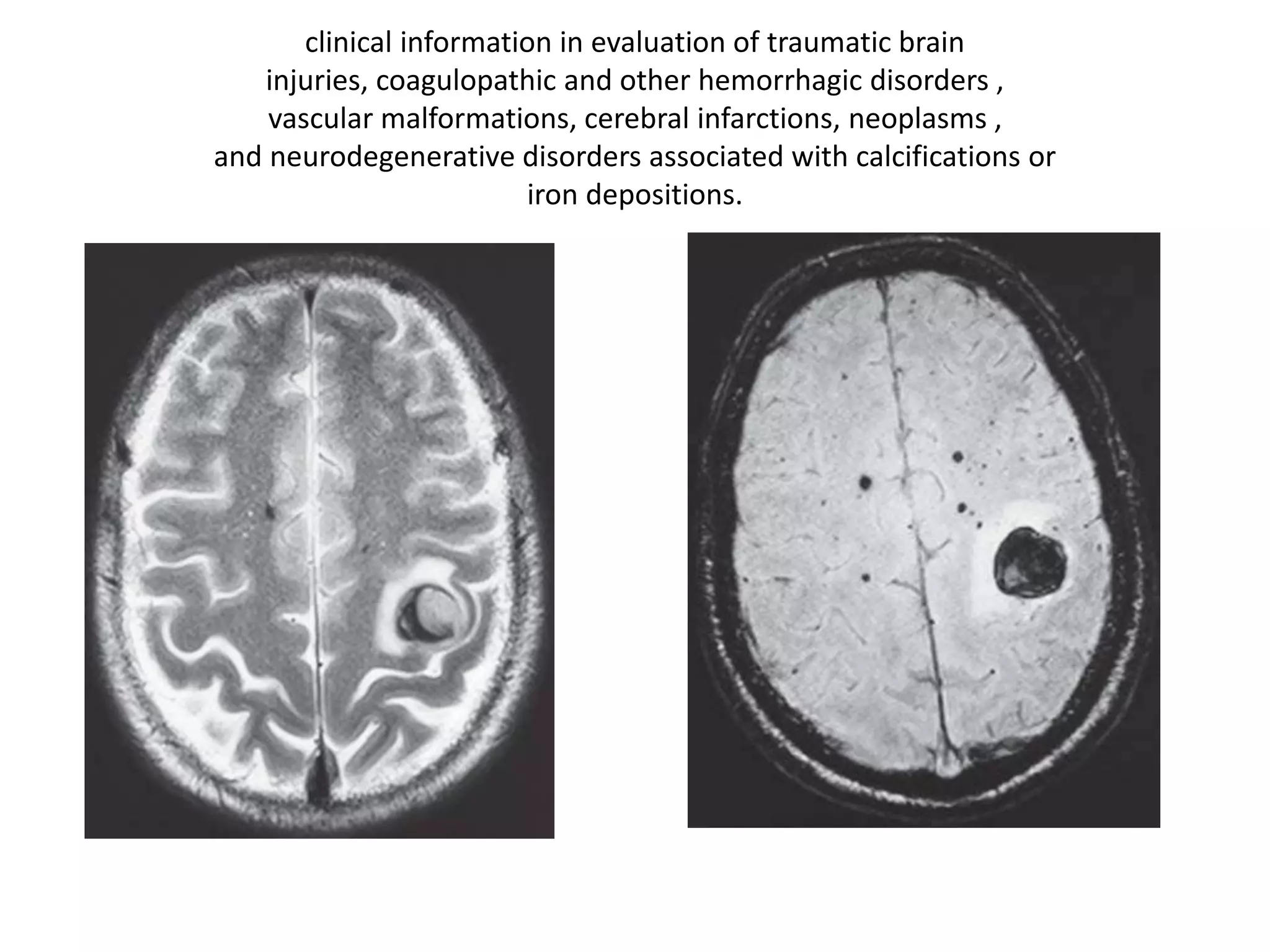
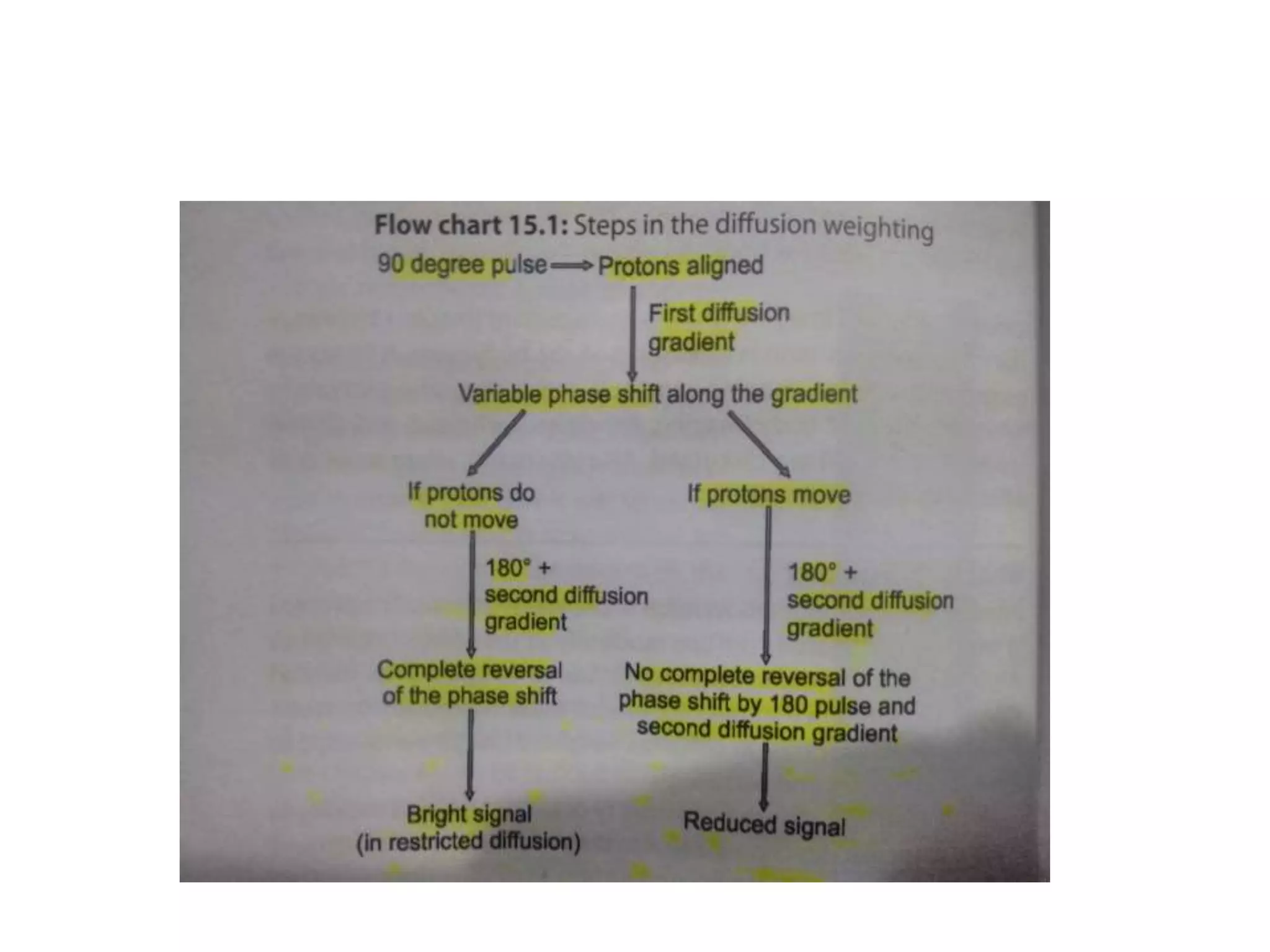
ADVANCES IN NEUROIMAGING:
MRI
Dr Fahad Shafi
Four basic steps in MRI
• PLACING THE PATIENT IN A MAGNET
• SENDING RF PULSES
• RECEIVING RF PULSES
• TRANSFORMATION OF SIGNALS INTO
IMAGES BY ACOMPLEX PROCESS
1. Large ‘Field of Viewing’ imaging.
2. High Field strength MR imaging.
3. Efficient Data processing techniques.
4. Improvement in Pulse sequences.
Improvements in MR hardware and
Soft ware technology
• LARGE FIELD OF VIEW IMAGING
Development of sliding or rolling table
platform or a multi-coil technique using a
combination of surface coils in position
allows
unlimited field of view (FOV) for whole
body imaging
T2-weighted sagittal section of spine with excellent artifact free
depiction
HIGH FIELD STRENGTH MR IMAGING
(3T AND BEYOND)
• A major advantage is improved signal to
noise ratio (SNR) which increases linearly
with field strength, thus increasing signal.
• Speed and resolution can be traded
judiciously, i.e. imaging time can be
reduced for a similar SNR as 1.5 T or spatial
resolution increased for an equal imaging
time on both
• small lesion detection, i.e. multiple
sclerosis, evaluation of epilepsy, diffusion
tensor imaging, MR angiography, fast
spectroscopic imaging and techniques
exploiting susceptibility effects, i.e. BOLD
and perfusion imaging.
IMPROVEMENTS IN PULSE SEQUENCES
• ‘Scan time = TR × number of phase encode
steps × signal averages’.
• Development of newer pulse sequences was
directed towards overcoming these
problems
• Low flip angle imaging (Gradient echo) was
the first major development towards
reducing scan time.
• Reduction in flip angle and use of gradient
recall of echo instead of 180º rephasing
pulse allowed shorter TR and significant
reduction in acquisition time.
EFFICIENT DATA PROCESSING
TECHNIQUES
• Along with MR hardware, more efficient
methods of data processing were also
developed simultaneously
• unprocessed 2D data set prior to Fourier
transformation referred to as K space map is
a stacked plot of horizontally oriented
phase encoded views (Ky), the vertical arm
(Kx) being the frequency axis.
• Multiple lines of K space in the same TR can be
acquired by using differently phase encoded
echoes as in fast spin echo (FSE) and/or by use
of oscillating gradients as in single shot
techniques like EPI
• two halves of the K space data (top to bottom
and left to right) are symmetrical. Thus, less
than full data can be acquired and the
remaining part interpolated from it as is used
in HASTE sequence
(A) Axial T2 FSE section in an uncooperative child.
HASTE imaging allows diagnostic good quality images in spite of
movements (B)
• ‘keyhole technique
• PROPELLER
• BLADE
CLASSIFICATION OF THE PULSE
SEQUENCES
FAST SPIN ECHO (FSE)
• Originally called rapid acquisition with relaxation enhancement
(RARE) by Henning in 1986;
• FSE was one of the most important advances in MRI.
• In FSE a train of multiple spin echoes with different phase encoding
steps are generated from multiple closely applied 180° RF pulses to fill
up the K space. The number of echoes (ETL) utilized are directly
proportional to the reduction of time.
• As the number of echoes is increased the SNR falls, however larger
matrix size and more signal averaging compensate to improve the
SNR even at small FOV.
Single Shot Techniques of FSE
• HASTE
• SSFSE
FLUID ATTENUATED INVERSION
RECOVERY (FLAIR)
• Brain pathologies with intermediate T2
times are poorly visualized if they are
located near the CSF interface. FLAIR being
heavily T2 weighted improves conspicuity of
such lesions after suppressing the CSF.
• Major indication of FLAIR imaging is in
evaluation of multiple sclerosis plaques
particularly those situated near CSF interface,
• useful in imaging neonatal hypoxic brain
injury, epidermoid cysts, (differentiates them
from arachnoid cysts), dysplasias, subcortical
diffuse axonal injuries (superior to GRE for
nonhemorrhagic lesions), encephalitis and
brain tumors
T2 coronal FLAIR section (A) hyperintense signal in the
sylvian fissures on both sides due to unsuspected subarachnoid
hemorrhage in a patient with anterior communicating artery aneurysm. T1
SE axial section (B) does not give any clue to the hemorrhage
MEDIUM INVERSION MPRAGE
Gradient Echo Imaging (GRE) and its
Variants
• Instead of using a 180 degree refocusing
pulse, a gradient echo is formed by
reversing the polarity of the frequency
encoded gradient. This prototype fast
sequence using short TR and TE, yielded
images at less than one second per slice
SPOILED/INCOHERENT GRE
• FLASH/SPGR
• Useful in in phase and out phase imaging
• TOF MR angio sequences
• 3D versions like FLASH and VIBE in
multiphase post contrast T1 weighted
images
STEADYSTATE SEQUENCES
• Post excitation refocussed FISP
• Pre excitation refocussed PSIF
• Fully refocussed `True FISP
SS sequences can acquired within single
breath hold.Useful for cardiac imaging.
Highest possible SNR but lack internal spatial
resolution.
SS variants of GRE
CISS CP angles
Susceptibility Weighted Imaging (SWI)
• tissues of higher susceptibility distort the
magnetic field and become out of phase
• Unlike initial experience with Spoiled GRE,
with advent of 3T and parallel imaging, it is
now possible to image the entire brain with
SWI in a short time.
clinical information in evaluation of traumatic brain
injuries, coagulopathic and other hemorrhagic disorders ,
vascular malformations, cerebral infarctions, neoplasms ,
and neurodegenerative disorders associated with calcifications or
iron depositions.
Cranial and Extracranial MR
Angiography (MRA)
• MRA uses inflow effects of blood in 2D
and 3D TOF angiography or phase contrast
technique in PC MRA.
• TOF MRA provides satisfactory images of
extra and intracranial vasculature and is
recommended for screening of aneurysms
in asymptomatic patients
Time of flight MRA; Excellent angiogram due to background
suppression and better flow related signal leading to better distal smaller
vessel visibility
CEMRA 0.2mmol/kg
Echo Planar Imaging
• Ultrafast imaging technique EPI was
originally described by Mansfield
• EPI technique involves very rapid gradient
reversal (instead of the 180° pulse used in
FSE) to acquire multiple phase encoding
echoes that form a complete image in one
TR during a single T2* decay (approximately
20-100 ms in brain)
• Being extremely fast allow study of dynamic
processes and motion free images, i.e. brain
scan of uncooperative patients, breath hold
imaging of the abdomen and heart
Diffusion Studies
• Depends on molecular motion of water.
• As initially described by Stejskal and Tanner
in 1965, spin echo T2 EPI sequences can be
sensitized to random diffusion of water
molecules using bipolar gradients of equal
magnitude and opposite polarity
The primary use of DWI has been in brain imaging due
to its
exquisitely unique sensitivity for ischemic stroke
• diagnosing abscesses ,encephalitides and
diffuse axonal injuries
• characterizing tumors, i.e. differentiating
epidermoids from cysts, by showing restricted
diffusion in hypercellular tumors, i.e.
lymphoma, malignant meningioma,
differentiating necrotic from solid enhancing
components, radiation necrosis from recurrent
tumor
Diffusion Tensor Imaging (DTI, Tractography or
Fiber Tracking)
• DTI measures magnitude of ADC in the
preferred direction of water diffusion and
also perpendicular to it.
• The resultant image shows white matter
tracts very well hence called tractography.
Color-encoded maps
Red: left to right; Blue: Cranial to caudal
Green: Anterior to posterior.
• Useful for assessment of relationship of
tracts with tumors , tumor invasion of
tracts , and preoperative planning.
• Also used to evaluate white matter tracts in
various congenital anomalies and dysplasia.
Perfusion Weighted Imaging (PWI)
• Perfusion imaging measures signal reduction
induced in the brain during passage of injected
paramagnetic contrast agents which
induce magnetic susceptibility effects (T2*).
• Yields regional cerebral blood volume (rCBV).
Similarly mean transit time (MTT), total blood
flow (rCBF), time to arrival (TTA) or time to
peak (TPP)
PWI in Stroke
• ischemic penumbra, i.e. ‘functionally
impaired but not irreversibly damaged’ area
around an established infarction is
identified when areas of PWI and DWI
defect are compared
• MRI stroke protocol should include T2 FSE
and FLAIR sections of brain followed by
MRA, DWI, PWI and a GRE sequence for
hemorrhage.
• This comprehensive protocol should take
less than 15 minutes on a modern state of
the art MR scans
MR P in brain tumours
• Grading tumours
• Differentiating therapy based necrosis and
residual/recurrent lesions
• Differentiating metastases from primary
tumours
MR SPECTROSCOPY
• An exciting application to non inasively
assess various metabolites or biochemicals
from body tissues
• This metabolite information is than used to
diagnose disease,monitoring and assessing
response to treatment.
• In Clinical use 1H and 31P spectroscopy .
• Chemical shift forms the basics.The
precessional frequency of protons is
determined by chemical environment.
• Proton in water will precess at different
frequency than proton in fat or same proton
in another metabolite like NAA
METABOLITES OF 1H
• NAA, Cr , Cho , Mi, Lac , Glx, Lipids,
Aminoacids , Glucose, GABA
• Clinical uses;
• Brain tumours
• Neonatal hypoxia
• Metabolic disorders
• Stroke
• Epilepsy, MS to name a few
(A) Post-contrast T1W MR image of a postoperative and postradiotherapy of infiltrating
astrocytoma showing irregular ring
enhancing lesion in the postoperative location (B) CSI 1H-MRS(TE = 135 ms) of the lesion shows
marked increase in the Choline/NAA ratios
with no significant lactate peaks suggestive of recurrent tumor
THANK YOU

Advances in neuroimaging

  • 1.
  • 2.
    Four basic stepsin MRI • PLACING THE PATIENT IN A MAGNET • SENDING RF PULSES • RECEIVING RF PULSES • TRANSFORMATION OF SIGNALS INTO IMAGES BY ACOMPLEX PROCESS
  • 3.
    1. Large ‘Fieldof Viewing’ imaging. 2. High Field strength MR imaging. 3. Efficient Data processing techniques. 4. Improvement in Pulse sequences.
  • 4.
    Improvements in MRhardware and Soft ware technology • LARGE FIELD OF VIEW IMAGING Development of sliding or rolling table platform or a multi-coil technique using a combination of surface coils in position allows unlimited field of view (FOV) for whole body imaging
  • 5.
    T2-weighted sagittal sectionof spine with excellent artifact free depiction
  • 6.
    HIGH FIELD STRENGTHMR IMAGING (3T AND BEYOND) • A major advantage is improved signal to noise ratio (SNR) which increases linearly with field strength, thus increasing signal. • Speed and resolution can be traded judiciously, i.e. imaging time can be reduced for a similar SNR as 1.5 T or spatial resolution increased for an equal imaging time on both
  • 7.
    • small lesiondetection, i.e. multiple sclerosis, evaluation of epilepsy, diffusion tensor imaging, MR angiography, fast spectroscopic imaging and techniques exploiting susceptibility effects, i.e. BOLD and perfusion imaging.
  • 9.
    IMPROVEMENTS IN PULSESEQUENCES • ‘Scan time = TR × number of phase encode steps × signal averages’. • Development of newer pulse sequences was directed towards overcoming these problems
  • 10.
    • Low flipangle imaging (Gradient echo) was the first major development towards reducing scan time. • Reduction in flip angle and use of gradient recall of echo instead of 180º rephasing pulse allowed shorter TR and significant reduction in acquisition time.
  • 11.
    EFFICIENT DATA PROCESSING TECHNIQUES •Along with MR hardware, more efficient methods of data processing were also developed simultaneously • unprocessed 2D data set prior to Fourier transformation referred to as K space map is a stacked plot of horizontally oriented phase encoded views (Ky), the vertical arm (Kx) being the frequency axis.
  • 12.
    • Multiple linesof K space in the same TR can be acquired by using differently phase encoded echoes as in fast spin echo (FSE) and/or by use of oscillating gradients as in single shot techniques like EPI • two halves of the K space data (top to bottom and left to right) are symmetrical. Thus, less than full data can be acquired and the remaining part interpolated from it as is used in HASTE sequence
  • 13.
    (A) Axial T2FSE section in an uncooperative child. HASTE imaging allows diagnostic good quality images in spite of movements (B)
  • 14.
    • ‘keyhole technique •PROPELLER • BLADE
  • 15.
    CLASSIFICATION OF THEPULSE SEQUENCES
  • 16.
    FAST SPIN ECHO(FSE) • Originally called rapid acquisition with relaxation enhancement (RARE) by Henning in 1986; • FSE was one of the most important advances in MRI. • In FSE a train of multiple spin echoes with different phase encoding steps are generated from multiple closely applied 180° RF pulses to fill up the K space. The number of echoes (ETL) utilized are directly proportional to the reduction of time. • As the number of echoes is increased the SNR falls, however larger matrix size and more signal averaging compensate to improve the SNR even at small FOV.
  • 17.
    Single Shot Techniquesof FSE • HASTE • SSFSE
  • 18.
    FLUID ATTENUATED INVERSION RECOVERY(FLAIR) • Brain pathologies with intermediate T2 times are poorly visualized if they are located near the CSF interface. FLAIR being heavily T2 weighted improves conspicuity of such lesions after suppressing the CSF.
  • 20.
    • Major indicationof FLAIR imaging is in evaluation of multiple sclerosis plaques particularly those situated near CSF interface, • useful in imaging neonatal hypoxic brain injury, epidermoid cysts, (differentiates them from arachnoid cysts), dysplasias, subcortical diffuse axonal injuries (superior to GRE for nonhemorrhagic lesions), encephalitis and brain tumors
  • 21.
    T2 coronal FLAIRsection (A) hyperintense signal in the sylvian fissures on both sides due to unsuspected subarachnoid hemorrhage in a patient with anterior communicating artery aneurysm. T1 SE axial section (B) does not give any clue to the hemorrhage
  • 22.
  • 23.
    Gradient Echo Imaging(GRE) and its Variants • Instead of using a 180 degree refocusing pulse, a gradient echo is formed by reversing the polarity of the frequency encoded gradient. This prototype fast sequence using short TR and TE, yielded images at less than one second per slice
  • 24.
    SPOILED/INCOHERENT GRE • FLASH/SPGR •Useful in in phase and out phase imaging • TOF MR angio sequences • 3D versions like FLASH and VIBE in multiphase post contrast T1 weighted images
  • 25.
    STEADYSTATE SEQUENCES • Postexcitation refocussed FISP • Pre excitation refocussed PSIF • Fully refocussed `True FISP SS sequences can acquired within single breath hold.Useful for cardiac imaging. Highest possible SNR but lack internal spatial resolution.
  • 26.
    SS variants ofGRE CISS CP angles
  • 27.
    Susceptibility Weighted Imaging(SWI) • tissues of higher susceptibility distort the magnetic field and become out of phase • Unlike initial experience with Spoiled GRE, with advent of 3T and parallel imaging, it is now possible to image the entire brain with SWI in a short time.
  • 28.
    clinical information inevaluation of traumatic brain injuries, coagulopathic and other hemorrhagic disorders , vascular malformations, cerebral infarctions, neoplasms , and neurodegenerative disorders associated with calcifications or iron depositions.
  • 29.
    Cranial and ExtracranialMR Angiography (MRA) • MRA uses inflow effects of blood in 2D and 3D TOF angiography or phase contrast technique in PC MRA. • TOF MRA provides satisfactory images of extra and intracranial vasculature and is recommended for screening of aneurysms in asymptomatic patients
  • 30.
    Time of flightMRA; Excellent angiogram due to background suppression and better flow related signal leading to better distal smaller vessel visibility
  • 31.
  • 32.
    Echo Planar Imaging •Ultrafast imaging technique EPI was originally described by Mansfield • EPI technique involves very rapid gradient reversal (instead of the 180° pulse used in FSE) to acquire multiple phase encoding echoes that form a complete image in one TR during a single T2* decay (approximately 20-100 ms in brain)
  • 33.
    • Being extremelyfast allow study of dynamic processes and motion free images, i.e. brain scan of uncooperative patients, breath hold imaging of the abdomen and heart
  • 34.
    Diffusion Studies • Dependson molecular motion of water. • As initially described by Stejskal and Tanner in 1965, spin echo T2 EPI sequences can be sensitized to random diffusion of water molecules using bipolar gradients of equal magnitude and opposite polarity
  • 36.
    The primary useof DWI has been in brain imaging due to its exquisitely unique sensitivity for ischemic stroke
  • 37.
    • diagnosing abscesses,encephalitides and diffuse axonal injuries • characterizing tumors, i.e. differentiating epidermoids from cysts, by showing restricted diffusion in hypercellular tumors, i.e. lymphoma, malignant meningioma, differentiating necrotic from solid enhancing components, radiation necrosis from recurrent tumor
  • 38.
    Diffusion Tensor Imaging(DTI, Tractography or Fiber Tracking) • DTI measures magnitude of ADC in the preferred direction of water diffusion and also perpendicular to it. • The resultant image shows white matter tracts very well hence called tractography.
  • 39.
    Color-encoded maps Red: leftto right; Blue: Cranial to caudal Green: Anterior to posterior.
  • 40.
    • Useful forassessment of relationship of tracts with tumors , tumor invasion of tracts , and preoperative planning. • Also used to evaluate white matter tracts in various congenital anomalies and dysplasia.
  • 41.
    Perfusion Weighted Imaging(PWI) • Perfusion imaging measures signal reduction induced in the brain during passage of injected paramagnetic contrast agents which induce magnetic susceptibility effects (T2*). • Yields regional cerebral blood volume (rCBV). Similarly mean transit time (MTT), total blood flow (rCBF), time to arrival (TTA) or time to peak (TPP)
  • 42.
    PWI in Stroke •ischemic penumbra, i.e. ‘functionally impaired but not irreversibly damaged’ area around an established infarction is identified when areas of PWI and DWI defect are compared
  • 45.
    • MRI strokeprotocol should include T2 FSE and FLAIR sections of brain followed by MRA, DWI, PWI and a GRE sequence for hemorrhage. • This comprehensive protocol should take less than 15 minutes on a modern state of the art MR scans
  • 46.
    MR P inbrain tumours • Grading tumours • Differentiating therapy based necrosis and residual/recurrent lesions • Differentiating metastases from primary tumours
  • 47.
    MR SPECTROSCOPY • Anexciting application to non inasively assess various metabolites or biochemicals from body tissues • This metabolite information is than used to diagnose disease,monitoring and assessing response to treatment.
  • 48.
    • In Clinicaluse 1H and 31P spectroscopy . • Chemical shift forms the basics.The precessional frequency of protons is determined by chemical environment. • Proton in water will precess at different frequency than proton in fat or same proton in another metabolite like NAA
  • 49.
    METABOLITES OF 1H •NAA, Cr , Cho , Mi, Lac , Glx, Lipids, Aminoacids , Glucose, GABA • Clinical uses; • Brain tumours • Neonatal hypoxia • Metabolic disorders • Stroke • Epilepsy, MS to name a few
  • 50.
    (A) Post-contrast T1WMR image of a postoperative and postradiotherapy of infiltrating astrocytoma showing irregular ring enhancing lesion in the postoperative location (B) CSI 1H-MRS(TE = 135 ms) of the lesion shows marked increase in the Choline/NAA ratios with no significant lactate peaks suggestive of recurrent tumor
  • 51.