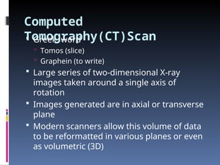
Computed
Tomography(CT)Scan
Greek word
Tomos (slice)
Graphein (to write)
Large series of two-dimensional X-ray
images taken around a single axis of
rotation
Images generated are in axial or transverse
plane
Modern scanners allow this volume of data
to be reformatted in various planes or even
as volumetric (3D)
3.
History
Invented byGodfrey Newbold Hounsfield
in Hayes, England using X-rays in 1972
“The greatest legacy” of the Beatles (the
massive profits from their record sales
enabled EMI to fund scientific research)
Allan McLeod Cormack of Tufts University
independently invented a similar process
and they shared a Nobel Prize in medicine
in 1979
4.
Prototype
Took 160parallel readings through 180
angles, each 1° apart, with each scan taking
a little over five minutes
The images from these scans took 2.5
hours to be processed
This scanner required the use of a water-
filled Perspex tank with a pre-shaped
rubber “head-cap” at the front, which
enclosed the patient's head
The water-tank was used to reduce the
dynamic range of the radiation reaching
the detectors
6.
History (Cont..)
FirstEMI-Scanner was installed in Atkinson
Morley's Hospital in Wimbledon, England
First patient brain-scan was made in 1972
In the U.S., the first installation was at the
Mayo Clinic
The images were relatively low resolution,
being composed of a matrix of only 80 x 80
pixels
CVA & Hemorrhages
Diagnosis of cerebrovascular accidents
and intracranial hemorrhage
Is the most frequent reason for a "head
CT" or "CT brain
Scanning is done with or without
intravenous contrast agents
Does not exclude infarct in the acute
stage of a stroke, but is useful to
exclude a bleed
9.
Tumors
Detection oftumors
CT scanning with IV contrast is
occasionally used but is less sensitive
than magnetic resonance imaging
10.
ed Intra-Cranial
Pressure
Canalso be used to detect increases in
intracranial pressure
Before lumbar puncture or to evaluate
the functioning of a
ventriculoperitoneal shunt
11.
Vascular & BBBarrier
IV contrast helps reveal vascular
anomalies like
Arteriovenous malformations
Aneurysms
Lesions producing abnormalities of Blood
Brain Barrier like
Abscesses
Certain Tumors
Demyelinating Lesions
Other Indications
Settingof trauma for evaluating facial and
skull fractures.
In the head/neck/mouth area, CT scanning
is used for
Surgical planning for craniofacial and
dentofacial deformities
Evaluation of cysts and some tumors of the
jaws/paranasal sinuses/nasal cavity/orbits
Diagnosis of the causes of chronic sinusitis,
and for planning of dental implant
reconstruction.
Advantages over
projection Radiography
Completely eliminates the superimposition of
images of structures outside the area of interest
Because of the inherent high-contrast resolution
of CT, differences between tissues that differ in
physical density by less than 1% can be
distinguished
Data from a single CT imaging procedure can be
viewed as images in the axial, coronal, or
sagittal planes, depending on the diagnostic
task
16.
Radiation exposure
Regardedas a moderate to high
radiation diagnostic technique
radiation dose depends on:
Volume scanned
Patient build
Number and type of scan sequences
Desired resolution and image quality
In children, produces increases in the
probability of lifetime cancer mortality
17.
Adverse reactions to
contrastagents
Certain patients may experience severe
and potentially life-threatening allergic
reactions to the contrast dye
May induce kidney damage (contrast
nephropathy)
Contraindicated in Moderate Renal
Failure
However can be done in severe renal
failure requiring dialysis
Limitations Of OldCT-
Scan
Unable to visualize structures immediately
adjacent to bone because of artifacts
Inferior surface of frontal and temporal
lobes as well as entire posterior fossa is
not best visualized
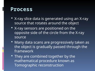
Process
X-ray slicedata is generated using an X-ray
source that rotates around the object
X-ray sensors are positioned on the
opposite side of the circle from the X-ray
source
Many data scans are progressively taken as
the object is gradually passed through the
framework
They are combined together by the
mathematical procedure known as
Tomographic reconstruction
22.
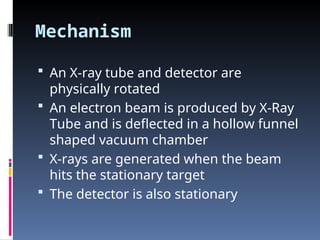
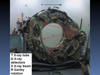
Mechanism
An X-raytube and detector are
physically rotated
An electron beam is produced by X-Ray
Tube and is deflected in a hollow funnel
shaped vacuum chamber
X-rays are generated when the beam
hits the stationary target
The detector is also stationary
24.
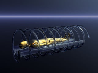
Spiral CT
Canprocess not only individual cross
sections but continuously changing cross
sections as the gantry, with the object to
be imaged, is slowly and smoothly slid
through the X-ray circle.
Their computer systems integrate the data
of the moving individual slices to generate
three dimensional volumetric information
(3D-CT scan)
26.
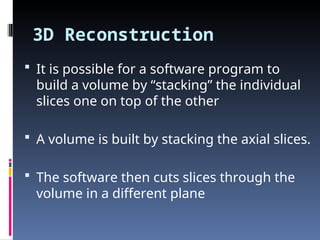
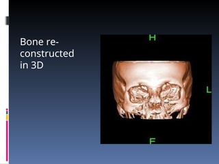
3D Reconstruction
Itis possible for a software program to
build a volume by “stacking” the individual
slices one on top of the other
A volume is built by stacking the axial slices.
The software then cuts slices through the
volume in a different plane
27.
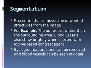
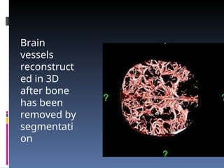
Segmentation
Procedure thatremoves the unwanted
structures from the image
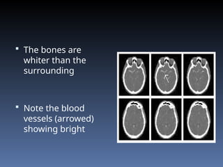
For Example, The bones are whiter than
the surrounding area. Blood vessels
also show brightly when injected with
iodine-based contrast agent
By segmentation, bone can be removed
and blood vessels can be seen in detail
28.
The bonesare
whiter than the
surrounding
Note the blood
vessels (arrowed)
showing bright
History
Inventor ofMRI was Paul Lauterbur
Named it – “zeugmatography”
A Greek term meaning “that which is used
for joining”
The term referred to the interaction
between the static and the gradient
magnetic fields necessary to create an
image
But, this name never caught on
33.
History (Cont..)
Laternamed as Nuclear Magnetic
Resonance Imaging (NMRI)
But, the word “nuclear” has been
associated with ionizing radiation
exposure
So, to prevent patients from making a
negative association between MRI and
ionizing radiation, the word has been
almost universally removed
34.
Technique
Relies onthe relaxation properties of excited
hydrogen nuclei (proton) in water and lipids.
Patient head is placed in a magnetic field that
forces protons in the brain to align
Series of radio frequency pulses are then applied
perpendicular to this field
Causes protons to change their alignment briefly
Then, emit energy as they relax back to previous
alignment
As the high-energy nuclei relax and realign they
emit energy at rates which are recorded to
provide information about the material they are
in
35.
Parameters of MRI
Image is created by using a selection of
image gaining parameters which include
1.The Spin proton Density – {No Relaxation
time}
2.T1 or Spin-Lattice relaxation time
3.T2 or Spin-Spin Relaxation time
36.
Proton densityreflects the numbers of
protons present in a tissue sample
T1 and T2 values are tissue specific
Imaging sequences can be varied to
highlight the T1 or T2 component of
signal
37.
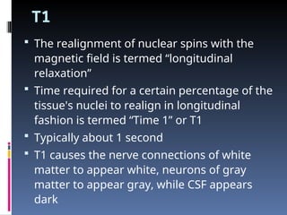
T1
The realignmentof nuclear spins with the
magnetic field is termed “longitudinal
relaxation”
Time required for a certain percentage of the
tissue's nuclei to realign in longitudinal
fashion is termed “Time 1” or T1
Typically about 1 second
T1 causes the nerve connections of white
matter to appear white, neurons of gray
matter to appear gray, while CSF appears
dark
38.
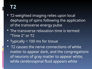
T2
T2-weighted imagingrelies upon local
dephasing of spins following the application
of the transverse energy pulse
The transverse relaxation time is termed
"Time 2" or T2
Typically < 100 ms for tissue
T2 causes the nerve connections of white
matter to appear dark, and the congregations
of neurons of gray matter to appear white,
while cerebrospinal fluid appears white
Inversion recovery
Relyheavily on the T1 component
Produce the best gray-white resolution
Best for visualizing anatomical detail
Very sensitive to differences between
normal and abnormal tissue
41.
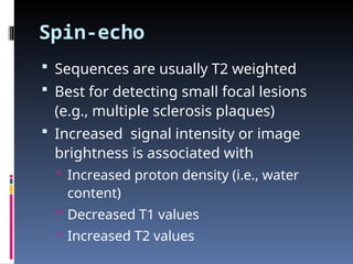
Spin-echo
Sequences areusually T2 weighted
Best for detecting small focal lesions
(e.g., multiple sclerosis plaques)
Increased signal intensity or image
brightness is associated with
Increased proton density (i.e., water
content)
Decreased T1 values
Increased T2 values
42.
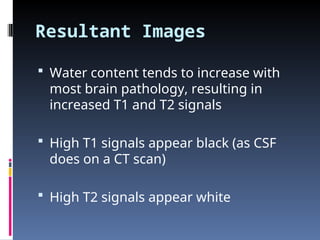
Resultant Images
Watercontent tends to increase with
most brain pathology, resulting in
increased T1 and T2 signals
High T1 signals appear black (as CSF
does on a CT scan)
High T2 signals appear white
43.
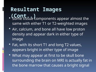
Resultant Images
(Cont..)
Sometissue components appear almost the
same with either T1 or T2-weighted images
Air, calcium, and bone all have low proton
density and appear dark in either type of
image
Fat, with its short T1 and long T2 values,
appears bright in either type of image
What may appear at first to be skull bone
surrounding the brain on MRI is actually fat in
the bone marrow that causes a bright signal
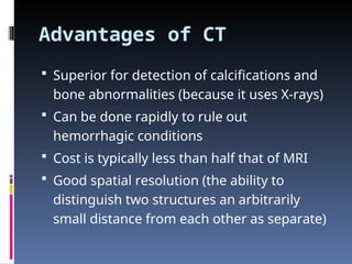
Advantages of CT
Superior for detection of calcifications and
bone abnormalities (because it uses X-rays)
Can be done rapidly to rule out
hemorrhagic conditions
Cost is typically less than half that of MRI
Good spatial resolution (the ability to
distinguish two structures an arbitrarily
small distance from each other as separate)
46.
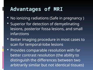
Advantages of MRI
No ionizing radiations (Safe in pregnancy )
Superior for detection of demyelinating
lesions, posterior fossa lesions, and small
infarctions
Better imaging procedure in most cases to
scan for temporal-lobe lesions
Provides comparable resolution with far
better contrast resolution (the ability to
distinguish the differences between two
arbitrarily similar but not identical tissues)
Principal
Diffusion MRImeasures the diffusion of
water molecules in biological tissues
Water Molecule inside the axon of a
neuron have a low probability of
crossing the myelin membrane
Therefore the molecule will move
principally along the axis of the neural
fiber
49.
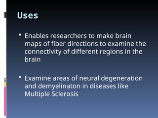
Uses
Enables researchersto make brain
maps of fiber directions to examine the
connectivity of different regions in the
brain
Examine areas of neural degeneration
and demyelinaton in diseases like
Multiple Sclerosis
50.
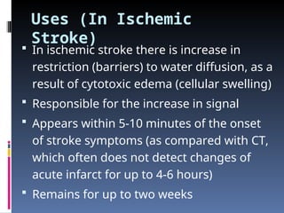
Uses (In Ischemic
Stroke)
In ischemic stroke there is increase in
restriction (barriers) to water diffusion, as a
result of cytotoxic edema (cellular swelling)
Responsible for the increase in signal
Appears within 5-10 minutes of the onset
of stroke symptoms (as compared with CT,
which often does not detect changes of
acute infarct for up to 4-6 hours)
Remains for up to two weeks
51.
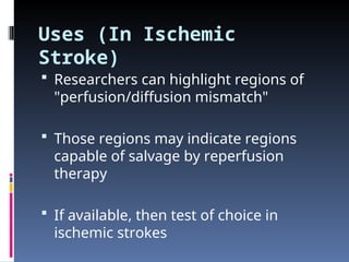
Uses (In Ischemic
Stroke)
Researchers can highlight regions of
"perfusion/diffusion mismatch"
Those regions may indicate regions
capable of salvage by reperfusion
therapy
If available, then test of choice in
ischemic strokes
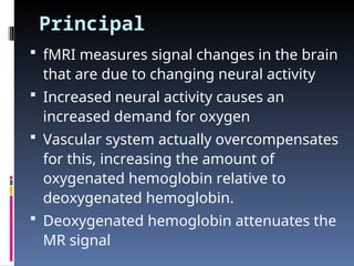
Principal
fMRI measuressignal changes in the brain
that are due to changing neural activity
Increased neural activity causes an
increased demand for oxygen
Vascular system actually overcompensates
for this, increasing the amount of
oxygenated hemoglobin relative to
deoxygenated hemoglobin.
Deoxygenated hemoglobin attenuates the
MR signal
54.
Principal (Cont..)
Vascularresponse leads to a signal
increase that is related to the neural
activity.
This mechanism is referred to as the
BOLD (blood-oxygen-level dependent)
effect
55.
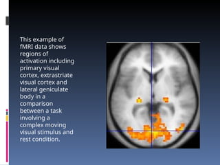
This example of
fMRIdata shows
regions of
activation including
primary visual
cortex, extrastriate
visual cortex and
lateral geniculate
body in a
comparison
between a task
involving a
complex moving
visual stimulus and
rest condition.
Contra-Indications
Pacemakers –(they can cause arrhythmia)
Other forms of medical or bio-stimulation
implants (Vagus nerve stimulators,
implanted cardio-defibrillators, insulin
pumps, cochlear implants etc.)
Ferromagnetic foreign bodies (e.g. shell
fragments)
Metallic implants (e.g. surgical prostheses,
aneurysm clips)
58.
Side-Effects
Hyperthermia –A powerful radio transmitter
is needed for excitation of proton spins. This
can heat the body significantly
Twitching in extremities - The rapid
switching (on and off) of the magnetic field
is capable of causing nerve stimulation
High acoustic noise – may reach 130 dB
(equivalent to a jet engine at take-off)
Asphyxia – If Helium is not properly
dissipated through vents
59.
Use in Pregnancy
Safe in pregnancy
However, as a precaution, pregnant
women undergo MRI only when
essential (particularly in the first
trimester )
Contrast agents (like gadolinium
compounds) should be avoided
60.
Claustrophobia
Potentially unpleasantto lie in (which is
often a long, narrow tube even up to one
hour)
Potential solutions include
Visiting the scanner to see the room and
practice lying on the table
Watching DVDs with a Head-mounted display
while in the machine
The use of open MRI
Use of sedation
For the most severe cases, general anesthesia
PET Scan
Itis a Nuclear medicine medical
imaging technique which produces a
three-dimensional image or map of
functional processes in the body
64.
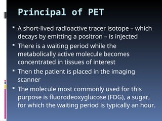
Principal of PET
A short-lived radioactive tracer isotope – which
decays by emitting a positron – is injected
There is a waiting period while the
metabolically active molecule becomes
concentrated in tissues of interest
Then the patient is placed in the imaging
scanner
The molecule most commonly used for this
purpose is fluorodeoxyglucose (FDG), a sugar,
for which the waiting period is typically an hour.
65.
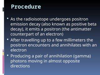
Procedure
As theradioisotope undergoes positron
emission decay (also known as positive beta
decay), it emits a positron (the antimatter
counterpart of an electron)
After travelling up to a few millimeters the
positron encounters and annihilates with an
electron
Producing a pair of annihilation (gamma)
photons moving in almost opposite
directions
66.
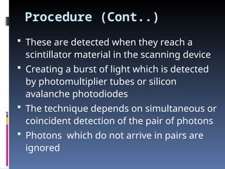
Procedure (Cont..)
Theseare detected when they reach a
scintillator material in the scanning device
Creating a burst of light which is detected
by photomultiplier tubes or silicon
avalanche photodiodes
The technique depends on simultaneous or
coincident detection of the pair of photons
Photons which do not arrive in pairs are
ignored
68.
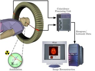
This figureshows how during the
annihilation process two photons are
emitted in diametrically opposing directions.
These photons are registered by the PET as
soon as they arrive at the detector ring
After the registration, the data is forwarded
to a processing unit which decides if two
registrered events are selected as a so-
called coincidence event.
All coincidences are fowarded to the image
processing unit where the final image data
is produced via image reconstruction
procedures
69.
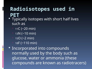
Radioisotopes used in
PET
Typically isotopes with short half lives
such as
11C (~20 min)
13N (~10 min)
15O (~2 min)
18F (~110 min)
Incorporated into compounds
normally used by the body such as
glucose, water or ammonia (these
compounds are known as radiotracers)
70.
Uses
Clinical oncology(medical imaging of
tumors and the search for metastases)
Various types of dementias
Tool to map normal human brain function
Changing of regional blood flow in various
anatomic structures
71.
Use in Neurology
Brain is normally a rapid user of glucose
Brain pathologies such as Alzheimer's
disease greatly decrease brain metabolism
of both glucose and oxygen in tandem
Standard FDG-PET of the brain (which
measures regional glucose use) is used to
differentiate Alzheimer's disease from other
dementing processes, and also to make
early diagnosis of Alzheimer's disease
72.
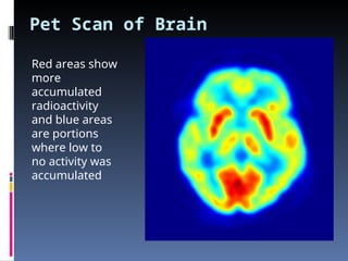
Pet Scan ofBrain
Red areas show
more
accumulated
radioactivity
and blue areas
are portions
where low to
no activity was
accumulated
73.
Use in Neuropsychology
To examine links between specific
psychological processes or disorders
and brain activity
74.
Use in psychiatry
Numerous compounds that bind selectively to
neuro-receptors of interest in biological
psychiatry have been radiolabeled with 11C or
18F
Radioligands that bind to dopamine receptors ,
serotonin receptors, opioid receptors (mu) and
other sites have been studied
Studies have been performed examining the
state of these receptors in patients of
schizophrenia, substance abuse, mood
disorders and other psychiatric conditions
75.
Safety
PET scanningis non-invasive, but it
does involve exposure to ionizing
radiation
So should be avoided in pregnancy
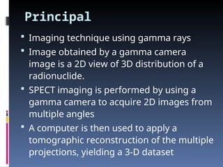
Principal
Imaging techniqueusing gamma rays
Image obtained by a gamma camera
image is a 2D view of 3D distribution of a
radionuclide.
SPECT imaging is performed by using a
gamma camera to acquire 2D images from
multiple angles
A computer is then used to apply a
tomographic reconstruction of the multiple
projections, yielding a 3-D dataset
79.
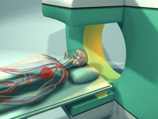
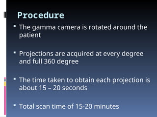
Procedure
The gammacamera is rotated around the
patient
Projections are acquired at every degree
and full 360 degree
The time taken to obtain each projection is
about 15 – 20 seconds
Total scan time of 15-20 minutes
80.
Uses
Tumor imaging
Infection (leukocyte) imaging
Thyroid imaging
Bone imaging
Information about localized function in
internal organs
Functional cardiac (diagnosis of ischemic
heart disease )
Brain imaging (dementia)


![MAGNETIC_RESONANCE.._IMAGING[MRI][1].pptx](https://cdn.slidesharecdn.com/ss_thumbnails/magneticresonanceimagingmri1-240903182728-4f857936-thumbnail.jpg?width=640&height=640&fit=bounds)


