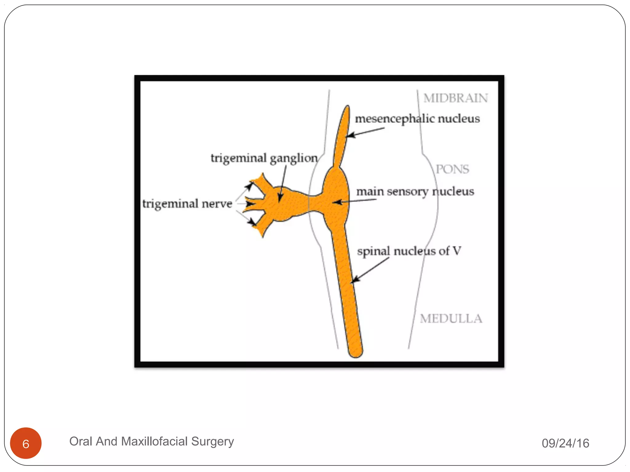
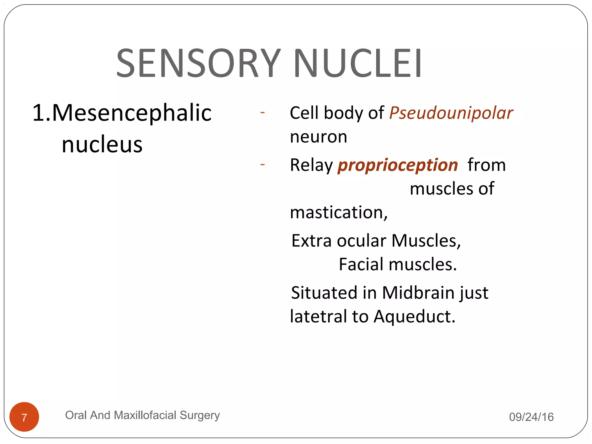
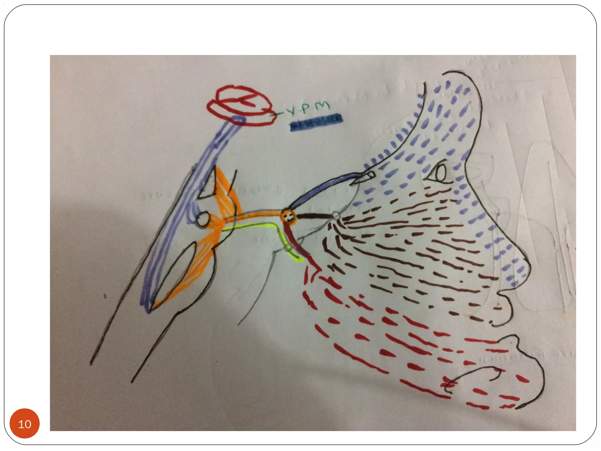
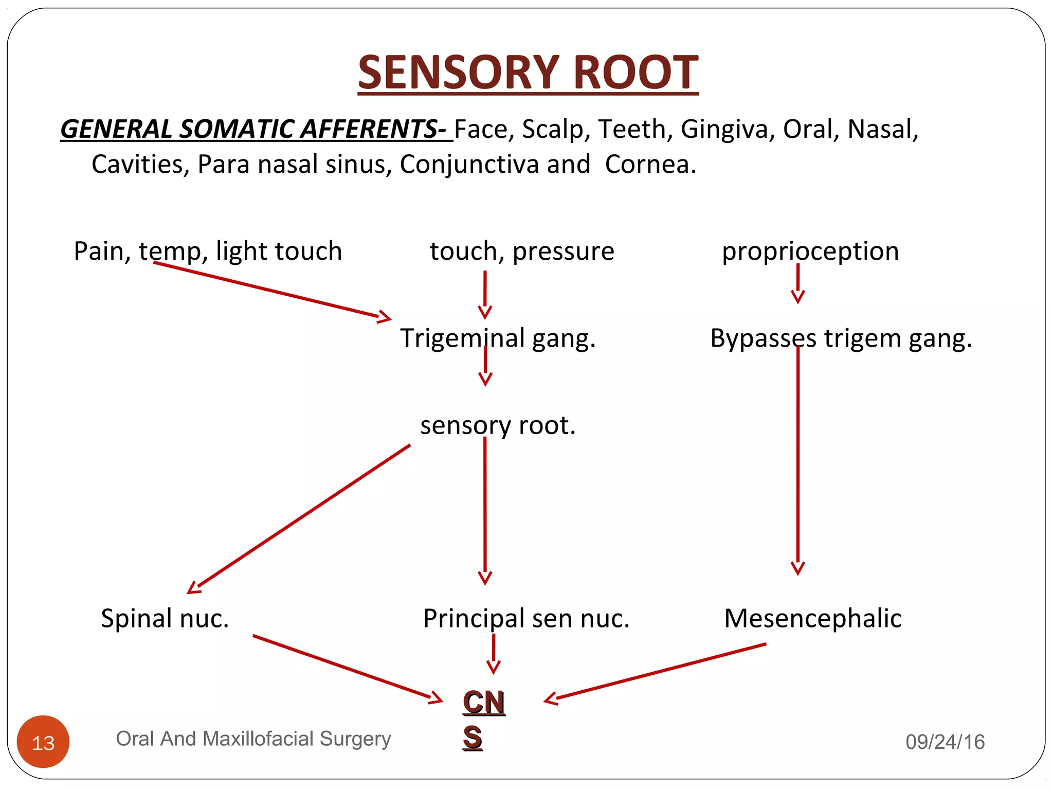
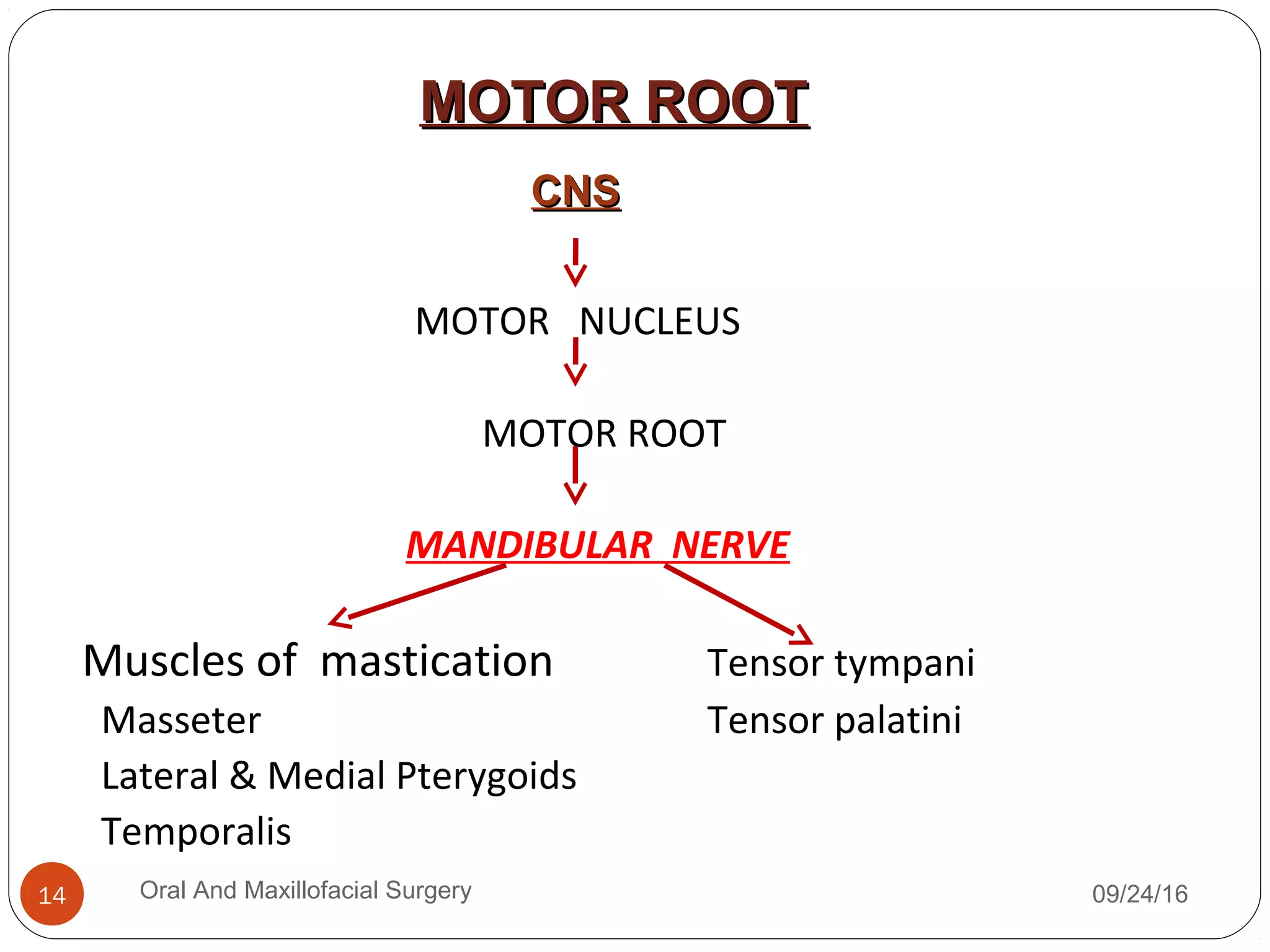
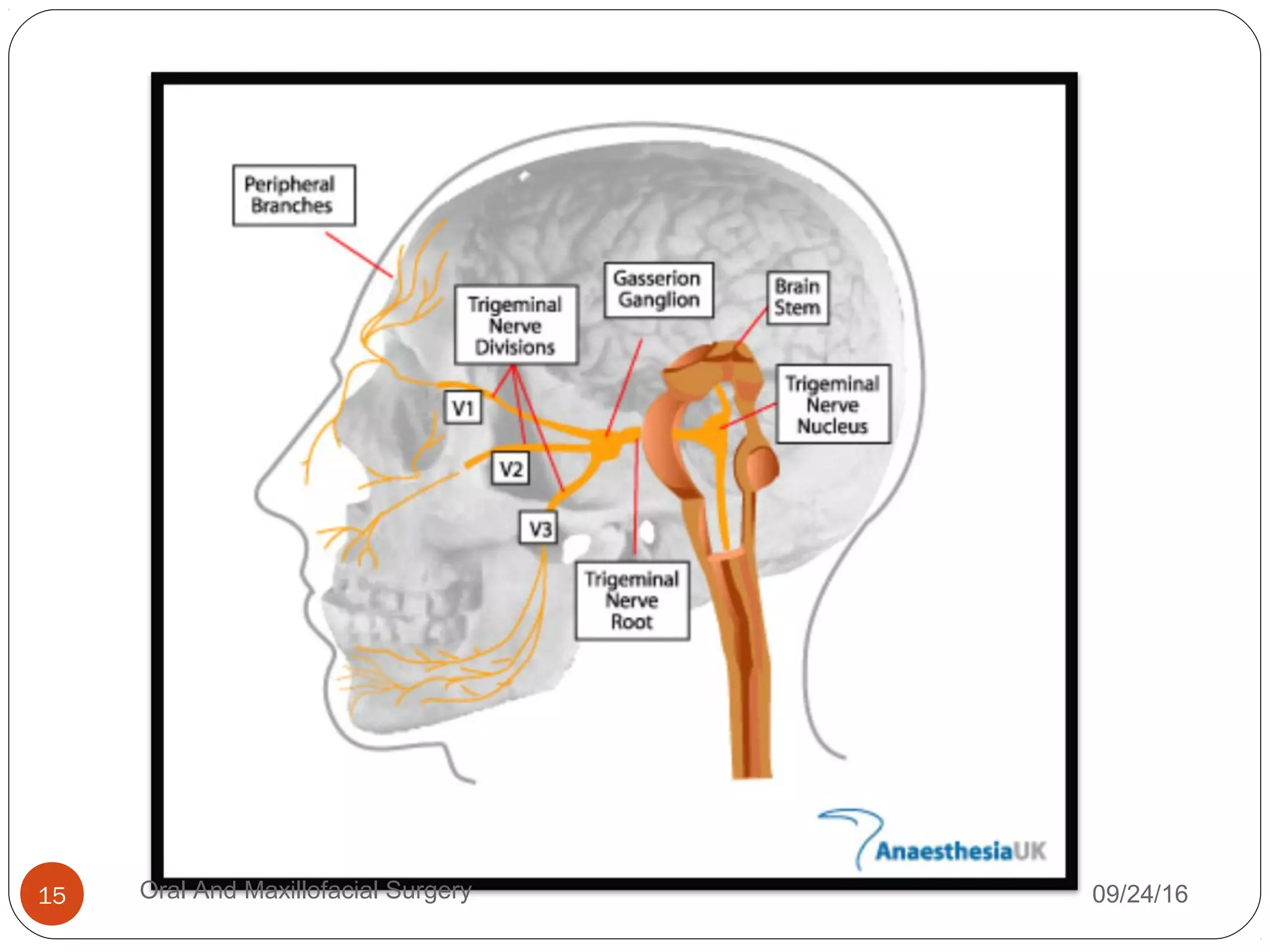
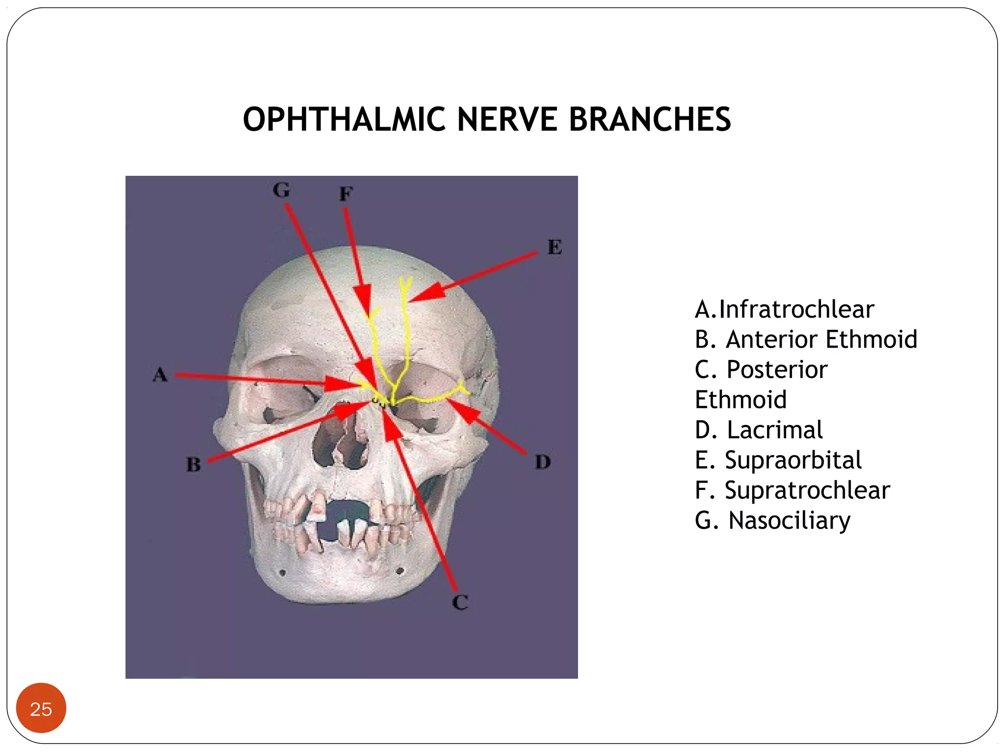
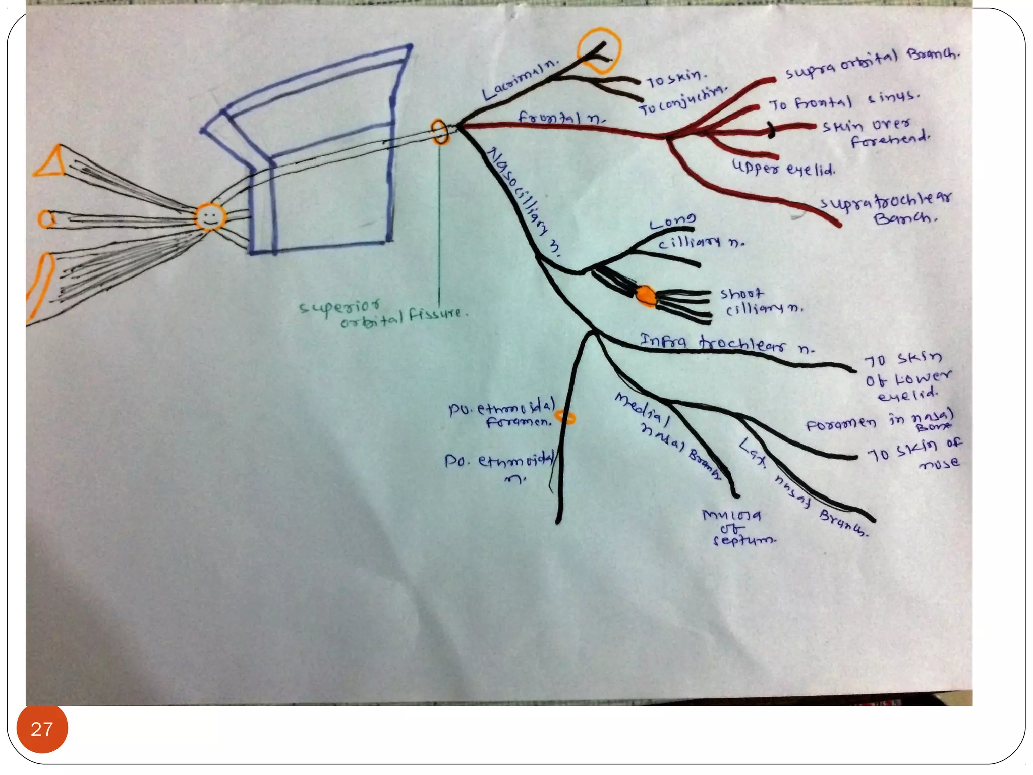
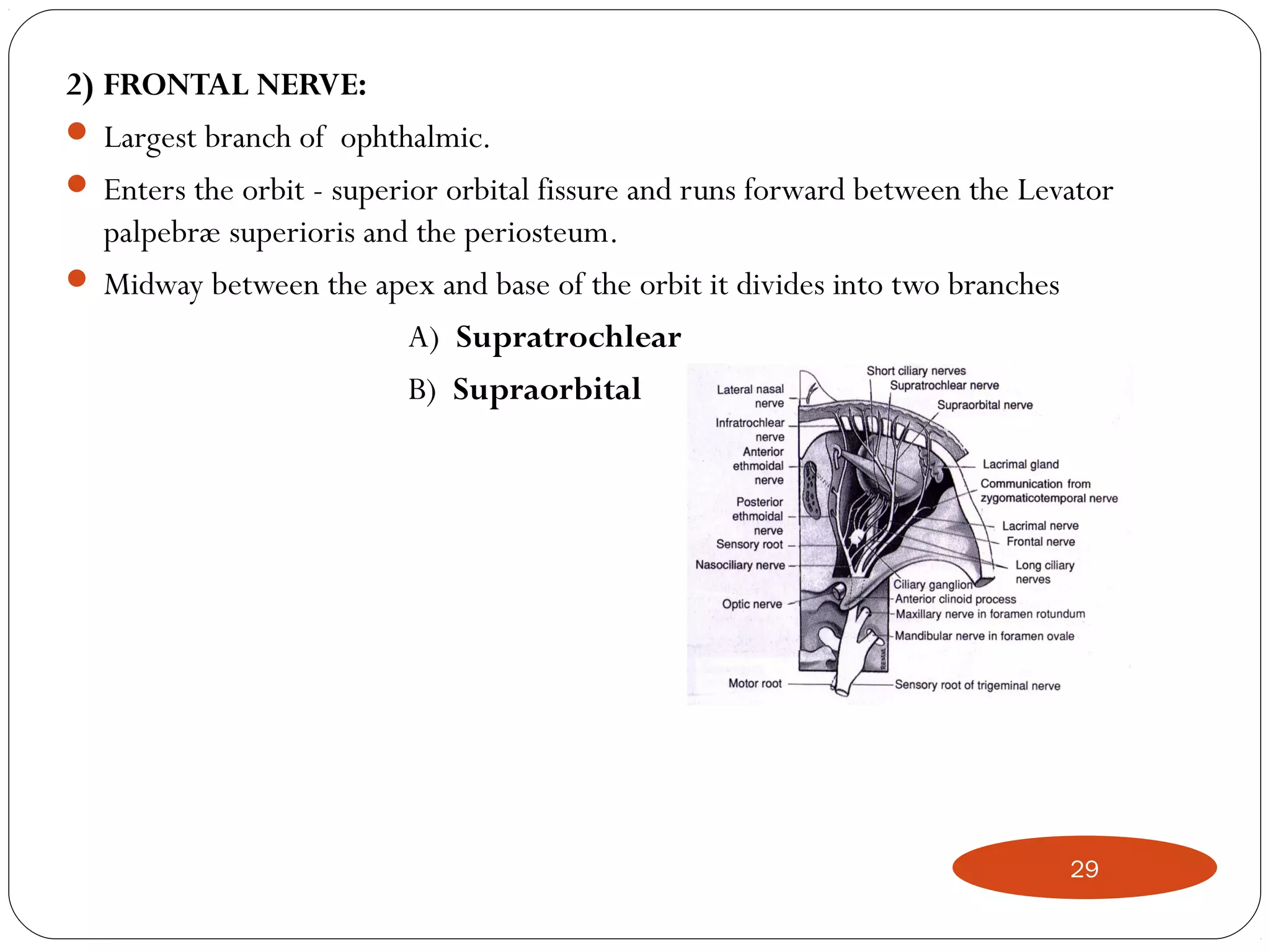
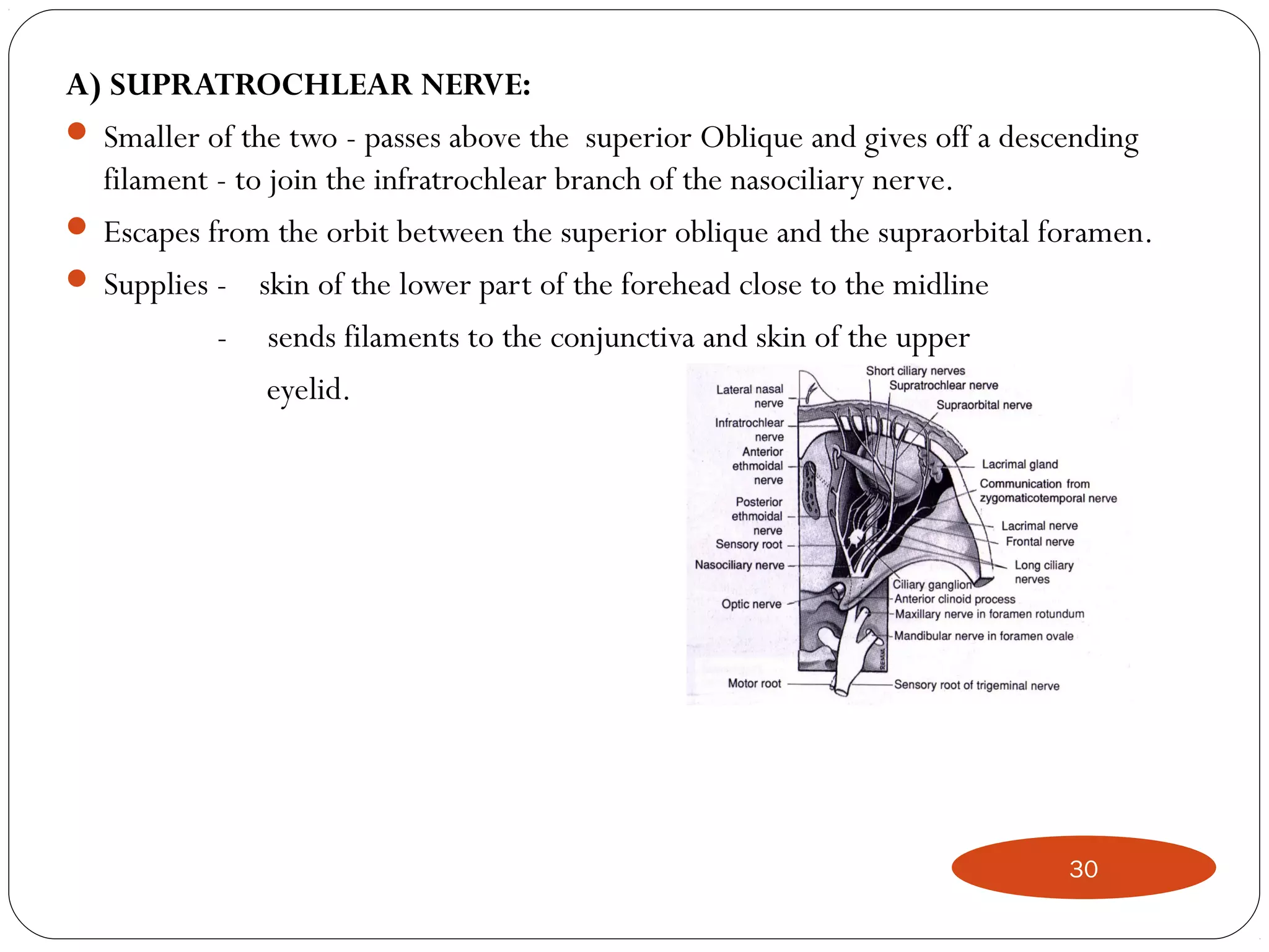
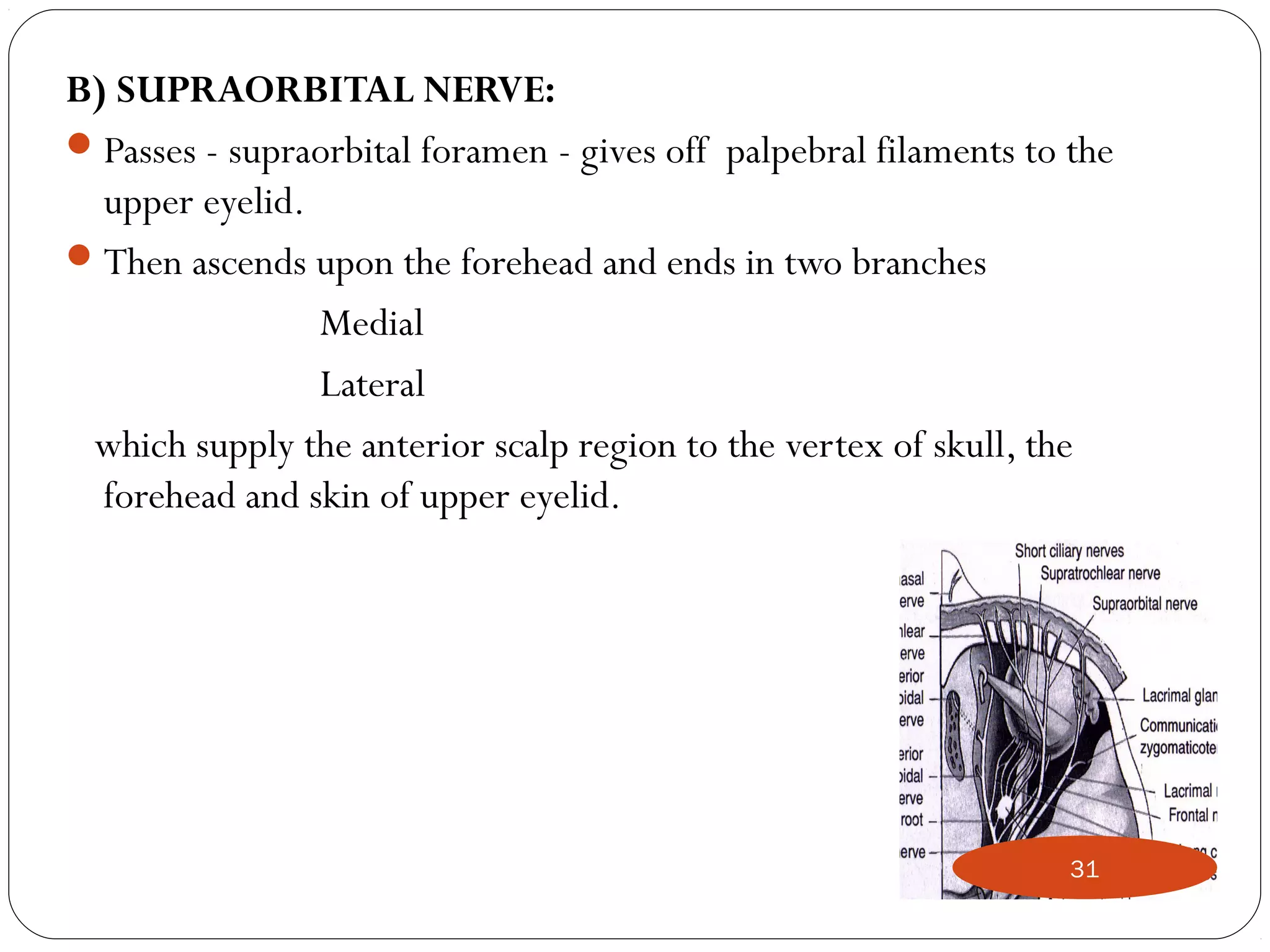
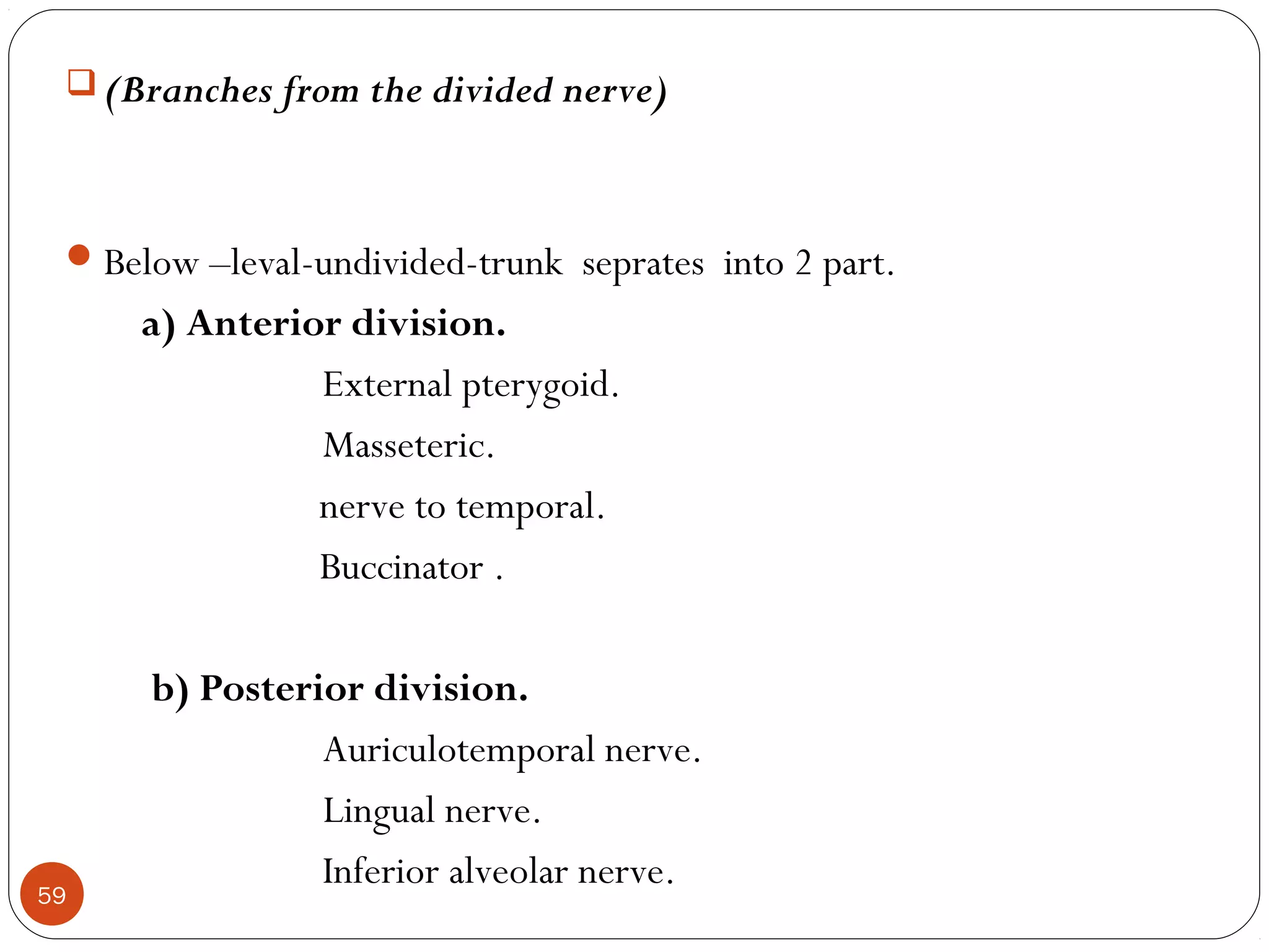
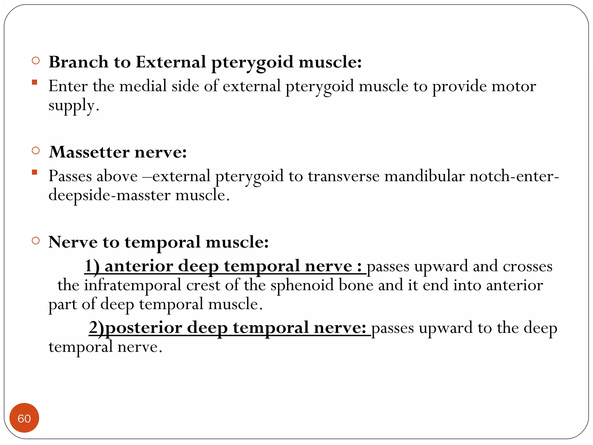
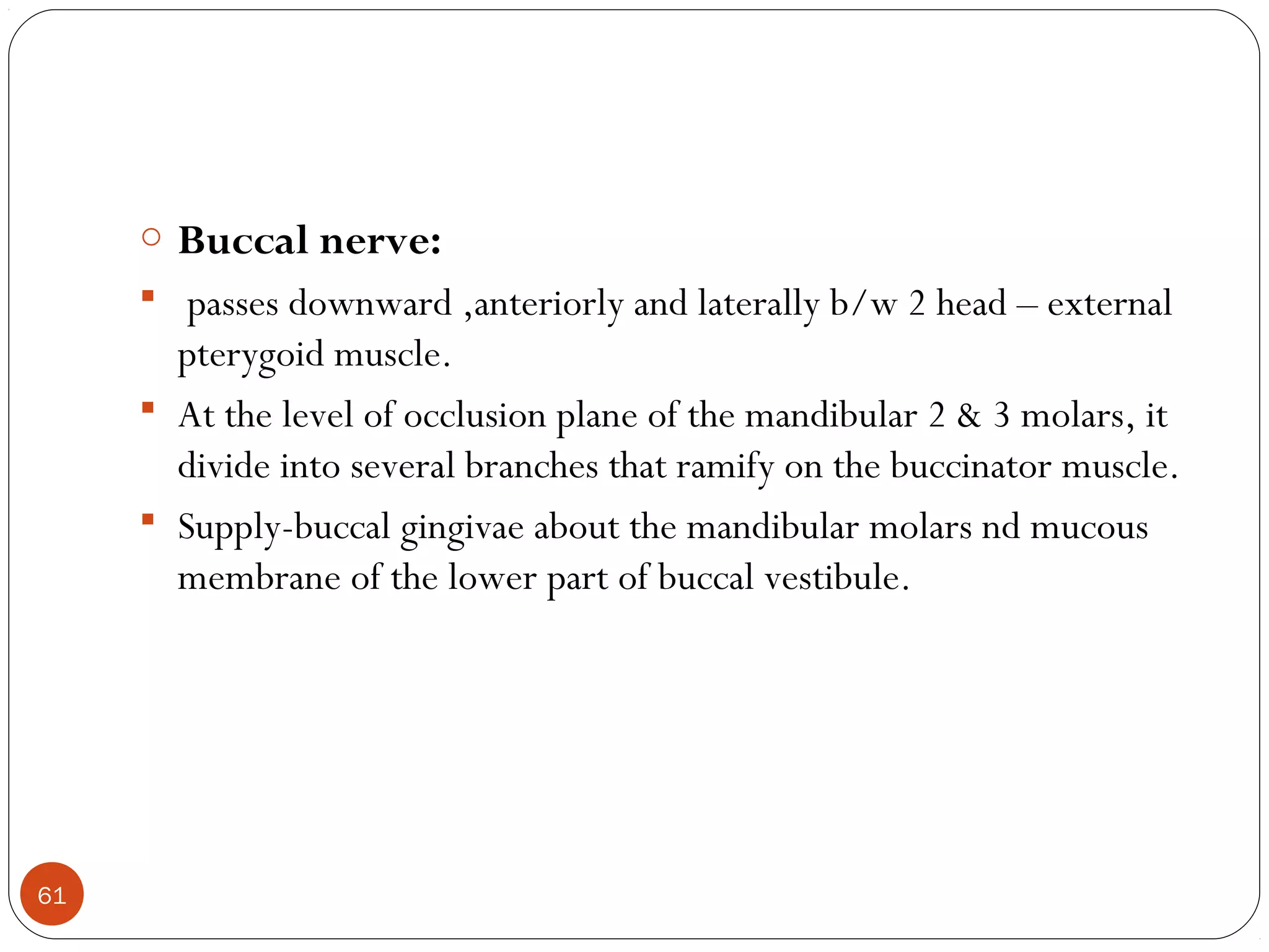
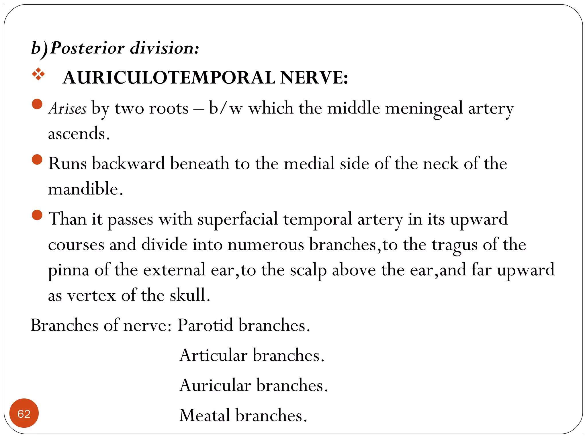
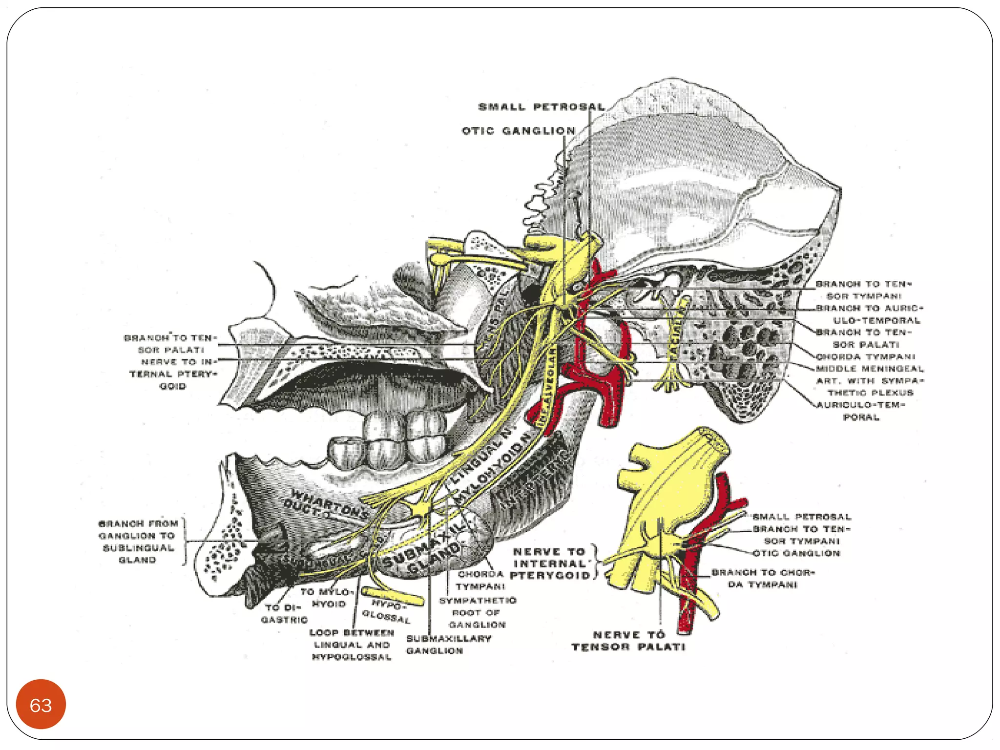
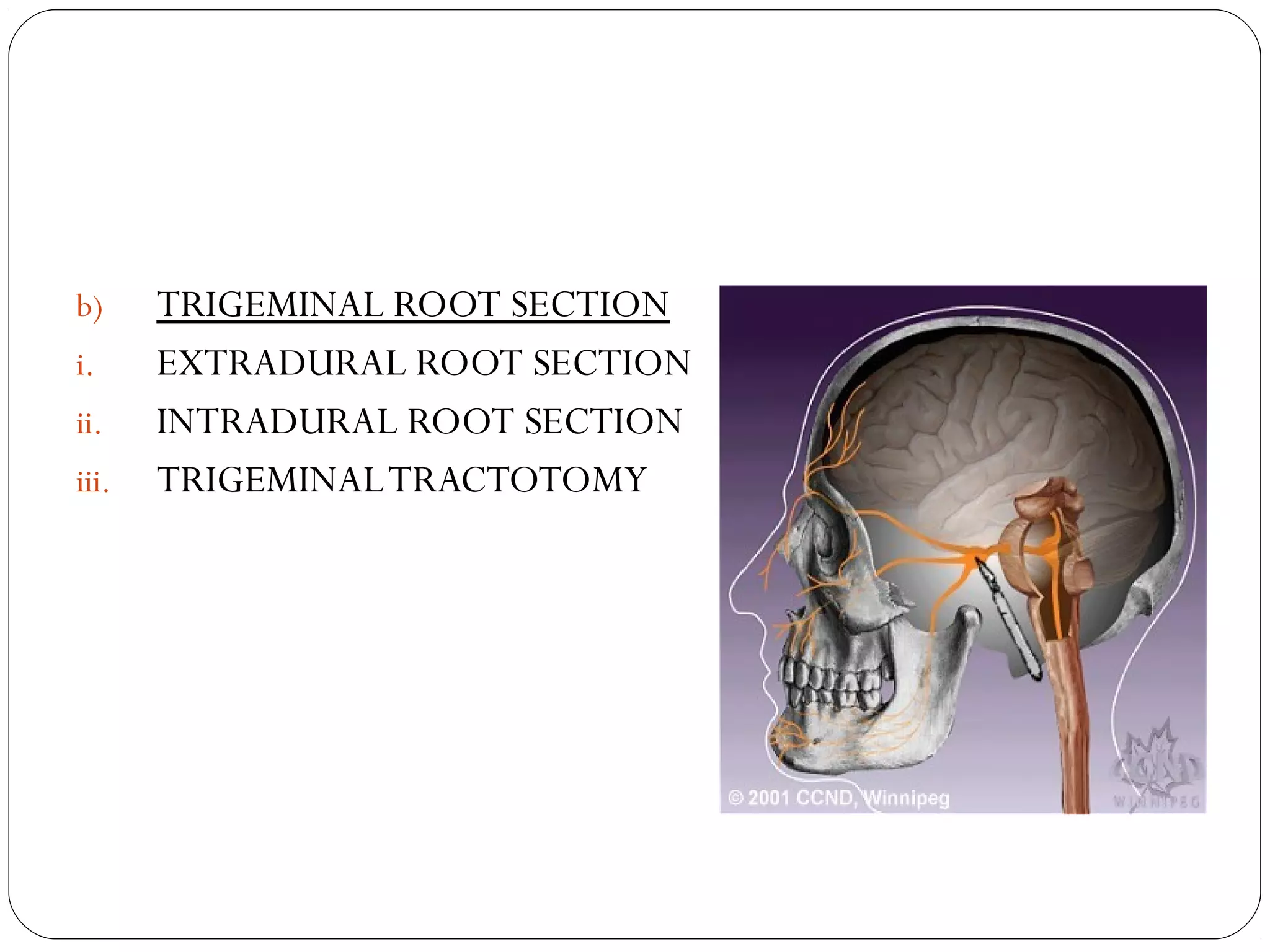
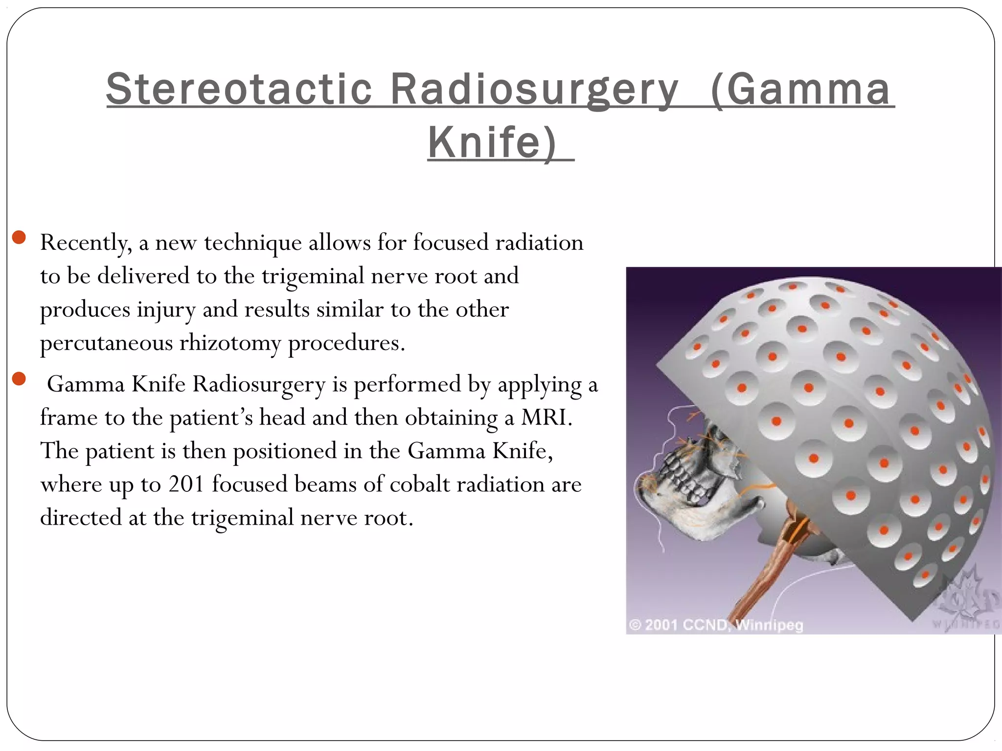
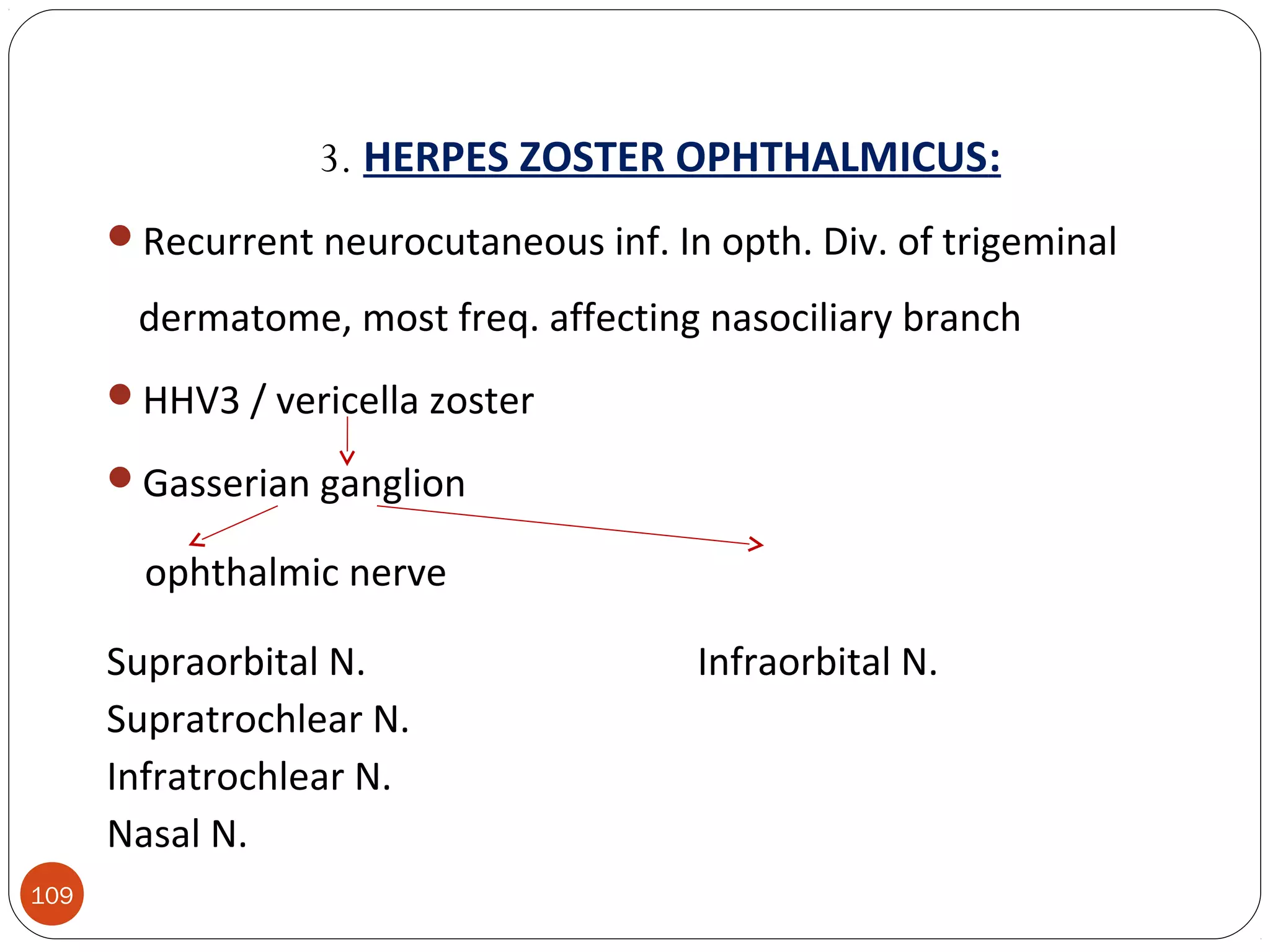
Department of Oral & Maxillofacial
Surgery
NARSINHBHAI PATEL DENTAL COLLEGE AND HOSPITALVISNAGAR
1
THE TRIGEMINAL NERVE
Guided By :
Dr. Anil managutti , HOD andDr. Anil managutti , HOD and
ProfessorProfessor
Dr.shailesh menat , ProfessorDr.shailesh menat , Professor
Presented by: Dr. Nirav Patel
1st
year PG
TRIGEMINAL NERVE
INTRODUCTION
MOTOR FUNCTION
SENSORY FUNCTION
GANGLION
OPTHALMIC NERVE
MAXILLARY NERVE
MANDIBULAR NERVE
EXAMINATION OF TRIGEMINAL NERVE
APPLIED ANATOMY
2
INTRODUCTION
The largestlargest cranial nerve
It is mixed nervemixed nerve ( sensory and motor )
Sensory to – Skin of face
-Mucosa of cranial viscera
-Except base of tongue and pharynx
Motor to –Muscles of Mastication
-Tensor ville palatini,Tensor tympany
-Anterior belly of digastric
-Mylohyoid
09/24/16Oral And Maxillofacial Surgery3
NUCLEI
09/24/16Oral And Maxillofacial Surgery4
TRIGEMINAL NUCLEI
o A cranial nerve nucleus is a collection of neurons (
gray matter) in the brain stem that is associated with one
or more cranial nerves. 
o Axons carrying information to and from the cranial
nerves form a synapse first at these nuclei.
o Lesions occurring at these nuclei can lead to effects
resembling those seen by the severing of nerve(s) they
are associated with.
09/24/16Oral And Maxillofacial Surgery5
09/24/16Oral And Maxillofacial Surgery6
SENSORY NUCLEI
1.Mesencephalic
nucleus
- Cell body of Pseudounipolar
neuron
- Relay proprioception from
muscles of
mastication,
Extra ocular Muscles,
Facial muscles.
Situated in Midbrain just
latetral to Aqueduct.
09/24/16Oral And Maxillofacial Surgery7
2.Principal sensory
nucleus-
Lies in Pons lateral to
Motor nucleus
Relays touch sensation
09/24/16Oral And Maxillofacial Surgery8
3.Spinal nucleus- Extends from caudal end of principal
sensory Nucles
in pons to 2nd
or 3rd
spinal
segment
It relys Pain and Temperature
09/24/16Oral And Maxillofacial Surgery9
10
MOTOR NUCLEUS :
 Innervates muscles of masticationmuscles of mastication and tensor tympanitensor tympani
andand tensor palatinitensor palatini
 Derived from first branchial arch.
 Located in pons medial to principle sensory nucleus.
09/24/16Oral And Maxillofacial Surgery11
FUNCTIONALFUNCTIONAL
COMPONENTSCOMPONENTS
Sensory RootSensory Root
Motor RootMotor Root
09/24/16Oral And Maxillofacial Surgery12
SENSORY ROOT
GENERAL SOMATIC AFFERENTS- Face, Scalp, Teeth, Gingiva, Oral, Nasal,
Cavities, Para nasal sinus, Conjunctiva and Cornea.
Pain, temp, light touch touch, pressure proprioception
Trigeminal gang. Bypasses trigem gang.
sensory root.
09/24/16Oral And Maxillofacial Surgery13
Spinal nuc. Principal sen nuc. Mesencephalic
CNCN
SS
MOTOR NUCLEUS
MOTOR ROOT
MANDIBULAR NERVE
Muscles of mastication Tensor tympani
Masseter Tensor palatini
Lateral & Medial Pterygoids
Temporalis
09/24/16Oral And Maxillofacial Surgery14
CNSCNS
MOTOR ROOTMOTOR ROOT
09/24/16Oral And Maxillofacial Surgery15
COURSE & DISTRIBUTIONCOURSE & DISTRIBUTION
Both motor and sensory root are attached ventrally to
junction of pons and middle cerebellar peduncle with motor
root lying ventromedially to the sensory root.
Pass anteriorly in middle cranial fossa to lie below tentorium
cerebelli in cavum trigeminale, here motor root lies inferior
to sensory root.
09/24/16Oral And Maxillofacial Surgery16
Sensory root connected to postromedial concave
border of the trigeminal ganglion.
Convex antrolatateral margin of the ganglion gives
attachment to the 3 div. Of the trigeminal nerve.
09/24/16Oral And Maxillofacial Surgery17
Motor root turns further inferior with sensory component of
V3 to emerge out of foramen Ovale as Mandibular
nerve.
 Ophthalmic and Maxillary division emerges through
Superior orbital fissure and foramen Rotundum
respectively.
09/24/16Oral And Maxillofacial Surgery18
GANGLIONGANGLION
09/24/16Oral And Maxillofacial Surgery19
THETHE TRIGEMINALTRIGEMINAL
GANGLIONGANGLION
SEMILUNAR OR GASSERIAN GANGLION.
Cresentric in shape with convexity anterolaterally.
Contains cell bodies of pseudounipolar neurons.
LOCATION: lies in a bony fossa at apex of the petrous
temporal bone on floor of middle cranial fossa, just
lateral to posterior part of lateral wall of the cavernous
sinus.
09/24/16Oral And Maxillofacial Surgery20
COVERINGS: covered by dural pouch = MECKLES CAVE or
CAVUM TRIGEMINALE.
cave lined by pia and arachnoid thus the
ganglion is bathed in CSFCSF.
ARTERIAL SUPPLY: Ganglionic branches of Internal Carotid
Artery, middle meningeal artery and accessory
meningeal artery.
09/24/16Oral And Maxillofacial Surgery21
RELATIONS:
SUPERIORLY: *superior petrosal sinus
*free margin of tentorium cerebelli
INFERIORLY: *motor root
*greater petrosal nerve
*petrous apex
*foramen lacerum
MEDIALLY: *posterior part of lateral wall of cavernous sinus
*Internal Carotid Artery with its sympathetic plexus
LATERALLY: *uncus of temporal lobe
*middle meningeal artery and vein
*nervous spinosum
09/24/16Oral And Maxillofacial Surgery22
DIVISIONS OFDIVISIONS OF
TRIGEMINAL NERVETRIGEMINAL NERVE
1. Ophthalmic nerve
2. Maxillary nerve
3. Mandibular nerve
09/24/16Oral And Maxillofacial Surgery23
OPHTHALMIC BRANCH OF TN
24
OPHTHALMIC NERVE BRANCHES
25
A.Infratrochlear
B. Anterior Ethmoid
C. Posterior
Ethmoid
D. Lacrimal
E. Supraorbital
F. Supratrochlear
G. Nasociliary
First division of the trigeminal.
Is a sensory nerve.
Smallest of the three divisions of the trigeminal.
Arises - upper part of the semi lunar ganglion as a short, flattened band,
about 2.5 cm. Long - passes forward along the lateral wall of the
cavernous sinus - below the oculomotor and trochlear nerves.
Before entering the orbit through superior orbital fissure, it divides into
three branches,
Lacrimal.
Frontal.
Nasociliary.
26
27
1)LACRIMAL NERVE:
Smallest of the three branches.
Derived – possibly from branch which goes from the ophthalmic to the
trochlear nerve.
Enters orbit through the lateral angle of the superior orbital fissure.
In orbit - runs along the upper border of the lateral rectus with the
lacrimal artery - communicates with the zygomatic branch of the
maxillary nerve.
Enters the lacrimal gland - gives off several filaments, which supply the
gland and the conjunctiva.
Finally - pierces the orbital septum - ends in the skin of the upper
eyelid, joining with filaments of the facial nerve.
28
2) FRONTAL NERVE:
 Largest branch of ophthalmic.
 Enters the orbit - superior orbital fissure and runs forward between the Levator
palpebræ superioris and the periosteum.
 Midway between the apex and base of the orbit it divides into two branches
A) Supratrochlear
B) Supraorbital
29
A) SUPRATROCHLEAR NERVE:
 Smaller of the two - passes above the superior Oblique and gives off a descending
filament - to join the infratrochlear branch of the nasociliary nerve.
 Escapes from the orbit between the superior oblique and the supraorbital foramen.
 Supplies - skin of the lower part of the forehead close to the midline
- sends filaments to the conjunctiva and skin of the upper
eyelid.
30
B) SUPRAORBITAL NERVE:
Passes - supraorbital foramen - gives off palpebral filaments to the
upper eyelid.
Then ascends upon the forehead and ends in two branches
Medial
Lateral
which supply the anterior scalp region to the vertex of skull, the
forehead and skin of upper eyelid.
31
3) Nasocillary nerve.
 Third main branch of ophthalmic division.
 Enters the orbit through superior orbital fissure.
 Divide into: those arising in the orbit, in the nasal, and on
the face.
32
1) Branches in the orbit
a) Long root of the ciliary ganglion
b) Long ciliary nerves.
c) Posterior ethmoid nerve.
d) Anterior Ethmoidal nerves.
33
a) LONG ROOT OF CILIARY GANGLION:
Arises – from nasociliary b/W the two heads of the lateral Rectus.
It contains sensory fibers-pass through the ganglion without synapsing
and continue on to the eyeball.
b) LONG CILIARY NERVE:
Two or three in number - given off from the nasociliary, as it crosses
the optic nerve.
Distributed to the Irish and cornea.
34
c) POSTERIOR ETHMOIDAL BRANCH:
Posterior branch leaves the orbital cavity through the posterior
ethmoidal foramen and gives some filaments to the sphenoidal sinus.
d) ANTERIOR ETHMOID NERVE :
Nerve continues anteriorly along the medial wall of the orbit.
In its course, the nerve gives of fillament -supply the ant.ethmoid cell &
frontal sinus.
Upper part of nasal cavity divide into 2 set of nasal branch :
1) internal nasal branch.
2) external nasal branch.
35
1) Internal nasal branches:
- 2 branches
: Medial or septal branches – travel downward to supply
sensory innervation to the mucous membrane of that area.
: lateral branches- supply to the mucous membrane of the
anterior ends of the superior and middle nasal conchae.
2) external nasal branches:
-At the border b/w the lower edge of the nasal bone and
the upper edge of the lateral nasal cartiladge.
-Supply- skin over the tip of the nose & ala of the nose.
36
B) Branches arising in the nasal cavity:
o Supply – mucous membrane lining the cavity.
C) Terminal branches of the ophthalmic division on
the face:
o Supply – medial part of the both eyelids, lacrimal sac.
37
MAXILLARY BRANCH OF TN
Second division of the trigeminal
Is a sensory nerve.
It is intermediate - both in position and size b/w the ophthalmic and
mandibular.
It begins - middle of semilunar ganglion as a flattened plexiform band -
passing horizontally forward - leaves the skull - foramen rotundum -
becomes more cylindrical in form and firmer in texture.
Then crosses - pterygopalatine fossa - enters the orbit through the
inferior orbital fissure - it traverses the infraorbital groove and canal in
the floor of the orbit and appears on the face - infraorbital foramen
38
At its termination - the nerve lies beneath the Quadratus labii superioris
& divides into branches which spread out upon the
side of the nose
the lower eyelid
the upper lip
joining with filaments of the facial nerve.
BRANCHES OF MAXILLARY NERVE:
May be divided into four groups, according as they are given
off
In the cranium
In the pterygopalatine fossa
In the infraorbital canal
On the face.
39
In the cranium Middle Meningeal Nerve
In the Pterygopalatine
fossa
Zygomatic
Sphenopalatine
Posterior Superior
Alveolar
In the Infraorbital Canal Anterior Superior
Alveolar
Middle Superior Alveolar
On the Face Inferior Palpebral
External Nasal
Superior Labial
40
MAXILLARY NERVE BRANCHES
A. Zygoticaticotemporal
B. Zygomaticofacial
C. Post. Sup. Alveolar Brs
D. Nasopalatine
E. Greater Palatine
F. Lesser Palatine
G. Mid. & Ant. Alveolar Brs
H. Infraorbital
41
42
 (Branches given off in the middle cranial foss)
MIDDLE
MENINGEAL NERVE:
Given off from the maxillary nerve directly after its origin from the
semilunar ganglion.
It accompanies the middle meningeal artery and supplies the dura
mater.
(Branches in the pterygopaltine fossa)
A) ZYGOMATIC NERVE:
Arises - pterygopalatine fossa.
Enters the orbit - inferior orbital fissure - into two branches,
Zygomaticotemporal
Zygomaticofacial
43
 ZYGOMATICO-TEMPORAL BRANCH:
Runs along - lateral wall of the orbit in a groove in the Zygomatic bone.
Receives - communication from the lacrimal and passing through a
foramen in the Zygomatic bone - enters the temporal fossa.
Ascends b/w the bone and substance of the Temporalis muscle -
pierces the temporal fascia about 2.5 cm. above the Zygomatic arch -
distributed to the skin of the side of the forehead - communicates with
the facial nerve.
44
 ZYGOMATICO-FACIAL BRANCH:
Passes along the infero-lateral angle of the orbit.
Emerges - face through a foramen in the Zygomatic bone.
Perforating the Orbicularis oculi - supplies the skin on the prominence
of the cheek.
B) SPHENOPALATINE NERVE (PTERYGOPALATINE NERVE):
Two short nerve trunk unite the pterygopalatine ganglion.
Descend to the sphenopalatine ganglion.
Branches of distribution of ptery.pal. Nerve divide in 3 group:
1)orbital branch
2)nasal branch
3)palatine branch45
46
1) ORBITAL BRANCH:
Two or three delicate filaments - enter the orbit by the inferior orbital
fissure - supply the periosteum.
2) PALATINE NERVES:
Distributed to the roof of the mouth, soft palate, tonsil, and lining
membrane of the nasal cavity.
Most of their fibers are derived - sphenopalatine branches of the
maxillary nerve.
They are three in number:
Anterior
Middle
Posterior.
47
ANTERIOR PALATINE NERVE:
Descends - pterygopalatine canal - emerges upon the hard palate -
greater palatine foramen.
Supplies the gums, the mucous membrane and glands of the hard palate
In the pterygopalatine canal - gives off posterior inferior nasal branches -
enter the nasal cavity through openings in the palatine bone.
MIDDLE PALATINE NERVE:
 emerge from lesser palatine foramen.
Emerges through one of the minor palatine canals and distributes
branches to the uvula, tonsil, and soft palate.
48
POSTERIOR PALATINE NERVES:
Descends - pterygopalatine canal – emerges – lesser palatine formen.
Supplies the tonsil.
49
3) NASAL BRANCHES :
• 2 branches divided:
• a) posterior superior latral nasal branch:
Transmit sensory impulses from the mucous membrane of the nasal
septum and posterior ethmoid cell.
b) Medial or septal branch:
Transmit sensory impulses from the mocous membrane over the
vomer.
50
POSTERIOR SUPERIOR ALVEOLAR NERVE:
Arise - trunk of the nerve just before it enters the infraorbital groove.
Descend - tuberosity of the maxilla - give off several twigs to the gums
and mucous membrane of the cheek.
Then enter - posterior alveolar canals on the infratemporal surface of
the maxilla - communicate with the middle superior alveolar nerve -
give off branches to the lining membrane of the maxillary sinus and
three twigs to each molar tooth.
51
(In the infraorbital groove and canal)
MIDDLE SUPERIOR ALVEOLAR NERVE:
Given off from the nerve in the posterior part of the infraorbital canal.
Runs and forward in a canal - lateral wall of the maxillary sinus to
supply - two premolar teeth.
It forms - superior dental plexus with the anterior and posterior
superior alveolar branches.
ANTERIOR SUPERIOR ALVEOLAR NERVE:
Given off from the nerve just before its exit from the infraorbital
foramen.
It descends in a canal in the anterior wall of the maxillary sinus - divides
into branches - supply the incisor and canine teeth.
52
53
(Terminal branches of the maxillary division on the face)
INFERIOR PALPEBRAL BRANCH:
Ascend behind the Orbicularis oculi.
Supply - skin and conjunctiva of the lower eyelid.
Join at the lateral angle of the orbit with the facial and zygomaticofacial
nerves.
54
EXTERNAL NASAL BRANCHES:
 Supply - skin of the side of the nose and of the septum mobile nasi - join with the
terminal twigs of the nasociliary nerve.
SUPERIOR LABIAL BRANCHES:
 The largest and most numerous - descend behind the Quadratus labii superioris.
 Distributed - skin of the upper lip, the mucous membrane of the mouth and labial
glands.
 They are joined - immediately beneath the orbit, by filaments from the facial nerve -
forming with them the infraorbital plexus.
55
MANDIBULAR BRANCH OF TN
Largest of the three divisions of the fifth nerve.
Made up of two roots:
A large - sensory root proceeding from the inferior angle of the
semilunar ganglion.
A small motor root which passes beneath the ganglion -
unites with the sensory root - just after -its exit - foramen ovale.
Branches divided into 2 group:
1)undivided nerve.
2)divided nerve.
56
Supplies - Teeth and gums of the mandible
- The skin of the temporal
- The lower lip
- The lower part of the face
- The muscles of mastication
- It also supplies the mucous membrane of the
anterior two-thirds of the tongue.
57
 (Branches from the undivided nerve)
NERVOUS
SPINOSUS:
Enters the skull through the foramen spinosum with the middle meningeal artery. It
divides into two branches, anterior and posterior
 Supply the dura mater.
 The posterior branch - supplies the mucous lining of the mastoid cells.
 The anterior communicates with the meningeal branch of the maxillary nerve.
INTERNAL PTERYGOID NERVE:
 Nerve to the Pterygoideus internus - slender branch - enters the deep surface of the
muscle.
 It gives off one or two filaments to the otic ganglion.
58
 (Branches from the divided nerve)
Below –leval-undivided-trunk seprates into 2 part.
a) Anterior division.
External pterygoid.
Masseteric.
nerve to temporal.
Buccinator .
b) Posterior division.
Auriculotemporal nerve.
Lingual nerve.
Inferior alveolar nerve.59
o Branch to External pterygoid muscle:
 Enter the medial side of external pterygoid muscle to provide motor
supply.
o Massetter nerve:
 Passes above –external pterygoid to transverse mandibular notch-enter-
deepside-masster muscle.
o Nerve to temporal muscle:
1) anterior deep temporal nerve : passes upward and crosses
the infratemporal crest of the sphenoid bone and it end into anterior
part of deep temporal muscle.
2)posterior deep temporal nerve: passes upward to the deep
temporal nerve.
60
o Buccal nerve:
 passes downward ,anteriorly and laterally b/w 2 head – external
pterygoid muscle.
 At the level of occlusion plane of the mandibular 2 & 3 molars, it
divide into several branches that ramify on the buccinator muscle.
 Supply-buccal gingivae about the mandibular molars nd mucous
membrane of the lower part of buccal vestibule.
61
b)Posterior division:
 AURICULOTEMPORAL NERVE:
Arises by two roots – b/w which the middle meningeal artery
ascends.
Runs backward beneath to the medial side of the neck of the
mandible.
Than it passes with superfacial temporal artery in its upward
courses and divide into numerous branches,to the tragus of the
pinna of the external ear,to the scalp above the ear,and far upward
as vertex of the skull.
Branches of nerve: Parotid branches.
Articular branches.
Auricular branches.
Meatal branches.62
63
LINGUAL NERVE:
Supplies - mucous membrane of the anterior two-thirds of the tongue.
First it passes to medially to the external pterygoid muscle.
Than descends,lies b/w the internal pterygoid muscle and ramus in
pterygoid space.
Finally runs across the duct of the submaxillary gland and along the
tongue to its tip.
BRANCHES OF DISTRIBUTION:
 Supply - sublingual gland
- mucous membrane of the mouth, the gums,
and the anterior two-thirds of the tongue.
The terminal filaments communicate - at the tip of the tongue with the
hypoglossal nerve.
Communications of the lingual nerve with the corda tympani.
64
INFERIOR ALVEOLAR NERVE:
Largest branch of the mandibular nerve.
Descends with the inferior alveolar artery - at first beneath the External
pterygoid muscle – medial side of the ramus in pterygomandibular
space, it enters the mandibular foramen.
Then passes forward in the mandibular canal - beneath the teeth as far
as the mental foramen - where it divides into two terminal branches -
incisive and mental.
Branches of the inferior alveolar nerve are the
mylohyoid
Incisive
Mental.
65
MYLOHYOID NERVE:
Derived - inferior alveolar just before it enters the mandibular foramen.
It descends - groove on the deep surface of the ramus of the mandible -
reaching the under surface of the Mylohyoid - supplies this muscle and
the anterior belly of the Digastric.
66
INCISIVE BRANCH:
Continued onward within the bone and supplies the 1st
premolar, canine
and incisor teeth.
MENTAL NERVE:
Emerges at the mental foramen - divides beneath the Triangularis
muscle into three branches.
One descends to the skin of the chin.
Two ascend to the skin and mucous
membrane of the lower lip.
67
68
Examination of trigeminal nerve
69
Examination of trigeminal nerve
1- Sensation Function
2- Motor Function
3- Corneal reflex
4- Test jaw jerk
70
Sensation function
use sterile sharp item on forehead, cheek, and jaw
If any abnormality present we test the thermal
sensation and light touch
71
Corneal reflex
a clean piece of cotton wool and ask the patient
to look away gently touch the cornea with the
cotton wool and the patient will blink.
72
Test jaw jerk
Doctor finger on tip of jaw, grip patellar hammer
halfway up shaft and tap finger lightly usually
nothing happens, or just a slight closure.
APPLIED ANATOMY
Sensory distribution of the trigeminal nerve explains
why headache is a common symptom in involvements of
the nose, paranasal air sinuses, the teeth & gums, the
eyes & the meninges
Trigeminal neuralgia may involve one or more of the
three divisions of the V nerve
Congenital cutaneous naevi on the face (port wine
stains) map out accurately the areas supplied by one or
more divisions of the V nerve
73
APPLIED ANATOMY OF TRIGEMINAL
NERVE
The sensory distribution of the trigeminal nerve explains why
headache is a uniformly common symptom in
involvements of the nose (common cold,boils), the
paranasal air sinuses(sinusitis),the teeth and gums, the
eyes, the meninges and so on.
Trigeminal neuralgia. – it causes attacks of very severe ,
burning and scalding pain along the distribution of the affected
nerve.
TRIGEMINAL
NEURALGIA
DEFINATION
Trigeminal neuralgia (TN) is defined as sudden, usually unilateral,
severe, brief, stabbing, lancinating, recurring pain in the distribution of
one or more branches of 5th cranial nerve.
John Locke in 1677 gave the first full description with its treatment.
Nicholaus Andre in 1756 coined the term ‘Tic Doloureux.’
John Fothergill in 1773 published detailed description of TN, since then,
it has been referred to as ‘Fothergill’s disease’.
In spite of the condition being known since centuries, it still continues to
baffle the clinician and its pathogenesis remains as enigma to the medical
profession. Multiple views have been hypothecated regarding its etiology
generating nothing, but confusion and simultaneously opting for many
different therapies in an effort to treat this ongoing condition.
Types of Trigeminal Neuralgia
and Their Causes
We define seven forms of TN: typical TN, atypical TN, pre-TN,
multiple-sclerosis-related TN, secondary TN,
post-traumatic TN (trigeminal neuropathy), and failed TN. These
forms of TN should be distinguished from
idiopathic (atypical) facial pain, as well as other disorders causing
cranio-facial pain.
Typical Trigeminal Neuralgia (Tic
Douloureux)

     This is the most common form of TN, that has previously been termed Classical,
Idiopathic and Essential TN.  Nearly all cases of typical TN are caused by blood vessels
compressing the trigeminal nerve root as it enters the brain stem. This
neurovascular or microvascular compression at the trigeminal nerve root entry zone
may be caused by arteries of veins, large or small, that may simply contact or indent
the trigeminal nerve. In people without TN, blood vessels are usually not in contact
with the trigeminal nerve root entry zone. 
In people without TN, there is usually no vascular
compression upon the trigeminal nerve root.
In most sufferers of typical trigeminal neuralgia,
vessels compress the trigeminal nerve root.
Pulsation of vessels upon the trigeminal nerve root do
not visibly damage the nerve. However, irritation
from repeated pulsations may lead to changes of nerve
function, and delivery of abnormal signals to the
trigeminal nerve nucleus. Over time, this is thought to
cause hyperactivity of the trigeminal nerve nucleus,
resulting in the generation of TN pain.
The generation of TN pain is thought to result from
peripheral pathology (i.e. neurovascular compression) and
central pathophysiology (i.e. hyperactivity of the trigeminal
nerve nucleus).
The superior cerebellar artery is the vessel most often
responsible for neurovascular compression upon the
trigeminal nerve root, although other arteries or veins
may be the culprit vessels. TN may be cured by an
operation that effectively relieves the neurovascular
compression upon the trigeminal nerve root.  This
operation is called microvascular decompression, and
is described in
Part Two: Treatment of Trigeminal Neuralgia.
Atypical Trigeminal Neuralgia
      Atypical TN is characterized by a unilateral, prominent constant and severe aching,
boring or burning pain superimposed upon otherwise typical TN symptoms. This
should be differentiated from cases of typical TN that develop a minor aching or
burning pain within the affected distribution of the trigeminal nerve.
      Vascular compression, as described above in typical TN, is thought to be the cause
of many cases of atypical TN. Some believe atypical TN is due to vascular compression
upon a specific part of the trigeminal nerve (the portio minor), while others theorize that
atypical TN represents a more severe form or progression of typical TN.
  
Atypical TN pain can be at least partially
relieved with medications used for typical TN,
such as carbamazepine (Tegretol®). MVD
surgery is curative for many patients with
atypical TN, but not as reliably as for those with
typical TN. It is also important to note that
rhizotomy procedures may be effective in
treating atypical TN, but are more likely to be
complicated by annoying or even painful
numbness (i.e. deafferentation pain).
85
Pre-Trigeminal Neuralgia
     Days to years before the first attack of TN pain, some sufferers
experience odd sensations in the trigeminal distributions destined to
become affected by TN. These odd sensations of pain, (such as a
toothache) or discomfort (like "pins and needles", parasthesia), may be
symptoms of pre-trigeminal neuralgia. Pre-TN is most effectively
treated with medical therapy used for typical TN. When the first attack
of true TN occurs, it is very distinct from pre-TN symptoms.
MULTIPLE SCLEROSIS REALATED TRIGEMINAL
NEURALGIA
      The symptoms and characteristics of multiple sclerosis (MS)-related TN are identical to those
for typical TN.  Two to four percent of patients with TN have evidence of multiple sclerosis and
about 1% of patients suffering from multiple sclerosis develop TN.  Those with MS-related TN
tend to be younger when they experience their first attack of pain, and the pain progresses over
a shorter amount of time than in those with typical TN. Furthermore, bilateral TN is more
commonly seen in people with multiple sclerosis.
      MS involves the formation of demyelinating plaques within the brain. When these areas of
injury involve the trigeminal nerve system, TN may develop. MS-related TN is treated with the
same medications used for typical TN (see Medications). Trigeminal rhizotomies are employed
when medications fail to control the pain. For some individuals with MS and TN, neurovascular
compression of the trigeminal nerve root may be a rare cause and demonstrated with special
MRI or CT scans. In such cases, microvascular decompression surgery may be considered for
treating the MS-related TN.
Secondary or Tumor Related Trigeminal
Neuralgia
      Trigeminal neuralgia pain caused by a lesion, such as a tumor, is referred to as
secondary trigeminal neuralgia. A tumor that severely compresses or distorts the
trigeminal nerve may cause facial numbness, weakness of chewing muscles, and/or
constant aching pain (also see
Trigeminal Neuropathy or Post-Traumatic Trigeminal Neuralgia). Medications usually
help control secondary TN pain when first tried, although often become. Surgically
removing the tumor usually alleviates pain and trigeminal function may return. At the
time of surgery, after the removal of the tumor, the trigeminal nerve may be found to
also be compressed by an artery or vein that causes the typical features of TN. This
vessel must then be moved away from the nerve by microvascular decompression
techniques to cure TN.
In these MRI images, a tumor responsible for
compressing the trigeminal nerve is highlighted in red.
Trigeminal Neuropathy or
Post-Traumatic Trigeminal Neuralgia
  Injury to the trigeminal nerve may cause this severe pain condition. Trigeminal
Neuropathy or Post-Traumatic TN may develop following cranio-facial trauma (such as
from a car accident), dental trauma, sinus trauma (such as following Caldwell Luc
procedures) but most commonly following destructive procedures (rhizotomies) used
for treatment of TN. Following TN injury, numbness may become associated with
bothersome sensations or pain, sometimes called phantom pain or deafferentation pain.
These pain conditions are caused by irreparable damage to the trigeminal nerve and
secondary hyperactivity of the trigeminal nerve nucleus
 The pain of trigeminal neuropathy or post-traumatic TN is usually constant, aching or
burning, but may be worsened by exposure to triggers such as wind and cold. Such
deafferentation pain can start immediately or days to years following injury to the
trigeminal nerve. In the most extreme form, called anesthesia dolorosa, there is continuous
severe pain in areas of complete numbness.
     Unfortunately, treatment of post-traumatic TN is often ineffective and pain may not be
controlled with medications. There are some reports of pain relief associated with the use of
trigeminal nerve stimulation procedures. More invasive procedures such as brain surface
(pre-motor cortex) stimulation, or focused injuries in the brain stem (tractotomy) have also
been tried.
"Failed"
Trigeminal Neuralgia
  Not all cases of TN may be effectively controlled with any one form of medications or
surgical interventions. When medications are no longer effective, surgical interventions are
considered. If pain recurs or persists following surgery, medications are tried again and may then
work more effectively. Rarely, additional or repeated surgical interventions are necessary.
Unfortunately, in a very small proportion of sufferers, all medications,
microvascular decompression and destructive rhizotomy procedures prove ineffective in
controlling TN pain. This condition is called "failed" trigeminal neuralgia. Such individuals also
often suffer from additional trigeminal neuropathy or post-traumatic TN as a result of the
destructive interventions they underwent. Investigational treatments may be considered
including stimulation of the brain surface (pre-motor cortex stimulation), controlled lesioning of
the brain stem (tractotomy), or stimulation of the trigeminal nerve or Gasserion ganglion
(trigeminal nerve stimulation).
Treatment of Trigeminal
Neuralgia
 Medications are the first line of treatment for TN and include carbamazepine
(Tegretol®), phenytoin (Dilantin®), gabapentin (Neurontin®) and baclophen
(Lioresal®).
 As the disease progresses and pain becomes more frequent and severe, increased
doses of medications are required which may lead to intolerable side effects and/or
inadequate pain control.
 Each sufferer has differing tolerance to these medications and pain, but at least half
will eventually find that medications do not adequately control their progressively
worsening TN.
 The surgical procedures then considered are either microvascular decompression
surgery or some form of nerve injury procedure (rhizotomies).
MEDICINAL TREATMENT OF
TRIGEMINAL NEURALGIA
 
Carbamazepine (Tegretol®)

     Almost all typicalTN sufferers experience
significant pain relief with carbamazepine.The
starting daily dose is low, (one to two pills a
day), which is gradually increased until the
pain is completely alleviated or side effects
occur. Good relief of pain may be achieved at
low doses, but the usual effective dose ranges
from 200 to 1200 mg divided in three or four
doses per day. Even higher doses may be
required during severe attacks of pain.  Once
relief of the pain has been achieved, the same
dose is usually continued for at least two weeks
before trying to reduce to a minimal dosage
that provides pain relief.  As with
Carbamazepine (Tegretol®)
 As with all TN medications, Tegretol® may gradually be decreased during
periods of remission.
      Several dose-related side effects are often experienced including drowsiness,
mental confusion, dizziness, nystagmus (rapid movements of the eye), ataxia
(decreased coordination), diplopia (double vision), nausea, and anorexia (loss
of appetite). If side effects are severe, the daily dose of carbamazepine may be
decreased for 1 to 3 days, before trying to increase the daily dose again. 
     
Trileptal (Oxycarbazepine)
 Trileptal, or oxycarbemazepine, is a form of Tegretol® that is becoming more
widely prescribed for a variety of conditions. Like Tegretol®, it is an anti-
seizure drug, but the side effects are less severe and less frequently
experienced.
 The dose ::begins at 300 mg twice a day
gradually increased to achieve pain control. The maximum dose is
2400-3000 mg per day.
 Common side effects are nausea, vomiting, dizziness, fatigue and tremors. Less
frequent symptoms are rash, respiratory infections, double vision, and changes
in electrolytes in blood. If you have had an allergic reaction to Tegretol®
(carbemazepine), then you should not try Trileptal. As with other anti-seizure
medications, increasing and decreasing the dose should be gradual.
Phenytoin (Dilantin®)
 Phenytoin relieves tic pain in
over half ofTN sufferers at
doses of 300 to 500 mg,
divided into three doses per
day.
 Phenytoin may also be
administered intravenously to
treat severe exacerbations of
TN.
Baclophen (Lioresal®)
 not as effective as carbamazepine or
phenytoin forTN, but may be used in
combination with these medications.
 The starting dose is 5 mg two or three times
a day, and may be gradually increased.
 The usual dosage taken for complete pain
relief is between 50 and 60 mg per day.
Baclophen has a short duration of function so
sufferers with severeTN may need to take
doses every 3 to 4 hours.
Gabapentin (Neurontin®)
 Gabapentin is an anti-epileptic drug that is structurally related to
the neurotransmitter GABA.
 This drug is almost as effective as carbamazepine but involves
fewer side effects. 
 The starting dose is usually 300mg three times a day and this is
increased to a maximal dose. The most common adverse
reactions include somnolence (sleepiness), ataxia (decreased
coordination), fatigue, and nystagmus (rapid movements of the
eye). There is no known interaction withTegretol® or
Dilantin®, permitting usage of these drugs in combination with
Neurontin®.As with all of these drugs, rapid discontinuation
should be avoided as severe withdrawal reactions may occur.
SURGICAL TREATMENT OF
TRIGEMINAL NEURALGIA
1. PERIPHERAL NERVE SURGICAL
TREATMENT
A. PERIPHERAL INJECTIONS
a) LONG ACTING ANAESTHETIC AGENTS
b) ALCHOHOL INJECTIONS
B. PERIPHERAL NEURECTOMY
a) INFRAORBITAL
b) INFERIOR ALVEOLAR NERVE
c) LINGUAL NERVE
C. CRYOTHERAPY OR CRYONEUROLYSIS
D. PERIPHERAL RADIOFREQUENCY NEUROLYSIS
ORTHERMOCOAGULATION
2. GASSERIAN GANGLION
PROCEDURES
A. TECHNIQUES FOR PERCUTANEOUS
APPROACH
a) INJECTION METHOHEXITONE
b) INJECTION GLYCEROL
c) CONTROLLED
RADIOFREQUENCY
THERMOCOAGULA
TION
d) BALLOON COMPRESSION RHIZOTOMY
The introduced
cannula positioned
and balloon
catheter advanced.
The balloon is then
inflated, injuring
the nerve.
B. OPEN PROCEDURES
a) MICROVASCULAR
DECOMPRESSION OFTHE
SENSORY ROOT OR POSTERIOR
FOSSA DECOMPRESSION
During MVD, the vessel is mobilized
away from the nerve root entry zone.
b) TRIGEMINAL ROOT SECTION
i. EXTRADURAL ROOT SECTION
ii. INTRADURAL ROOT SECTION
iii. TRIGEMINALTRACTOTOMY
Stereotactic Radiosurgery (Gamma
Knife)
 Recently, a new technique allows for focused radiation
to be delivered to the trigeminal nerve root and
produces injury and results similar to the other
percutaneous rhizotomy procedures. 
 Gamma Knife Radiosurgery is performed by applying a
frame to the patient’s head and then obtaining a MRI. 
The patient is then positioned in the Gamma Knife,
where up to 201 focused beams of cobalt radiation are
directed at the trigeminal nerve root. 
108
2. TRIGEMINAL NEUROPATHY
• sensory loss of face or weakness of the jaw muscles
• causes- sjogren syndrome
• herpes zoster, leprosy
• meningioma,schwanomma
109
3. HERPES ZOSTER OPHTHALMICUS:
Recurrent neurocutaneous inf. In opth. Div. of trigeminal
dermatome, most freq. affecting nasociliary branch
HHV3 / vericella zoster
Gasserian ganglion
ophthalmic nerve
Supraorbital N. Infraorbital N.
Supratrochlear N.
Infratrochlear N.
Nasal N.
110
4. Cavernous sinus syndrome
111
• Cavernous sinus syndrome
• Multiple cranial neuropathies
• Exophthalmos, ocular motor defects, sensory loss in V1 and / or
V2.
• Pupils may be spared or involved.
causes: bacterial thrombophlebitis
actinomycosis
rhinocerebellar mucormycosis
aspergillosis
tolosa hunt syndrome
neoplasms
vascular lesions
5.Gradenigos syndrome
112
Petrous bone osteitis due to otitis media
Characterized by I/L trigeminal N palsy (Va, Vb)
retro orbital pain
I/L sixth N palsy.
Conclusion
113
Since Trigeminal nerve is mixed nerve, suplies mainly head
and neck region. Hence as a Oral and Maxillofacial surgeon
one should know throughly about intracranial and
extracranial course and distribution of Trigeminal nerve,to
diagnose the pathologies associated with Trigeminal nerve
and for appropriate treatment.
114
Refrences:
Greys anatomy
Snells anatomy
Head and Neck Anatomy-BD Chourasia
Textbook of Local Anesthesia-Stenly F Malamed
Dr najeeb lacture on trigeminal nerve.
115
THANK YOU

Dr Nirav patel seminar on trigeminal nerve

  • 1.
    Department of Oral& Maxillofacial Surgery NARSINHBHAI PATEL DENTAL COLLEGE AND HOSPITALVISNAGAR 1 THE TRIGEMINAL NERVE Guided By : Dr. Anil managutti , HOD andDr. Anil managutti , HOD and ProfessorProfessor Dr.shailesh menat , ProfessorDr.shailesh menat , Professor Presented by: Dr. Nirav Patel 1st year PG
  • 2.
    TRIGEMINAL NERVE INTRODUCTION MOTOR FUNCTION SENSORYFUNCTION GANGLION OPTHALMIC NERVE MAXILLARY NERVE MANDIBULAR NERVE EXAMINATION OF TRIGEMINAL NERVE APPLIED ANATOMY 2
  • 3.
    INTRODUCTION The largestlargest cranialnerve It is mixed nervemixed nerve ( sensory and motor ) Sensory to – Skin of face -Mucosa of cranial viscera -Except base of tongue and pharynx Motor to –Muscles of Mastication -Tensor ville palatini,Tensor tympany -Anterior belly of digastric -Mylohyoid 09/24/16Oral And Maxillofacial Surgery3
  • 4.
  • 5.
    TRIGEMINAL NUCLEI o A cranialnerve nucleus is a collection of neurons ( gray matter) in the brain stem that is associated with one or more cranial nerves.  o Axons carrying information to and from the cranial nerves form a synapse first at these nuclei. o Lesions occurring at these nuclei can lead to effects resembling those seen by the severing of nerve(s) they are associated with. 09/24/16Oral And Maxillofacial Surgery5
  • 6.
  • 7.
    SENSORY NUCLEI 1.Mesencephalic nucleus - Cellbody of Pseudounipolar neuron - Relay proprioception from muscles of mastication, Extra ocular Muscles, Facial muscles. Situated in Midbrain just latetral to Aqueduct. 09/24/16Oral And Maxillofacial Surgery7
  • 8.
    2.Principal sensory nucleus- Lies inPons lateral to Motor nucleus Relays touch sensation 09/24/16Oral And Maxillofacial Surgery8
  • 9.
    3.Spinal nucleus- Extendsfrom caudal end of principal sensory Nucles in pons to 2nd or 3rd spinal segment It relys Pain and Temperature 09/24/16Oral And Maxillofacial Surgery9
  • 10.
  • 11.
    MOTOR NUCLEUS : Innervates muscles of masticationmuscles of mastication and tensor tympanitensor tympani andand tensor palatinitensor palatini  Derived from first branchial arch.  Located in pons medial to principle sensory nucleus. 09/24/16Oral And Maxillofacial Surgery11
  • 12.
    FUNCTIONALFUNCTIONAL COMPONENTSCOMPONENTS Sensory RootSensory Root MotorRootMotor Root 09/24/16Oral And Maxillofacial Surgery12
  • 13.
    SENSORY ROOT GENERAL SOMATICAFFERENTS- Face, Scalp, Teeth, Gingiva, Oral, Nasal, Cavities, Para nasal sinus, Conjunctiva and Cornea. Pain, temp, light touch touch, pressure proprioception Trigeminal gang. Bypasses trigem gang. sensory root. 09/24/16Oral And Maxillofacial Surgery13 Spinal nuc. Principal sen nuc. Mesencephalic CNCN SS
  • 14.
    MOTOR NUCLEUS MOTOR ROOT MANDIBULARNERVE Muscles of mastication Tensor tympani Masseter Tensor palatini Lateral & Medial Pterygoids Temporalis 09/24/16Oral And Maxillofacial Surgery14 CNSCNS MOTOR ROOTMOTOR ROOT
  • 15.
  • 16.
    COURSE & DISTRIBUTIONCOURSE& DISTRIBUTION Both motor and sensory root are attached ventrally to junction of pons and middle cerebellar peduncle with motor root lying ventromedially to the sensory root. Pass anteriorly in middle cranial fossa to lie below tentorium cerebelli in cavum trigeminale, here motor root lies inferior to sensory root. 09/24/16Oral And Maxillofacial Surgery16
  • 17.
    Sensory root connectedto postromedial concave border of the trigeminal ganglion. Convex antrolatateral margin of the ganglion gives attachment to the 3 div. Of the trigeminal nerve. 09/24/16Oral And Maxillofacial Surgery17
  • 18.
    Motor root turnsfurther inferior with sensory component of V3 to emerge out of foramen Ovale as Mandibular nerve.  Ophthalmic and Maxillary division emerges through Superior orbital fissure and foramen Rotundum respectively. 09/24/16Oral And Maxillofacial Surgery18
  • 19.
  • 20.
    THETHE TRIGEMINALTRIGEMINAL GANGLIONGANGLION SEMILUNAR ORGASSERIAN GANGLION. Cresentric in shape with convexity anterolaterally. Contains cell bodies of pseudounipolar neurons. LOCATION: lies in a bony fossa at apex of the petrous temporal bone on floor of middle cranial fossa, just lateral to posterior part of lateral wall of the cavernous sinus. 09/24/16Oral And Maxillofacial Surgery20
  • 21.
    COVERINGS: covered bydural pouch = MECKLES CAVE or CAVUM TRIGEMINALE. cave lined by pia and arachnoid thus the ganglion is bathed in CSFCSF. ARTERIAL SUPPLY: Ganglionic branches of Internal Carotid Artery, middle meningeal artery and accessory meningeal artery. 09/24/16Oral And Maxillofacial Surgery21
  • 22.
    RELATIONS: SUPERIORLY: *superior petrosalsinus *free margin of tentorium cerebelli INFERIORLY: *motor root *greater petrosal nerve *petrous apex *foramen lacerum MEDIALLY: *posterior part of lateral wall of cavernous sinus *Internal Carotid Artery with its sympathetic plexus LATERALLY: *uncus of temporal lobe *middle meningeal artery and vein *nervous spinosum 09/24/16Oral And Maxillofacial Surgery22
  • 23.
    DIVISIONS OFDIVISIONS OF TRIGEMINALNERVETRIGEMINAL NERVE 1. Ophthalmic nerve 2. Maxillary nerve 3. Mandibular nerve 09/24/16Oral And Maxillofacial Surgery23
  • 24.
  • 25.
    OPHTHALMIC NERVE BRANCHES 25 A.Infratrochlear B.Anterior Ethmoid C. Posterior Ethmoid D. Lacrimal E. Supraorbital F. Supratrochlear G. Nasociliary
  • 26.
    First division ofthe trigeminal. Is a sensory nerve. Smallest of the three divisions of the trigeminal. Arises - upper part of the semi lunar ganglion as a short, flattened band, about 2.5 cm. Long - passes forward along the lateral wall of the cavernous sinus - below the oculomotor and trochlear nerves. Before entering the orbit through superior orbital fissure, it divides into three branches, Lacrimal. Frontal. Nasociliary. 26
  • 27.
  • 28.
    1)LACRIMAL NERVE: Smallest ofthe three branches. Derived – possibly from branch which goes from the ophthalmic to the trochlear nerve. Enters orbit through the lateral angle of the superior orbital fissure. In orbit - runs along the upper border of the lateral rectus with the lacrimal artery - communicates with the zygomatic branch of the maxillary nerve. Enters the lacrimal gland - gives off several filaments, which supply the gland and the conjunctiva. Finally - pierces the orbital septum - ends in the skin of the upper eyelid, joining with filaments of the facial nerve. 28
  • 29.
    2) FRONTAL NERVE: Largest branch of ophthalmic.  Enters the orbit - superior orbital fissure and runs forward between the Levator palpebræ superioris and the periosteum.  Midway between the apex and base of the orbit it divides into two branches A) Supratrochlear B) Supraorbital 29
  • 30.
    A) SUPRATROCHLEAR NERVE: Smaller of the two - passes above the superior Oblique and gives off a descending filament - to join the infratrochlear branch of the nasociliary nerve.  Escapes from the orbit between the superior oblique and the supraorbital foramen.  Supplies - skin of the lower part of the forehead close to the midline - sends filaments to the conjunctiva and skin of the upper eyelid. 30
  • 31.
    B) SUPRAORBITAL NERVE: Passes- supraorbital foramen - gives off palpebral filaments to the upper eyelid. Then ascends upon the forehead and ends in two branches Medial Lateral which supply the anterior scalp region to the vertex of skull, the forehead and skin of upper eyelid. 31
  • 32.
    3) Nasocillary nerve. Third main branch of ophthalmic division.  Enters the orbit through superior orbital fissure.  Divide into: those arising in the orbit, in the nasal, and on the face. 32
  • 33.
    1) Branches inthe orbit a) Long root of the ciliary ganglion b) Long ciliary nerves. c) Posterior ethmoid nerve. d) Anterior Ethmoidal nerves. 33
  • 34.
    a) LONG ROOTOF CILIARY GANGLION: Arises – from nasociliary b/W the two heads of the lateral Rectus. It contains sensory fibers-pass through the ganglion without synapsing and continue on to the eyeball. b) LONG CILIARY NERVE: Two or three in number - given off from the nasociliary, as it crosses the optic nerve. Distributed to the Irish and cornea. 34
  • 35.
    c) POSTERIOR ETHMOIDALBRANCH: Posterior branch leaves the orbital cavity through the posterior ethmoidal foramen and gives some filaments to the sphenoidal sinus. d) ANTERIOR ETHMOID NERVE : Nerve continues anteriorly along the medial wall of the orbit. In its course, the nerve gives of fillament -supply the ant.ethmoid cell & frontal sinus. Upper part of nasal cavity divide into 2 set of nasal branch : 1) internal nasal branch. 2) external nasal branch. 35
  • 36.
    1) Internal nasalbranches: - 2 branches : Medial or septal branches – travel downward to supply sensory innervation to the mucous membrane of that area. : lateral branches- supply to the mucous membrane of the anterior ends of the superior and middle nasal conchae. 2) external nasal branches: -At the border b/w the lower edge of the nasal bone and the upper edge of the lateral nasal cartiladge. -Supply- skin over the tip of the nose & ala of the nose. 36
  • 37.
    B) Branches arisingin the nasal cavity: o Supply – mucous membrane lining the cavity. C) Terminal branches of the ophthalmic division on the face: o Supply – medial part of the both eyelids, lacrimal sac. 37
  • 38.
    MAXILLARY BRANCH OFTN Second division of the trigeminal Is a sensory nerve. It is intermediate - both in position and size b/w the ophthalmic and mandibular. It begins - middle of semilunar ganglion as a flattened plexiform band - passing horizontally forward - leaves the skull - foramen rotundum - becomes more cylindrical in form and firmer in texture. Then crosses - pterygopalatine fossa - enters the orbit through the inferior orbital fissure - it traverses the infraorbital groove and canal in the floor of the orbit and appears on the face - infraorbital foramen 38
  • 39.
    At its termination- the nerve lies beneath the Quadratus labii superioris & divides into branches which spread out upon the side of the nose the lower eyelid the upper lip joining with filaments of the facial nerve. BRANCHES OF MAXILLARY NERVE: May be divided into four groups, according as they are given off In the cranium In the pterygopalatine fossa In the infraorbital canal On the face. 39
  • 40.
    In the craniumMiddle Meningeal Nerve In the Pterygopalatine fossa Zygomatic Sphenopalatine Posterior Superior Alveolar In the Infraorbital Canal Anterior Superior Alveolar Middle Superior Alveolar On the Face Inferior Palpebral External Nasal Superior Labial 40
  • 41.
    MAXILLARY NERVE BRANCHES A.Zygoticaticotemporal B. Zygomaticofacial C. Post. Sup. Alveolar Brs D. Nasopalatine E. Greater Palatine F. Lesser Palatine G. Mid. & Ant. Alveolar Brs H. Infraorbital 41
  • 42.
  • 43.
     (Branches givenoff in the middle cranial foss) MIDDLE MENINGEAL NERVE: Given off from the maxillary nerve directly after its origin from the semilunar ganglion. It accompanies the middle meningeal artery and supplies the dura mater. (Branches in the pterygopaltine fossa) A) ZYGOMATIC NERVE: Arises - pterygopalatine fossa. Enters the orbit - inferior orbital fissure - into two branches, Zygomaticotemporal Zygomaticofacial 43
  • 44.
     ZYGOMATICO-TEMPORAL BRANCH: Runsalong - lateral wall of the orbit in a groove in the Zygomatic bone. Receives - communication from the lacrimal and passing through a foramen in the Zygomatic bone - enters the temporal fossa. Ascends b/w the bone and substance of the Temporalis muscle - pierces the temporal fascia about 2.5 cm. above the Zygomatic arch - distributed to the skin of the side of the forehead - communicates with the facial nerve. 44
  • 45.
     ZYGOMATICO-FACIAL BRANCH: Passesalong the infero-lateral angle of the orbit. Emerges - face through a foramen in the Zygomatic bone. Perforating the Orbicularis oculi - supplies the skin on the prominence of the cheek. B) SPHENOPALATINE NERVE (PTERYGOPALATINE NERVE): Two short nerve trunk unite the pterygopalatine ganglion. Descend to the sphenopalatine ganglion. Branches of distribution of ptery.pal. Nerve divide in 3 group: 1)orbital branch 2)nasal branch 3)palatine branch45
  • 46.
  • 47.
    1) ORBITAL BRANCH: Twoor three delicate filaments - enter the orbit by the inferior orbital fissure - supply the periosteum. 2) PALATINE NERVES: Distributed to the roof of the mouth, soft palate, tonsil, and lining membrane of the nasal cavity. Most of their fibers are derived - sphenopalatine branches of the maxillary nerve. They are three in number: Anterior Middle Posterior. 47
  • 48.
    ANTERIOR PALATINE NERVE: Descends- pterygopalatine canal - emerges upon the hard palate - greater palatine foramen. Supplies the gums, the mucous membrane and glands of the hard palate In the pterygopalatine canal - gives off posterior inferior nasal branches - enter the nasal cavity through openings in the palatine bone. MIDDLE PALATINE NERVE:  emerge from lesser palatine foramen. Emerges through one of the minor palatine canals and distributes branches to the uvula, tonsil, and soft palate. 48
  • 49.
    POSTERIOR PALATINE NERVES: Descends- pterygopalatine canal – emerges – lesser palatine formen. Supplies the tonsil. 49
  • 50.
    3) NASAL BRANCHES: • 2 branches divided: • a) posterior superior latral nasal branch: Transmit sensory impulses from the mucous membrane of the nasal septum and posterior ethmoid cell. b) Medial or septal branch: Transmit sensory impulses from the mocous membrane over the vomer. 50
  • 51.
    POSTERIOR SUPERIOR ALVEOLARNERVE: Arise - trunk of the nerve just before it enters the infraorbital groove. Descend - tuberosity of the maxilla - give off several twigs to the gums and mucous membrane of the cheek. Then enter - posterior alveolar canals on the infratemporal surface of the maxilla - communicate with the middle superior alveolar nerve - give off branches to the lining membrane of the maxillary sinus and three twigs to each molar tooth. 51
  • 52.
    (In the infraorbitalgroove and canal) MIDDLE SUPERIOR ALVEOLAR NERVE: Given off from the nerve in the posterior part of the infraorbital canal. Runs and forward in a canal - lateral wall of the maxillary sinus to supply - two premolar teeth. It forms - superior dental plexus with the anterior and posterior superior alveolar branches. ANTERIOR SUPERIOR ALVEOLAR NERVE: Given off from the nerve just before its exit from the infraorbital foramen. It descends in a canal in the anterior wall of the maxillary sinus - divides into branches - supply the incisor and canine teeth. 52
  • 53.
  • 54.
    (Terminal branches ofthe maxillary division on the face) INFERIOR PALPEBRAL BRANCH: Ascend behind the Orbicularis oculi. Supply - skin and conjunctiva of the lower eyelid. Join at the lateral angle of the orbit with the facial and zygomaticofacial nerves. 54
  • 55.
    EXTERNAL NASAL BRANCHES: Supply - skin of the side of the nose and of the septum mobile nasi - join with the terminal twigs of the nasociliary nerve. SUPERIOR LABIAL BRANCHES:  The largest and most numerous - descend behind the Quadratus labii superioris.  Distributed - skin of the upper lip, the mucous membrane of the mouth and labial glands.  They are joined - immediately beneath the orbit, by filaments from the facial nerve - forming with them the infraorbital plexus. 55
  • 56.
    MANDIBULAR BRANCH OFTN Largest of the three divisions of the fifth nerve. Made up of two roots: A large - sensory root proceeding from the inferior angle of the semilunar ganglion. A small motor root which passes beneath the ganglion - unites with the sensory root - just after -its exit - foramen ovale. Branches divided into 2 group: 1)undivided nerve. 2)divided nerve. 56
  • 57.
    Supplies - Teethand gums of the mandible - The skin of the temporal - The lower lip - The lower part of the face - The muscles of mastication - It also supplies the mucous membrane of the anterior two-thirds of the tongue. 57
  • 58.
     (Branches fromthe undivided nerve) NERVOUS SPINOSUS: Enters the skull through the foramen spinosum with the middle meningeal artery. It divides into two branches, anterior and posterior  Supply the dura mater.  The posterior branch - supplies the mucous lining of the mastoid cells.  The anterior communicates with the meningeal branch of the maxillary nerve. INTERNAL PTERYGOID NERVE:  Nerve to the Pterygoideus internus - slender branch - enters the deep surface of the muscle.  It gives off one or two filaments to the otic ganglion. 58
  • 59.
     (Branches fromthe divided nerve) Below –leval-undivided-trunk seprates into 2 part. a) Anterior division. External pterygoid. Masseteric. nerve to temporal. Buccinator . b) Posterior division. Auriculotemporal nerve. Lingual nerve. Inferior alveolar nerve.59
  • 60.
    o Branch toExternal pterygoid muscle:  Enter the medial side of external pterygoid muscle to provide motor supply. o Massetter nerve:  Passes above –external pterygoid to transverse mandibular notch-enter- deepside-masster muscle. o Nerve to temporal muscle: 1) anterior deep temporal nerve : passes upward and crosses the infratemporal crest of the sphenoid bone and it end into anterior part of deep temporal muscle. 2)posterior deep temporal nerve: passes upward to the deep temporal nerve. 60
  • 61.
    o Buccal nerve: passes downward ,anteriorly and laterally b/w 2 head – external pterygoid muscle.  At the level of occlusion plane of the mandibular 2 & 3 molars, it divide into several branches that ramify on the buccinator muscle.  Supply-buccal gingivae about the mandibular molars nd mucous membrane of the lower part of buccal vestibule. 61
  • 62.
    b)Posterior division:  AURICULOTEMPORALNERVE: Arises by two roots – b/w which the middle meningeal artery ascends. Runs backward beneath to the medial side of the neck of the mandible. Than it passes with superfacial temporal artery in its upward courses and divide into numerous branches,to the tragus of the pinna of the external ear,to the scalp above the ear,and far upward as vertex of the skull. Branches of nerve: Parotid branches. Articular branches. Auricular branches. Meatal branches.62
  • 63.
  • 64.
    LINGUAL NERVE: Supplies -mucous membrane of the anterior two-thirds of the tongue. First it passes to medially to the external pterygoid muscle. Than descends,lies b/w the internal pterygoid muscle and ramus in pterygoid space. Finally runs across the duct of the submaxillary gland and along the tongue to its tip. BRANCHES OF DISTRIBUTION:  Supply - sublingual gland - mucous membrane of the mouth, the gums, and the anterior two-thirds of the tongue. The terminal filaments communicate - at the tip of the tongue with the hypoglossal nerve. Communications of the lingual nerve with the corda tympani. 64
  • 65.
    INFERIOR ALVEOLAR NERVE: Largestbranch of the mandibular nerve. Descends with the inferior alveolar artery - at first beneath the External pterygoid muscle – medial side of the ramus in pterygomandibular space, it enters the mandibular foramen. Then passes forward in the mandibular canal - beneath the teeth as far as the mental foramen - where it divides into two terminal branches - incisive and mental. Branches of the inferior alveolar nerve are the mylohyoid Incisive Mental. 65
  • 66.
    MYLOHYOID NERVE: Derived -inferior alveolar just before it enters the mandibular foramen. It descends - groove on the deep surface of the ramus of the mandible - reaching the under surface of the Mylohyoid - supplies this muscle and the anterior belly of the Digastric. 66
  • 67.
    INCISIVE BRANCH: Continued onwardwithin the bone and supplies the 1st premolar, canine and incisor teeth. MENTAL NERVE: Emerges at the mental foramen - divides beneath the Triangularis muscle into three branches. One descends to the skin of the chin. Two ascend to the skin and mucous membrane of the lower lip. 67
  • 68.
  • 69.
    69 Examination of trigeminalnerve 1- Sensation Function 2- Motor Function 3- Corneal reflex 4- Test jaw jerk
  • 70.
    70 Sensation function use sterilesharp item on forehead, cheek, and jaw If any abnormality present we test the thermal sensation and light touch
  • 71.
    71 Corneal reflex a cleanpiece of cotton wool and ask the patient to look away gently touch the cornea with the cotton wool and the patient will blink.
  • 72.
    72 Test jaw jerk Doctorfinger on tip of jaw, grip patellar hammer halfway up shaft and tap finger lightly usually nothing happens, or just a slight closure.
  • 73.
    APPLIED ANATOMY Sensory distributionof the trigeminal nerve explains why headache is a common symptom in involvements of the nose, paranasal air sinuses, the teeth & gums, the eyes & the meninges Trigeminal neuralgia may involve one or more of the three divisions of the V nerve Congenital cutaneous naevi on the face (port wine stains) map out accurately the areas supplied by one or more divisions of the V nerve 73
  • 74.
    APPLIED ANATOMY OFTRIGEMINAL NERVE The sensory distribution of the trigeminal nerve explains why headache is a uniformly common symptom in involvements of the nose (common cold,boils), the paranasal air sinuses(sinusitis),the teeth and gums, the eyes, the meninges and so on. Trigeminal neuralgia. – it causes attacks of very severe , burning and scalding pain along the distribution of the affected nerve.
  • 75.
  • 76.
    DEFINATION Trigeminal neuralgia (TN)is defined as sudden, usually unilateral, severe, brief, stabbing, lancinating, recurring pain in the distribution of one or more branches of 5th cranial nerve. John Locke in 1677 gave the first full description with its treatment. Nicholaus Andre in 1756 coined the term ‘Tic Doloureux.’ John Fothergill in 1773 published detailed description of TN, since then, it has been referred to as ‘Fothergill’s disease’. In spite of the condition being known since centuries, it still continues to baffle the clinician and its pathogenesis remains as enigma to the medical profession. Multiple views have been hypothecated regarding its etiology generating nothing, but confusion and simultaneously opting for many different therapies in an effort to treat this ongoing condition.
  • 77.
    Types of TrigeminalNeuralgia and Their Causes We define seven forms of TN: typical TN, atypical TN, pre-TN, multiple-sclerosis-related TN, secondary TN, post-traumatic TN (trigeminal neuropathy), and failed TN. These forms of TN should be distinguished from idiopathic (atypical) facial pain, as well as other disorders causing cranio-facial pain.
  • 78.
    Typical Trigeminal Neuralgia(Tic Douloureux)       This is the most common form of TN, that has previously been termed Classical, Idiopathic and Essential TN.  Nearly all cases of typical TN are caused by blood vessels compressing the trigeminal nerve root as it enters the brain stem. This neurovascular or microvascular compression at the trigeminal nerve root entry zone may be caused by arteries of veins, large or small, that may simply contact or indent the trigeminal nerve. In people without TN, blood vessels are usually not in contact with the trigeminal nerve root entry zone. 
  • 79.
    In people withoutTN, there is usually no vascular compression upon the trigeminal nerve root.
  • 80.
    In most sufferersof typical trigeminal neuralgia, vessels compress the trigeminal nerve root.
  • 81.
    Pulsation of vesselsupon the trigeminal nerve root do not visibly damage the nerve. However, irritation from repeated pulsations may lead to changes of nerve function, and delivery of abnormal signals to the trigeminal nerve nucleus. Over time, this is thought to cause hyperactivity of the trigeminal nerve nucleus, resulting in the generation of TN pain.
  • 82.
    The generation ofTN pain is thought to result from peripheral pathology (i.e. neurovascular compression) and central pathophysiology (i.e. hyperactivity of the trigeminal nerve nucleus).
  • 83.
    The superior cerebellarartery is the vessel most often responsible for neurovascular compression upon the trigeminal nerve root, although other arteries or veins may be the culprit vessels. TN may be cured by an operation that effectively relieves the neurovascular compression upon the trigeminal nerve root.  This operation is called microvascular decompression, and is described in Part Two: Treatment of Trigeminal Neuralgia.
  • 84.
    Atypical Trigeminal Neuralgia      Atypical TN is characterized by a unilateral, prominent constant and severe aching, boring or burning pain superimposed upon otherwise typical TN symptoms. This should be differentiated from cases of typical TN that develop a minor aching or burning pain within the affected distribution of the trigeminal nerve.       Vascular compression, as described above in typical TN, is thought to be the cause of many cases of atypical TN. Some believe atypical TN is due to vascular compression upon a specific part of the trigeminal nerve (the portio minor), while others theorize that atypical TN represents a more severe form or progression of typical TN.   
  • 85.
    Atypical TN paincan be at least partially relieved with medications used for typical TN, such as carbamazepine (Tegretol®). MVD surgery is curative for many patients with atypical TN, but not as reliably as for those with typical TN. It is also important to note that rhizotomy procedures may be effective in treating atypical TN, but are more likely to be complicated by annoying or even painful numbness (i.e. deafferentation pain). 85
  • 86.
    Pre-Trigeminal Neuralgia      Days toyears before the first attack of TN pain, some sufferers experience odd sensations in the trigeminal distributions destined to become affected by TN. These odd sensations of pain, (such as a toothache) or discomfort (like "pins and needles", parasthesia), may be symptoms of pre-trigeminal neuralgia. Pre-TN is most effectively treated with medical therapy used for typical TN. When the first attack of true TN occurs, it is very distinct from pre-TN symptoms.
  • 87.
    MULTIPLE SCLEROSIS REALATEDTRIGEMINAL NEURALGIA       The symptoms and characteristics of multiple sclerosis (MS)-related TN are identical to those for typical TN.  Two to four percent of patients with TN have evidence of multiple sclerosis and about 1% of patients suffering from multiple sclerosis develop TN.  Those with MS-related TN tend to be younger when they experience their first attack of pain, and the pain progresses over a shorter amount of time than in those with typical TN. Furthermore, bilateral TN is more commonly seen in people with multiple sclerosis.       MS involves the formation of demyelinating plaques within the brain. When these areas of injury involve the trigeminal nerve system, TN may develop. MS-related TN is treated with the same medications used for typical TN (see Medications). Trigeminal rhizotomies are employed when medications fail to control the pain. For some individuals with MS and TN, neurovascular compression of the trigeminal nerve root may be a rare cause and demonstrated with special MRI or CT scans. In such cases, microvascular decompression surgery may be considered for treating the MS-related TN.
  • 88.
    Secondary or TumorRelated Trigeminal Neuralgia       Trigeminal neuralgia pain caused by a lesion, such as a tumor, is referred to as secondary trigeminal neuralgia. A tumor that severely compresses or distorts the trigeminal nerve may cause facial numbness, weakness of chewing muscles, and/or constant aching pain (also see Trigeminal Neuropathy or Post-Traumatic Trigeminal Neuralgia). Medications usually help control secondary TN pain when first tried, although often become. Surgically removing the tumor usually alleviates pain and trigeminal function may return. At the time of surgery, after the removal of the tumor, the trigeminal nerve may be found to also be compressed by an artery or vein that causes the typical features of TN. This vessel must then be moved away from the nerve by microvascular decompression techniques to cure TN.
  • 89.
    In these MRIimages, a tumor responsible for compressing the trigeminal nerve is highlighted in red.
  • 90.
    Trigeminal Neuropathy or Post-TraumaticTrigeminal Neuralgia   Injury to the trigeminal nerve may cause this severe pain condition. Trigeminal Neuropathy or Post-Traumatic TN may develop following cranio-facial trauma (such as from a car accident), dental trauma, sinus trauma (such as following Caldwell Luc procedures) but most commonly following destructive procedures (rhizotomies) used for treatment of TN. Following TN injury, numbness may become associated with bothersome sensations or pain, sometimes called phantom pain or deafferentation pain. These pain conditions are caused by irreparable damage to the trigeminal nerve and secondary hyperactivity of the trigeminal nerve nucleus
  • 91.
     The painof trigeminal neuropathy or post-traumatic TN is usually constant, aching or burning, but may be worsened by exposure to triggers such as wind and cold. Such deafferentation pain can start immediately or days to years following injury to the trigeminal nerve. In the most extreme form, called anesthesia dolorosa, there is continuous severe pain in areas of complete numbness.      Unfortunately, treatment of post-traumatic TN is often ineffective and pain may not be controlled with medications. There are some reports of pain relief associated with the use of trigeminal nerve stimulation procedures. More invasive procedures such as brain surface (pre-motor cortex) stimulation, or focused injuries in the brain stem (tractotomy) have also been tried.
  • 92.
    "Failed" Trigeminal Neuralgia   Notall cases of TN may be effectively controlled with any one form of medications or surgical interventions. When medications are no longer effective, surgical interventions are considered. If pain recurs or persists following surgery, medications are tried again and may then work more effectively. Rarely, additional or repeated surgical interventions are necessary. Unfortunately, in a very small proportion of sufferers, all medications, microvascular decompression and destructive rhizotomy procedures prove ineffective in controlling TN pain. This condition is called "failed" trigeminal neuralgia. Such individuals also often suffer from additional trigeminal neuropathy or post-traumatic TN as a result of the destructive interventions they underwent. Investigational treatments may be considered including stimulation of the brain surface (pre-motor cortex stimulation), controlled lesioning of the brain stem (tractotomy), or stimulation of the trigeminal nerve or Gasserion ganglion (trigeminal nerve stimulation).
  • 93.
    Treatment of Trigeminal Neuralgia Medications are the first line of treatment for TN and include carbamazepine (Tegretol®), phenytoin (Dilantin®), gabapentin (Neurontin®) and baclophen (Lioresal®).  As the disease progresses and pain becomes more frequent and severe, increased doses of medications are required which may lead to intolerable side effects and/or inadequate pain control.  Each sufferer has differing tolerance to these medications and pain, but at least half will eventually find that medications do not adequately control their progressively worsening TN.  The surgical procedures then considered are either microvascular decompression surgery or some form of nerve injury procedure (rhizotomies).
  • 94.
  • 95.
    Carbamazepine (Tegretol®)       Almost alltypicalTN sufferers experience significant pain relief with carbamazepine.The starting daily dose is low, (one to two pills a day), which is gradually increased until the pain is completely alleviated or side effects occur. Good relief of pain may be achieved at low doses, but the usual effective dose ranges from 200 to 1200 mg divided in three or four doses per day. Even higher doses may be required during severe attacks of pain.  Once relief of the pain has been achieved, the same dose is usually continued for at least two weeks before trying to reduce to a minimal dosage that provides pain relief.  As with
  • 96.
    Carbamazepine (Tegretol®)  Aswith all TN medications, Tegretol® may gradually be decreased during periods of remission.       Several dose-related side effects are often experienced including drowsiness, mental confusion, dizziness, nystagmus (rapid movements of the eye), ataxia (decreased coordination), diplopia (double vision), nausea, and anorexia (loss of appetite). If side effects are severe, the daily dose of carbamazepine may be decreased for 1 to 3 days, before trying to increase the daily dose again.       
  • 97.
    Trileptal (Oxycarbazepine)  Trileptal,or oxycarbemazepine, is a form of Tegretol® that is becoming more widely prescribed for a variety of conditions. Like Tegretol®, it is an anti- seizure drug, but the side effects are less severe and less frequently experienced.  The dose ::begins at 300 mg twice a day gradually increased to achieve pain control. The maximum dose is 2400-3000 mg per day.  Common side effects are nausea, vomiting, dizziness, fatigue and tremors. Less frequent symptoms are rash, respiratory infections, double vision, and changes in electrolytes in blood. If you have had an allergic reaction to Tegretol® (carbemazepine), then you should not try Trileptal. As with other anti-seizure medications, increasing and decreasing the dose should be gradual.
  • 98.
    Phenytoin (Dilantin®)  Phenytoin relievestic pain in over half ofTN sufferers at doses of 300 to 500 mg, divided into three doses per day.  Phenytoin may also be administered intravenously to treat severe exacerbations of TN.
  • 99.
    Baclophen (Lioresal®)  notas effective as carbamazepine or phenytoin forTN, but may be used in combination with these medications.  The starting dose is 5 mg two or three times a day, and may be gradually increased.  The usual dosage taken for complete pain relief is between 50 and 60 mg per day. Baclophen has a short duration of function so sufferers with severeTN may need to take doses every 3 to 4 hours.
  • 100.
    Gabapentin (Neurontin®)  Gabapentinis an anti-epileptic drug that is structurally related to the neurotransmitter GABA.  This drug is almost as effective as carbamazepine but involves fewer side effects.   The starting dose is usually 300mg three times a day and this is increased to a maximal dose. The most common adverse reactions include somnolence (sleepiness), ataxia (decreased coordination), fatigue, and nystagmus (rapid movements of the eye). There is no known interaction withTegretol® or Dilantin®, permitting usage of these drugs in combination with Neurontin®.As with all of these drugs, rapid discontinuation should be avoided as severe withdrawal reactions may occur.
  • 101.
  • 102.
    1. PERIPHERAL NERVESURGICAL TREATMENT A. PERIPHERAL INJECTIONS a) LONG ACTING ANAESTHETIC AGENTS b) ALCHOHOL INJECTIONS B. PERIPHERAL NEURECTOMY a) INFRAORBITAL b) INFERIOR ALVEOLAR NERVE c) LINGUAL NERVE C. CRYOTHERAPY OR CRYONEUROLYSIS D. PERIPHERAL RADIOFREQUENCY NEUROLYSIS ORTHERMOCOAGULATION
  • 103.
    2. GASSERIAN GANGLION PROCEDURES A.TECHNIQUES FOR PERCUTANEOUS APPROACH a) INJECTION METHOHEXITONE b) INJECTION GLYCEROL c) CONTROLLED RADIOFREQUENCY THERMOCOAGULA TION
  • 104.
    d) BALLOON COMPRESSIONRHIZOTOMY The introduced cannula positioned and balloon catheter advanced. The balloon is then inflated, injuring the nerve.
  • 105.
    B. OPEN PROCEDURES a)MICROVASCULAR DECOMPRESSION OFTHE SENSORY ROOT OR POSTERIOR FOSSA DECOMPRESSION During MVD, the vessel is mobilized away from the nerve root entry zone.
  • 106.
    b) TRIGEMINAL ROOTSECTION i. EXTRADURAL ROOT SECTION ii. INTRADURAL ROOT SECTION iii. TRIGEMINALTRACTOTOMY
  • 107.
    Stereotactic Radiosurgery (Gamma Knife) Recently, a new technique allows for focused radiation to be delivered to the trigeminal nerve root and produces injury and results similar to the other percutaneous rhizotomy procedures.   Gamma Knife Radiosurgery is performed by applying a frame to the patient’s head and then obtaining a MRI.  The patient is then positioned in the Gamma Knife, where up to 201 focused beams of cobalt radiation are directed at the trigeminal nerve root. 
  • 108.
    108 2. TRIGEMINAL NEUROPATHY •sensory loss of face or weakness of the jaw muscles • causes- sjogren syndrome • herpes zoster, leprosy • meningioma,schwanomma
  • 109.
    109 3. HERPES ZOSTEROPHTHALMICUS: Recurrent neurocutaneous inf. In opth. Div. of trigeminal dermatome, most freq. affecting nasociliary branch HHV3 / vericella zoster Gasserian ganglion ophthalmic nerve Supraorbital N. Infraorbital N. Supratrochlear N. Infratrochlear N. Nasal N.
  • 110.
  • 111.
    4. Cavernous sinussyndrome 111 • Cavernous sinus syndrome • Multiple cranial neuropathies • Exophthalmos, ocular motor defects, sensory loss in V1 and / or V2. • Pupils may be spared or involved. causes: bacterial thrombophlebitis actinomycosis rhinocerebellar mucormycosis aspergillosis tolosa hunt syndrome neoplasms vascular lesions
  • 112.
    5.Gradenigos syndrome 112 Petrous boneosteitis due to otitis media Characterized by I/L trigeminal N palsy (Va, Vb) retro orbital pain I/L sixth N palsy.
  • 113.
    Conclusion 113 Since Trigeminal nerveis mixed nerve, suplies mainly head and neck region. Hence as a Oral and Maxillofacial surgeon one should know throughly about intracranial and extracranial course and distribution of Trigeminal nerve,to diagnose the pathologies associated with Trigeminal nerve and for appropriate treatment.
  • 114.
    114 Refrences: Greys anatomy Snells anatomy Headand Neck Anatomy-BD Chourasia Textbook of Local Anesthesia-Stenly F Malamed Dr najeeb lacture on trigeminal nerve.
  • 115.