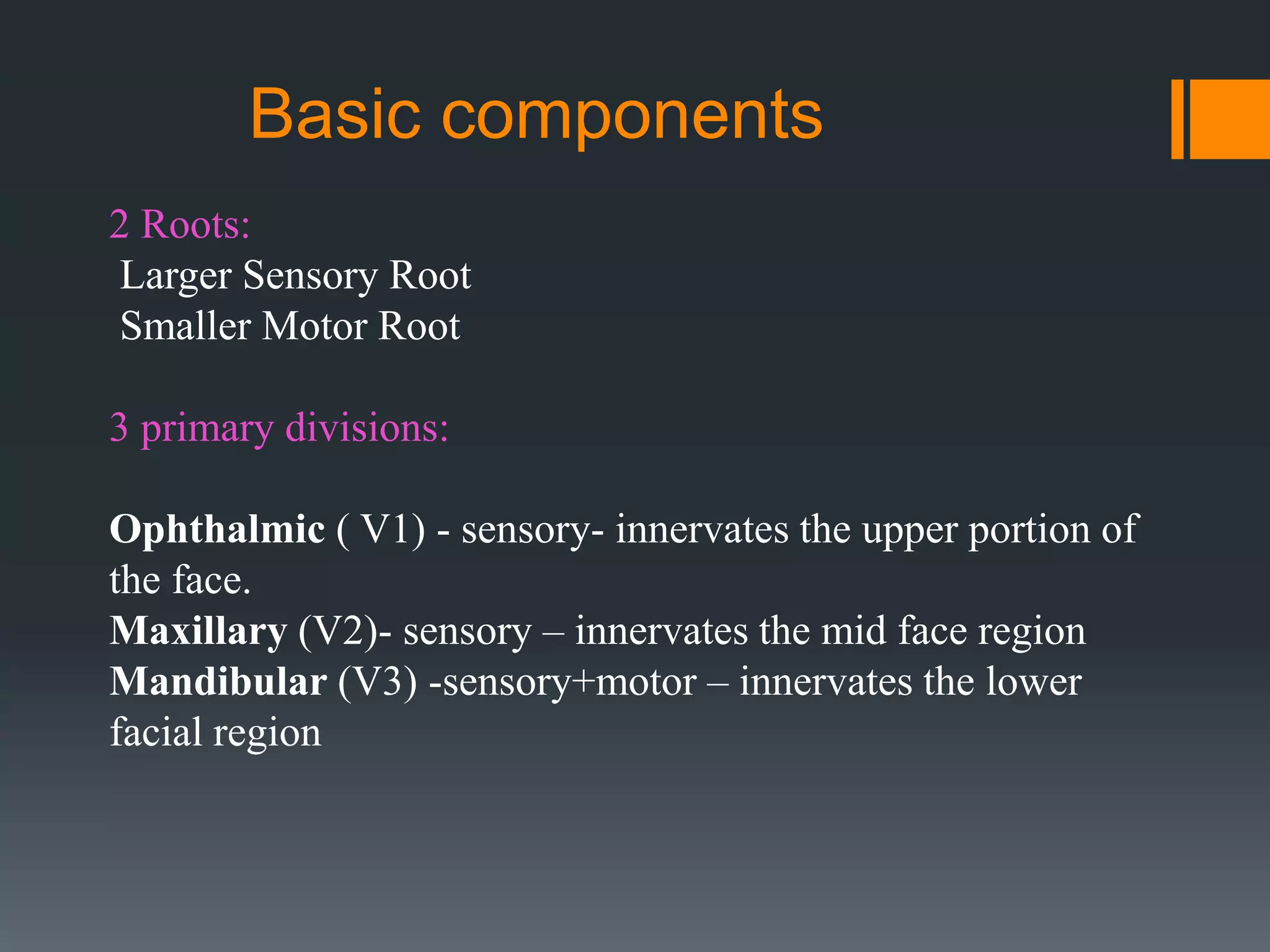
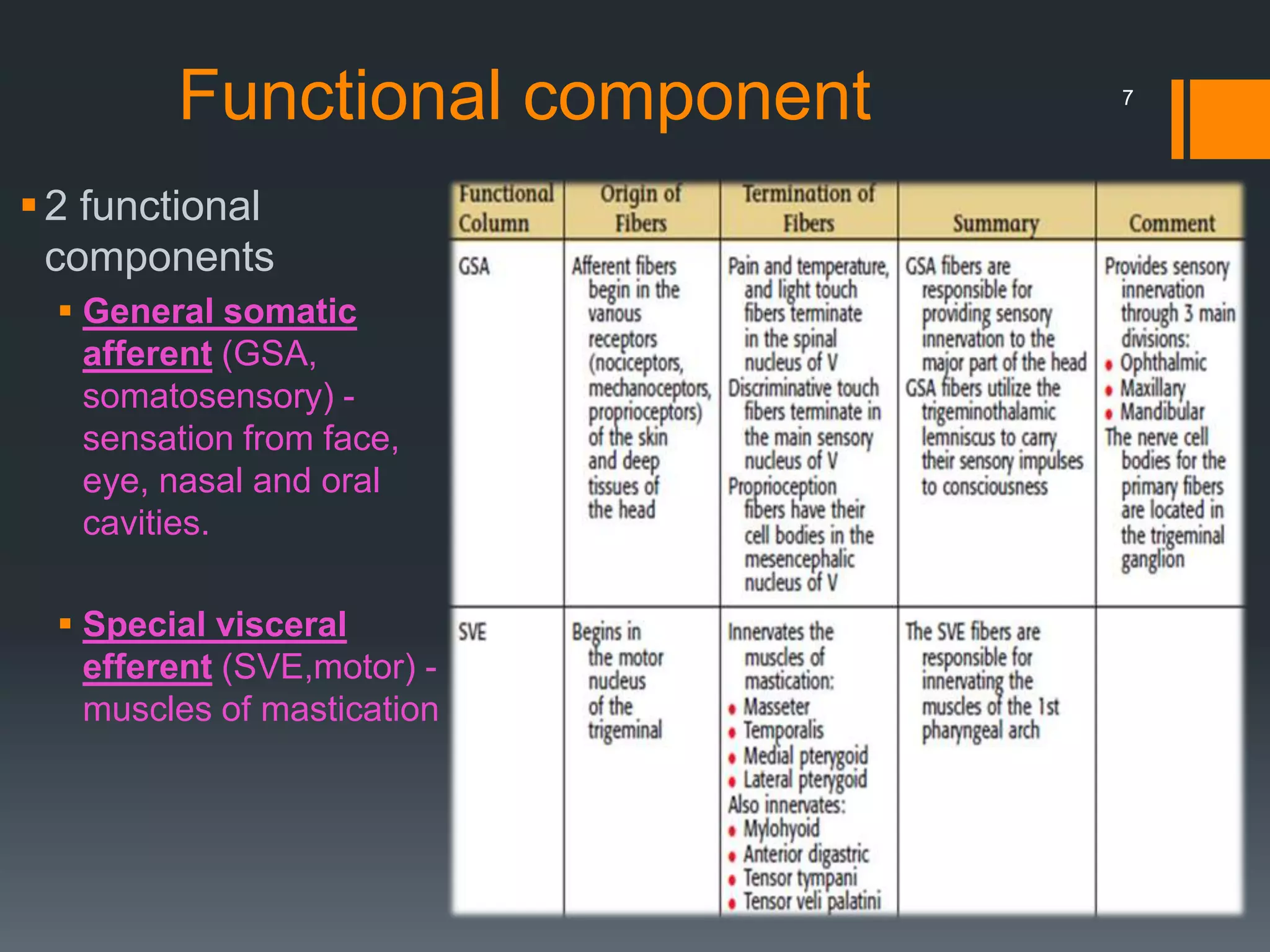
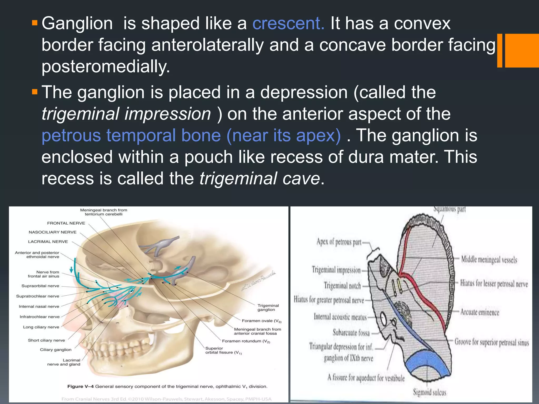
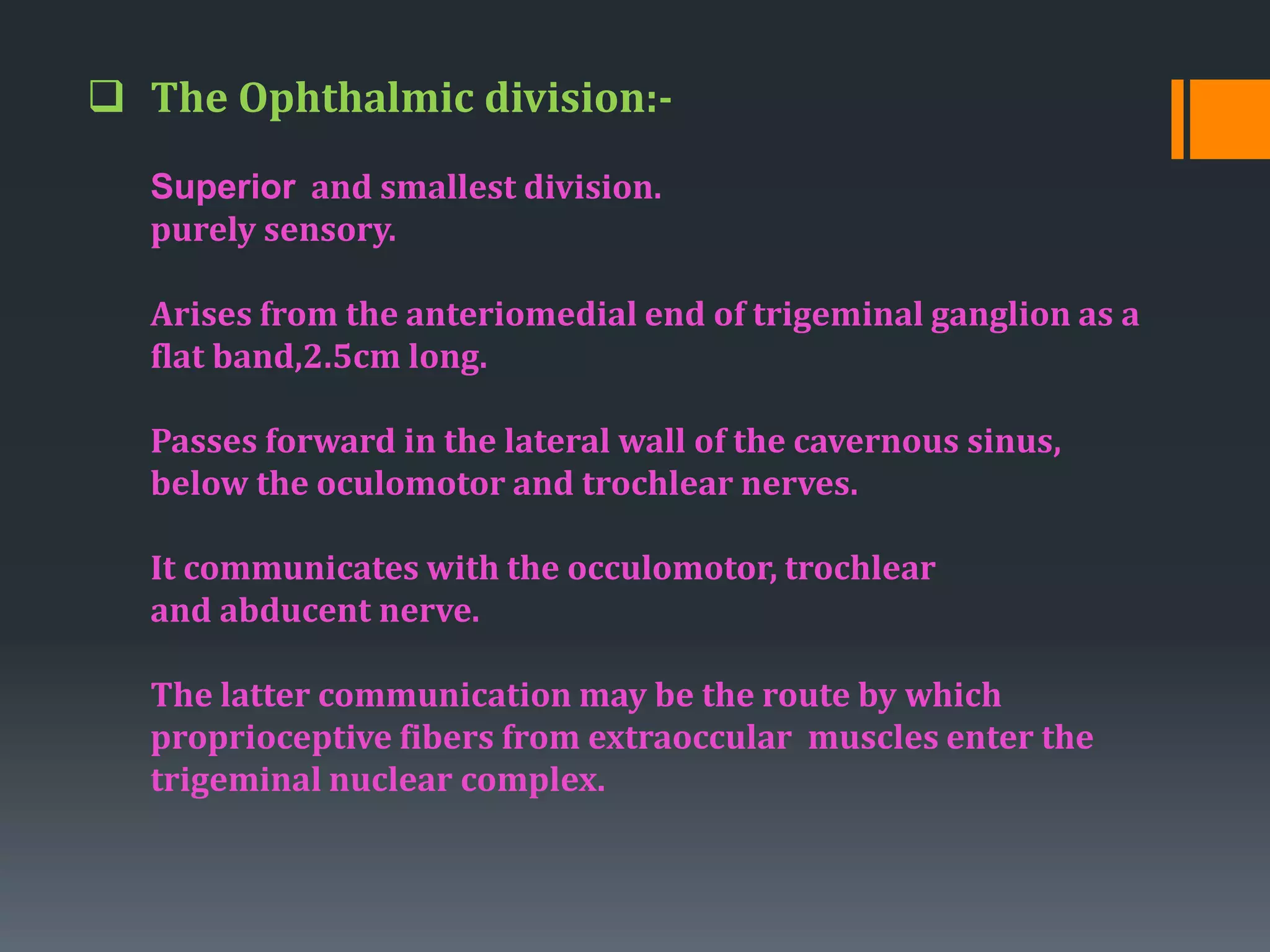
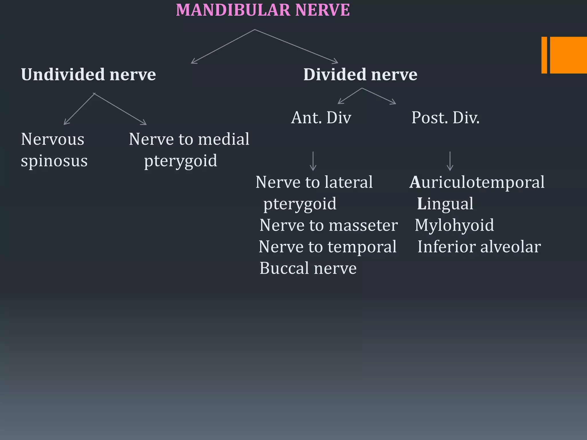
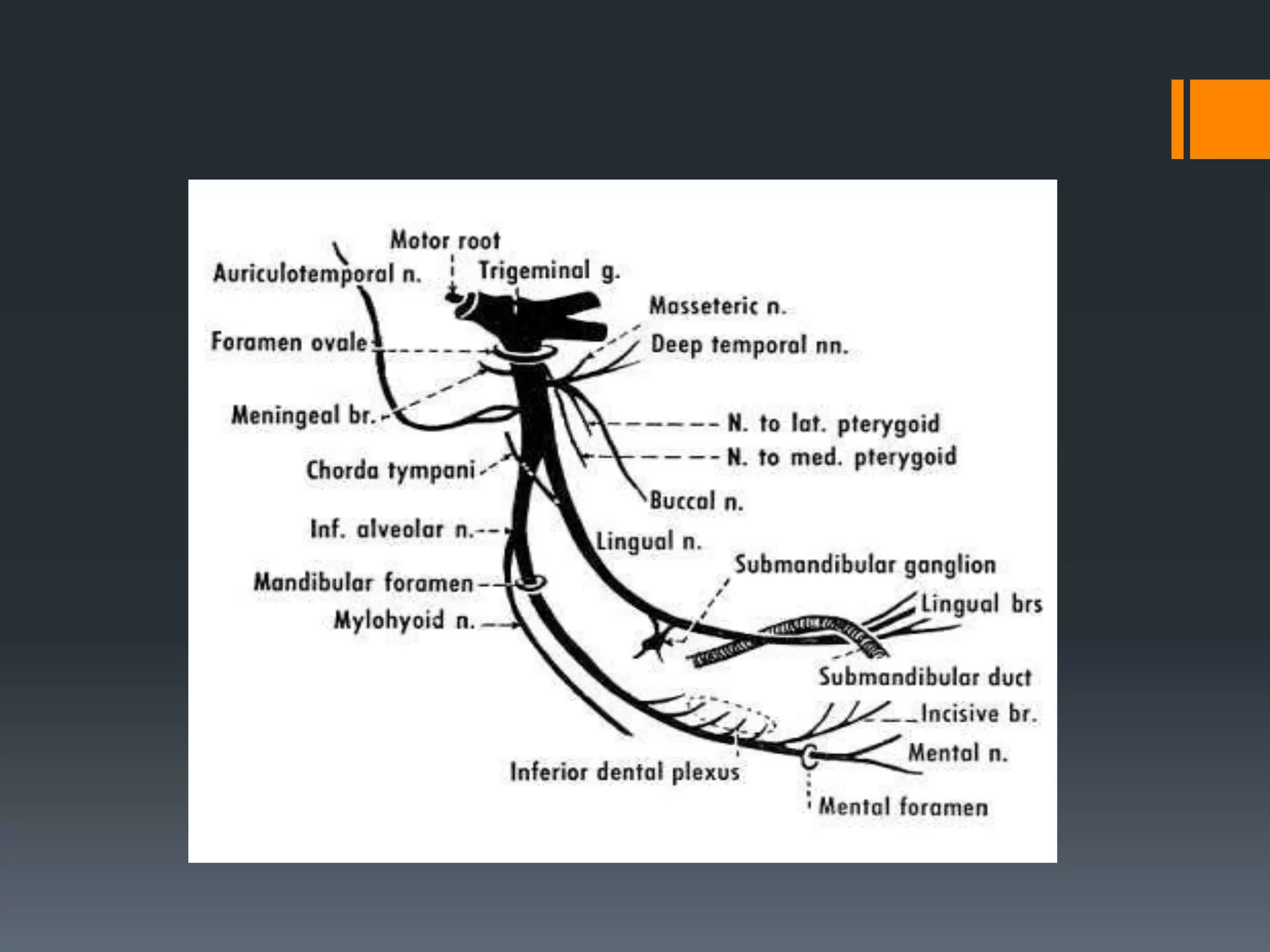
Trigeminal nerve
DR.ABHISHEK KARAN
Reader
Oral and maxillofacial
surgery
Rama Dental College and
Hospital
Kanpur
The cranial nerves are composed of twelve pairs of nerves
that emanate from the nervous tissue of the brain.
In order to reach their targets they must ultimately exit/enter
the cranium through openings in the skull.
Hence, their name is derived from their association with
the cranium.
Classification of cranial nerves
Sensory cranial nerves: contain only afferent (sensory) fibers
Motor cranial nerves: contain only efferent (motor) fibers
Mixed nerves: contain both sensory and motor fibers
Nerve in order
Cranial Nerve I - Olfactory (s)
Cranial Nerve II - Optic (s)
Cranial Nerve III - Occulomotor (mo)
Cranial Nerve IV - Trochlear (mo)
Cranial Nerve V - Trigeminal (m)
Cranial Nerve VI - Abducens (mo)
Cranial Nerve VII - Facial(m)
Cranial Nerve VIII- Vestibulocochlear (s)
Cranial Nerve IX - Glossopharyngeal (m)
Cranial Nerve X - Vagus (m)
Cranial Nerve XI - Spinal Accessory (mo)
Cranial Nerve XII - Hypoglossal (mo)
 The trigeminal is the fifth and largest cranial nerve
 It was first described by fallopius and meckle in 1748.
 It is the main sensory supply of the face and the head.
 The meaning of trigeminal in Latin tri-three; geminus-twin. This term
was given by winslow.
 The Trigeminal nerve is so called because it consists of three main
divisions. These are the ophthalmic nerve, the maxillary nerve and
the mandibular nerve.
Basic components
2 Roots:
Larger Sensory Root
Smaller Motor Root
3 primary divisions:
Ophthalmic ( V1) - sensory- innervates the upper portion of
the face.
Maxillary (V2)- sensory – innervates the mid face region
Mandibular (V3) -sensory+motor – innervates the lower
facial region
The trigeminal nerve has two roots
large sensory small motor
(middle of the pons)
fibers run forwards and laterally fibers run forwards and laterally
in the middle cranial fossa in the parallel to the sensory.
enters the gasserian ganglion passes immediately below gasserian
ganglion reaches foramen ovale
Divides into three divisions. Joins the mandibular nerve.
Functional component
2 functional
components
 General somatic
afferent (GSA,
somatosensory) -
sensation from face,
eye, nasal and oral
cavities.
 Special visceral
efferent (SVE,motor) -
muscles of mastication
7
Nuclei of trigeminal nerve:-
It has got 4 nuclei :
1) Main sensory nuclei
2) Spinal nuclei sensory
3) Mesencephalic nuclei
4) Motor nuclei
1.Mesencephalic nucleus in midbrain.
2.Main sensory nucleus situated in upper pons.
3.Spinal nucleus in upper pons to C2 segment of spinal cord.
4.Motor nucleus situated in upper pons.
.
Nucleus of trigeminal nerve
Trigeminal ganglion
 The ganglion is also called the semilunar ganglion or the Gasserian
ganglion.
 The ganglion is connected to the brain stem (pons) by a thick sensory
root.
 The trigeminal nerve also has a motor root, which emerges from the
pons medial to the sensory root. (Note: M for motor and M for medial).
 The motor root passes deep to the trigeminal ganglion and joins the
mandibular nerve
Ganglion is shaped like a crescent. It has a convex
border facing anterolaterally and a concave border facing
posteromedially.
The ganglion is placed in a depression (called the
trigeminal impression ) on the anterior aspect of the
petrous temporal bone (near its apex) . The ganglion is
enclosed within a pouch like recess of dura mater. This
recess is called the trigeminal cave.
Divisions of trigeminal nerve
 Ophthalmic nerve: smallest, enters orbit through superior orbital fissure
 Maxillary nerve : sensory, begins in the middle of semilunar ganglion
leaves skull through foramen rhotundum
 Mandibular nerve: consists two roots; a large sensory root from
semilunar ganglion and a smaller motor root passing beneath it to unite
with sensory root just after it emerges from foramen ovale.
 The Ophthalmic division:-
Superior and smallest division.
purely sensory.
Arises from the anteriomedial end of trigeminal ganglion as a
flat band,2.5cm long.
Passes forward in the lateral wall of the cavernous sinus,
below the oculomotor and trochlear nerves.
It communicates with the occulomotor, trochlear
and abducent nerve.
The latter communication may be the route by which
proprioceptive fibers from extraoccular muscles enter the
trigeminal nuclear complex.
Before entering the orbit by the superior orbital fissure it
divides into
Lacrimal Nasociliary Frontal
(smallest) (intermediate) (largest)
Internal External Supra Supra
nasal nasal Troclear Orbital
Long Infra Posterior
ciliary Trochlear Ethmoidal
The Maxillary Nerve:
It is intermediate division of trigeminal nerve.
purely sensory.
ORIGIN:
It leaves the trigeminal ganglion between the ophthalmic
and mandibular divisions as a flat plexiform band
Passes slightly medial to lateral wall of cavernous sinus
Leaves the cranium through foramen rhotundum, which is
located in the greater wing of sphenoid bone.
Once outside the cranium, it crosses the uppermost
part of the pterygopalatine fossa, between the
pterygoid plates of sphenoid bone and the palatine
bone
As it crosses the pterygopalatine fossa it gives of
branches
sphenopalatine ganglion zygomatic branches
posterior superior alveolar nerve
It then angles laterally in a groove on the posterior surface
of the maxilla,entering the orbit through the inferior
orbital fissure
Within the orbit it occupies the infraorbital groove and
becomes the infraorbital nerve,which courses anteriorly
into the infraorbital canal
The maxillary division emerges on the anterior surface of
face through the infraorbital foramen, where it divides into
its terminal branches, supplying the skin of the face, nose,
lower eyelid and upper lip
BRANCHES
Within cranium In pterygopalatine fossa In infraorbital canal On face
MAXILLARY NERVE
Middle meningeal
nerve
 Inferior palpebral
 Lateral nasal
 Superior labial
 MSA
(middle superior
alveolar nerve)
ASA
(anterior superior
alveolar nerve)
Zygomatic
PSA
(posterior
superior
alveolar)
Pterygopalatine
Zygomatico
temporal
zygomatico
facial
Orbital
Nasal
Palatine
Pharyngeal
Maxillary nerve
Terminal branches to the face
THE MANDIBULAR DIVISION:
Largest division of trigeminal nerve
Mixed in nature
Has a large sensory root and a small motor root
The sensory root originates from trigeminal ganglion
whereas the motor root originates in the pons and
medulla oblongata
The two roots emerge from the cranium separately
through the foramen ovale, the motor root lying
medial to sensory
they unite just outside the skull and form the main
trunk of 3rd division
MANDIBULAR NERVE
Undivided nerve Divided nerve
Ant. Div Post. Div.
Nervous Nerve to medial
spinosus pterygoid
Nerve to lateral Auriculotemporal
pterygoid Lingual
Nerve to masseter Mylohyoid
Nerve to temporal Inferior alveolar
Buccal nerve
mandibular nerve
Ganglions of the trigeminal nerve
GANGLIA ASSO WITH THE TRIGEMINAL
NERVE
1.CILLIARY GANGLION
connected with nasocilliary nerve by ganglionic
branches in orbit, non synapsing
sensory for orbit
2. PTERYGOPALATINE GANGLION:
connected to maxillary nerve in infratemporal fossa
sensory to orbital septum, orbital and nasal cavity, maxillary
sinus , palate , nasopharynx.
Branches of the pterygopalatine(sphenopalatine)
ganglion.
3. OTIC GANGLION: lies between trunk of mandibular
nerve and tensor palatini , nerve to med pterygoid passes
through but does not synapse in the ganglion.
4.SUBMANDIBULAR GANGLION: Related to lingual nerve,
rest on hypoglossus
Supplies posterior ganglionic Parasympathetic
secretomotor fibers to submandibular and sublingual
gland.
Commonest clinical application
 The most commonly anesthetized nerves in
dentistry are branches or nerve trunks associated
with the maxillary and mandibular divisions of the
trigeminal nerve.
 The maxilla’s relatively porous alveolar bone
allows for the use of straightforward local
anesthetic techniques of paraperiosteal field
blocks or infiltrations.
 The mandible is different. The outer layer of
cortical bone is thick and nonporous and thus
normally requires the use of a nerve block at a
site away from the teeth being treated.
CLINICAL APPLICATION OF
TRIGEMINAL GANGLION
Shingles and varicella-zoster: The trigeminal
ganglion, as any sensory ganglion, may be the site
of infection by the herpes zoster virus causing
shingles, a painful vesicular eruption in the sensory
distribution of the nerve.
Trigeminal neuralgia (tic douloureux): This is severe
pain in the distribution of the trigeminal nerve or one
of its branches, the cause often being unknown. It
may require partial destruction of the ganglion.
35
Trigeminal Neuralgia:-
also known as Fothergill’s disease
Tic douloureux (painful jerking)
it is defined as ,
sudden ,usually ,unilateral ,severe ,brief ,stabbing ,
lancinating, recurring pain in the distribution of one
or more branches of trigeminal nerve.
Mean age: 50 y onwards
Female predominance (male : female = 1:2 ~2:3)
TRIGGER ZONE
Provocated by obvious stimuli like
Touching face at particular site, Chewing, Speaking, Brushing, Shaving,
Washing the face
The characteristic of the disorder being that the attacks do not occur during
sleep
TREATMENT:-
Medical treatment
Surgical treatment:-
Peripheral injections
Peripheral neurectomy
Cryotherapy
Peripheral radiofrequency
Neurolysis(thermocoagulation)
Gasserian ganglion procedures
Wallenberg syndrome:-
A stroke which causes loss of pain/temperature sensation
from one side of the face and the other side of the body.
ETIOLOGY:-
In the medulla, the Ascending Spinothalamic Tract (which
carries pain/temperature information from the opposite side of the
body) is adjacent to the Descending Spinal Tract of the fifth nerve
(which carries pain /temperature information from the same side of the
face)
A stroke cuts off the blood supply to this area
Destroys both tracts simultaneously.
Results in loss of pain/temperature sensation in a unique
“checkerboard” pattern (ipsilateral face, contralateral body)
Characteristic diagnostic feature.
CLINICAL APPLICATION OF OPHTHALMIC NERVE
 Ethmoid tumours
Malignant tumors of the mucous lining of the
ethmoid air cells may expand into the orbits,
damaging branches of opthalmic nerve. This
may lead to displacement of the orbital contents
causing proptosis and squint, and sensory loss
over the anterior nasal skin.
 Nasal fractures
Trauma to the nose may damage the nasociliary
nerve. Sensory loss of the skin down to the tip of
the nose may result.
40
Corneal reflex: When the cornea is touched, usually with a wisp of
cotton, the subject blinks. This tests V and VII. The nerve impulses
pass through cornea and then through nasociliary nerve to the brain.
Supraorbital injuries
Trauma to the supraorbital margin may damage the supraorbital and
supratrochlear nerves causing sensory loss in the scalp.
41
CLINICAL APPLICATION OF MAXILLARY NERVE
Infraorbital injuries (malar fractures):
Trauma to infraorbital margin may cause sensory loss of
infraorbital skin.
Maxillary antrum tumours:
Malignant tumors of the mucous lining of the maxillary
antrum may expand into the orbit, damaging branches of
maxillary nerve, particularly the infraorbital. This may lead
to anesthesia over the facial skin.
42
Maxillary sinus infections:
Infections of the maxillary sinus may cause infraorbital pain or
may cause referred pain to other structures supplied by
maxillary nerve e.g. upper teeth.
Maxillary teeth abscesses:
The roots of the maxillary teeth (especially the second molars)
are intimately related to the maxillary sinus. Root abscesses
are painful.
CLINICAL APPLICATION OF MANDIBULAR NERVE
 Lingual nerve: Careless extractions of the third
lower molar, abscesses of its root, or fractures of
the angle of the mandible may all damage the
lingual nerve. This may result in loss of somatic
sensation from the anterior portion of the tongue
and loss of taste sensation.
 Protection of lingual nerve:: during surgical removal
of mandibular third molar- reflection of lingual
mucoperiosteal flap is raised and Howarth’s
periosteal elevetor is placed.
 Inferior alveolar nerve: Trauma to the mandible
may damage or tear the inferior alveolar nerve
in the mandibular canal leading to sensory loss
distal to the lesion.
 Mumps: Mumps is inflammation of the parotid gland causing tension in the
parotid capsule which is innervated by the auriculotemporal nerve. It gives
both local tenderness and referred ear ache.
 Submandibular duct: The intimate relationship between the submandibular
duct and the lingual nerve is significant in duct infections and surgery. If the
lingual nerve were damaged during a submandibular surgery, there would be
sensory loss, both somatic and taste, in the anterior portion of the tongue.
 Referred pain to the ear: Disease of the TMJ or swelling of the parotid gland
may cause ear ache because of referred pain. Also, pain from the lower teeth,
oral cavity and tongue may be referred to the ear.
 Superficial temporal artery biopsy: The auriculotemporal nerve accompanies
the superficial temporal artery on the temple. In cases of temporal arteritis, the
nerve is anaesthetized so that the overlying skin can be incised to obtain a
biopsy of the artery
LESIONS ASSOCIATED WITH INTRACRANIAL PART
OF TRIGEMINAL
Infections and neoplasia most commonly involve the peripheral divisions of the
trigeminal nerve rather than the intracranial part.
The Meckel’s cavity can be involved either by extrinsic or intrinsic disease. Extrinsic
lesions, usually bony metastasis, chordoma, or chondrosarcoma, destroy adjacent
bone as they extend toward the Meckel’s cavity. Intrinsic lesions simply expand the
Meckel’s cavity.
Pituitary fossa and cavernous sinus lesions may extend to the Meckel’s cavity or
involve the cavernous portion of the trigeminal nerve divisions as well.
The trigeminal nerve has three sensory and one motor nuclei. The sensory nuclei are
the principal, mesencephalic, and spinal sensory.
The cervical extension of the spinal sensory nucleus explains the relation of upper
cervical disk herniation and its association with trigeminal sensory neuropathy.
 Multiple sclerosis, glioma, and infarction are the most common
brainstem and upper cervical cord lesions resulting in fifth cranial
nerve symptom.
 Less common lesions include metastasis, cavernous hemangiomas,
hemorrhage, and arteriovenous malformation.
 Rarely, rhombencephalitis may develop as a result of retrograde
extension of herpes simplex virus type 1 from the trigeminal ganglion
into the brainstem.
46
THE AURICULOTEMPRAL NERVE SYNDROME
(FREY’s SYNDROME)
 Consists of flushing and sweating of the
ipsilateral face in the distribution of the
auriculotemporal nerve upon eating or tasting.
 It is occasionally seen following injury or
infection of the parotid gland area .
THE PARATRIGEMINAL SYNDROME
 It is also known as the Reader
Syndrome and is a rare disorder
produced by tumors arising in the
semilunar ganglion.
 Characterised by trigeminal
neuralgia at the onset , followed by
facial anesthesias on the affected
side.
 The muscles of mastication are
found weakened or paralysed.
Conclusion:-
Trigeminal nerve, its anatomic course and branches are
very important from a dentist point of view as inadvertant
surgical procedure may lead to trigeminal nerve injury.
Disorders of Trigeminal nerve are not rare ,knowing about
it will help in formulating appropriate diagnosis and
treatment thus achieving the best possible recovery of
Trigeminal nerve function.
Nerve blocks given for carrying various dental procedures
involves the various branches of Trigeminal nerve,hence to
avoid any complications ,one needs to have a knowledge
about the course and branches of the nerve .
Trigeminal nerve

Trigeminal nerve

  • 1.
    Trigeminal nerve DR.ABHISHEK KARAN Reader Oraland maxillofacial surgery Rama Dental College and Hospital Kanpur
  • 2.
    The cranial nervesare composed of twelve pairs of nerves that emanate from the nervous tissue of the brain. In order to reach their targets they must ultimately exit/enter the cranium through openings in the skull. Hence, their name is derived from their association with the cranium. Classification of cranial nerves Sensory cranial nerves: contain only afferent (sensory) fibers Motor cranial nerves: contain only efferent (motor) fibers Mixed nerves: contain both sensory and motor fibers
  • 3.
    Nerve in order CranialNerve I - Olfactory (s) Cranial Nerve II - Optic (s) Cranial Nerve III - Occulomotor (mo) Cranial Nerve IV - Trochlear (mo) Cranial Nerve V - Trigeminal (m) Cranial Nerve VI - Abducens (mo) Cranial Nerve VII - Facial(m) Cranial Nerve VIII- Vestibulocochlear (s) Cranial Nerve IX - Glossopharyngeal (m) Cranial Nerve X - Vagus (m) Cranial Nerve XI - Spinal Accessory (mo) Cranial Nerve XII - Hypoglossal (mo)
  • 4.
     The trigeminalis the fifth and largest cranial nerve  It was first described by fallopius and meckle in 1748.  It is the main sensory supply of the face and the head.  The meaning of trigeminal in Latin tri-three; geminus-twin. This term was given by winslow.  The Trigeminal nerve is so called because it consists of three main divisions. These are the ophthalmic nerve, the maxillary nerve and the mandibular nerve.
  • 5.
    Basic components 2 Roots: LargerSensory Root Smaller Motor Root 3 primary divisions: Ophthalmic ( V1) - sensory- innervates the upper portion of the face. Maxillary (V2)- sensory – innervates the mid face region Mandibular (V3) -sensory+motor – innervates the lower facial region
  • 6.
    The trigeminal nervehas two roots large sensory small motor (middle of the pons) fibers run forwards and laterally fibers run forwards and laterally in the middle cranial fossa in the parallel to the sensory. enters the gasserian ganglion passes immediately below gasserian ganglion reaches foramen ovale Divides into three divisions. Joins the mandibular nerve.
  • 7.
    Functional component 2 functional components General somatic afferent (GSA, somatosensory) - sensation from face, eye, nasal and oral cavities.  Special visceral efferent (SVE,motor) - muscles of mastication 7
  • 8.
    Nuclei of trigeminalnerve:- It has got 4 nuclei : 1) Main sensory nuclei 2) Spinal nuclei sensory 3) Mesencephalic nuclei 4) Motor nuclei 1.Mesencephalic nucleus in midbrain. 2.Main sensory nucleus situated in upper pons. 3.Spinal nucleus in upper pons to C2 segment of spinal cord. 4.Motor nucleus situated in upper pons.
  • 9.
  • 11.
    Trigeminal ganglion  Theganglion is also called the semilunar ganglion or the Gasserian ganglion.  The ganglion is connected to the brain stem (pons) by a thick sensory root.  The trigeminal nerve also has a motor root, which emerges from the pons medial to the sensory root. (Note: M for motor and M for medial).  The motor root passes deep to the trigeminal ganglion and joins the mandibular nerve
  • 12.
    Ganglion is shapedlike a crescent. It has a convex border facing anterolaterally and a concave border facing posteromedially. The ganglion is placed in a depression (called the trigeminal impression ) on the anterior aspect of the petrous temporal bone (near its apex) . The ganglion is enclosed within a pouch like recess of dura mater. This recess is called the trigeminal cave.
  • 13.
    Divisions of trigeminalnerve  Ophthalmic nerve: smallest, enters orbit through superior orbital fissure  Maxillary nerve : sensory, begins in the middle of semilunar ganglion leaves skull through foramen rhotundum  Mandibular nerve: consists two roots; a large sensory root from semilunar ganglion and a smaller motor root passing beneath it to unite with sensory root just after it emerges from foramen ovale.
  • 14.
     The Ophthalmicdivision:- Superior and smallest division. purely sensory. Arises from the anteriomedial end of trigeminal ganglion as a flat band,2.5cm long. Passes forward in the lateral wall of the cavernous sinus, below the oculomotor and trochlear nerves. It communicates with the occulomotor, trochlear and abducent nerve. The latter communication may be the route by which proprioceptive fibers from extraoccular muscles enter the trigeminal nuclear complex.
  • 15.
    Before entering theorbit by the superior orbital fissure it divides into Lacrimal Nasociliary Frontal (smallest) (intermediate) (largest) Internal External Supra Supra nasal nasal Troclear Orbital Long Infra Posterior ciliary Trochlear Ethmoidal
  • 18.
    The Maxillary Nerve: Itis intermediate division of trigeminal nerve. purely sensory. ORIGIN: It leaves the trigeminal ganglion between the ophthalmic and mandibular divisions as a flat plexiform band Passes slightly medial to lateral wall of cavernous sinus Leaves the cranium through foramen rhotundum, which is located in the greater wing of sphenoid bone.
  • 19.
    Once outside thecranium, it crosses the uppermost part of the pterygopalatine fossa, between the pterygoid plates of sphenoid bone and the palatine bone As it crosses the pterygopalatine fossa it gives of branches sphenopalatine ganglion zygomatic branches posterior superior alveolar nerve
  • 20.
    It then angleslaterally in a groove on the posterior surface of the maxilla,entering the orbit through the inferior orbital fissure Within the orbit it occupies the infraorbital groove and becomes the infraorbital nerve,which courses anteriorly into the infraorbital canal The maxillary division emerges on the anterior surface of face through the infraorbital foramen, where it divides into its terminal branches, supplying the skin of the face, nose, lower eyelid and upper lip
  • 21.
    BRANCHES Within cranium Inpterygopalatine fossa In infraorbital canal On face MAXILLARY NERVE Middle meningeal nerve  Inferior palpebral  Lateral nasal  Superior labial  MSA (middle superior alveolar nerve) ASA (anterior superior alveolar nerve) Zygomatic PSA (posterior superior alveolar) Pterygopalatine Zygomatico temporal zygomatico facial Orbital Nasal Palatine Pharyngeal
  • 22.
  • 23.
  • 24.
    THE MANDIBULAR DIVISION: Largestdivision of trigeminal nerve Mixed in nature Has a large sensory root and a small motor root The sensory root originates from trigeminal ganglion whereas the motor root originates in the pons and medulla oblongata The two roots emerge from the cranium separately through the foramen ovale, the motor root lying medial to sensory they unite just outside the skull and form the main trunk of 3rd division
  • 25.
    MANDIBULAR NERVE Undivided nerveDivided nerve Ant. Div Post. Div. Nervous Nerve to medial spinosus pterygoid Nerve to lateral Auriculotemporal pterygoid Lingual Nerve to masseter Mylohyoid Nerve to temporal Inferior alveolar Buccal nerve
  • 26.
  • 28.
    Ganglions of thetrigeminal nerve
  • 29.
    GANGLIA ASSO WITHTHE TRIGEMINAL NERVE 1.CILLIARY GANGLION connected with nasocilliary nerve by ganglionic branches in orbit, non synapsing sensory for orbit
  • 30.
    2. PTERYGOPALATINE GANGLION: connectedto maxillary nerve in infratemporal fossa sensory to orbital septum, orbital and nasal cavity, maxillary sinus , palate , nasopharynx.
  • 31.
    Branches of thepterygopalatine(sphenopalatine) ganglion.
  • 32.
    3. OTIC GANGLION:lies between trunk of mandibular nerve and tensor palatini , nerve to med pterygoid passes through but does not synapse in the ganglion.
  • 33.
    4.SUBMANDIBULAR GANGLION: Relatedto lingual nerve, rest on hypoglossus Supplies posterior ganglionic Parasympathetic secretomotor fibers to submandibular and sublingual gland.
  • 34.
    Commonest clinical application The most commonly anesthetized nerves in dentistry are branches or nerve trunks associated with the maxillary and mandibular divisions of the trigeminal nerve.  The maxilla’s relatively porous alveolar bone allows for the use of straightforward local anesthetic techniques of paraperiosteal field blocks or infiltrations.  The mandible is different. The outer layer of cortical bone is thick and nonporous and thus normally requires the use of a nerve block at a site away from the teeth being treated.
  • 35.
    CLINICAL APPLICATION OF TRIGEMINALGANGLION Shingles and varicella-zoster: The trigeminal ganglion, as any sensory ganglion, may be the site of infection by the herpes zoster virus causing shingles, a painful vesicular eruption in the sensory distribution of the nerve. Trigeminal neuralgia (tic douloureux): This is severe pain in the distribution of the trigeminal nerve or one of its branches, the cause often being unknown. It may require partial destruction of the ganglion. 35
  • 36.
    Trigeminal Neuralgia:- also knownas Fothergill’s disease Tic douloureux (painful jerking) it is defined as , sudden ,usually ,unilateral ,severe ,brief ,stabbing , lancinating, recurring pain in the distribution of one or more branches of trigeminal nerve. Mean age: 50 y onwards Female predominance (male : female = 1:2 ~2:3)
  • 37.
    TRIGGER ZONE Provocated byobvious stimuli like Touching face at particular site, Chewing, Speaking, Brushing, Shaving, Washing the face The characteristic of the disorder being that the attacks do not occur during sleep
  • 38.
    TREATMENT:- Medical treatment Surgical treatment:- Peripheralinjections Peripheral neurectomy Cryotherapy Peripheral radiofrequency Neurolysis(thermocoagulation) Gasserian ganglion procedures
  • 39.
    Wallenberg syndrome:- A strokewhich causes loss of pain/temperature sensation from one side of the face and the other side of the body. ETIOLOGY:- In the medulla, the Ascending Spinothalamic Tract (which carries pain/temperature information from the opposite side of the body) is adjacent to the Descending Spinal Tract of the fifth nerve (which carries pain /temperature information from the same side of the face) A stroke cuts off the blood supply to this area Destroys both tracts simultaneously. Results in loss of pain/temperature sensation in a unique “checkerboard” pattern (ipsilateral face, contralateral body) Characteristic diagnostic feature.
  • 40.
    CLINICAL APPLICATION OFOPHTHALMIC NERVE  Ethmoid tumours Malignant tumors of the mucous lining of the ethmoid air cells may expand into the orbits, damaging branches of opthalmic nerve. This may lead to displacement of the orbital contents causing proptosis and squint, and sensory loss over the anterior nasal skin.  Nasal fractures Trauma to the nose may damage the nasociliary nerve. Sensory loss of the skin down to the tip of the nose may result. 40
  • 41.
    Corneal reflex: Whenthe cornea is touched, usually with a wisp of cotton, the subject blinks. This tests V and VII. The nerve impulses pass through cornea and then through nasociliary nerve to the brain. Supraorbital injuries Trauma to the supraorbital margin may damage the supraorbital and supratrochlear nerves causing sensory loss in the scalp. 41
  • 42.
    CLINICAL APPLICATION OFMAXILLARY NERVE Infraorbital injuries (malar fractures): Trauma to infraorbital margin may cause sensory loss of infraorbital skin. Maxillary antrum tumours: Malignant tumors of the mucous lining of the maxillary antrum may expand into the orbit, damaging branches of maxillary nerve, particularly the infraorbital. This may lead to anesthesia over the facial skin. 42 Maxillary sinus infections: Infections of the maxillary sinus may cause infraorbital pain or may cause referred pain to other structures supplied by maxillary nerve e.g. upper teeth. Maxillary teeth abscesses: The roots of the maxillary teeth (especially the second molars) are intimately related to the maxillary sinus. Root abscesses are painful.
  • 43.
    CLINICAL APPLICATION OFMANDIBULAR NERVE  Lingual nerve: Careless extractions of the third lower molar, abscesses of its root, or fractures of the angle of the mandible may all damage the lingual nerve. This may result in loss of somatic sensation from the anterior portion of the tongue and loss of taste sensation.  Protection of lingual nerve:: during surgical removal of mandibular third molar- reflection of lingual mucoperiosteal flap is raised and Howarth’s periosteal elevetor is placed.  Inferior alveolar nerve: Trauma to the mandible may damage or tear the inferior alveolar nerve in the mandibular canal leading to sensory loss distal to the lesion.
  • 44.
     Mumps: Mumpsis inflammation of the parotid gland causing tension in the parotid capsule which is innervated by the auriculotemporal nerve. It gives both local tenderness and referred ear ache.  Submandibular duct: The intimate relationship between the submandibular duct and the lingual nerve is significant in duct infections and surgery. If the lingual nerve were damaged during a submandibular surgery, there would be sensory loss, both somatic and taste, in the anterior portion of the tongue.  Referred pain to the ear: Disease of the TMJ or swelling of the parotid gland may cause ear ache because of referred pain. Also, pain from the lower teeth, oral cavity and tongue may be referred to the ear.  Superficial temporal artery biopsy: The auriculotemporal nerve accompanies the superficial temporal artery on the temple. In cases of temporal arteritis, the nerve is anaesthetized so that the overlying skin can be incised to obtain a biopsy of the artery
  • 45.
    LESIONS ASSOCIATED WITHINTRACRANIAL PART OF TRIGEMINAL Infections and neoplasia most commonly involve the peripheral divisions of the trigeminal nerve rather than the intracranial part. The Meckel’s cavity can be involved either by extrinsic or intrinsic disease. Extrinsic lesions, usually bony metastasis, chordoma, or chondrosarcoma, destroy adjacent bone as they extend toward the Meckel’s cavity. Intrinsic lesions simply expand the Meckel’s cavity. Pituitary fossa and cavernous sinus lesions may extend to the Meckel’s cavity or involve the cavernous portion of the trigeminal nerve divisions as well. The trigeminal nerve has three sensory and one motor nuclei. The sensory nuclei are the principal, mesencephalic, and spinal sensory. The cervical extension of the spinal sensory nucleus explains the relation of upper cervical disk herniation and its association with trigeminal sensory neuropathy.
  • 46.
     Multiple sclerosis,glioma, and infarction are the most common brainstem and upper cervical cord lesions resulting in fifth cranial nerve symptom.  Less common lesions include metastasis, cavernous hemangiomas, hemorrhage, and arteriovenous malformation.  Rarely, rhombencephalitis may develop as a result of retrograde extension of herpes simplex virus type 1 from the trigeminal ganglion into the brainstem. 46
  • 47.
    THE AURICULOTEMPRAL NERVESYNDROME (FREY’s SYNDROME)  Consists of flushing and sweating of the ipsilateral face in the distribution of the auriculotemporal nerve upon eating or tasting.  It is occasionally seen following injury or infection of the parotid gland area .
  • 48.
    THE PARATRIGEMINAL SYNDROME It is also known as the Reader Syndrome and is a rare disorder produced by tumors arising in the semilunar ganglion.  Characterised by trigeminal neuralgia at the onset , followed by facial anesthesias on the affected side.  The muscles of mastication are found weakened or paralysed.
  • 49.
    Conclusion:- Trigeminal nerve, itsanatomic course and branches are very important from a dentist point of view as inadvertant surgical procedure may lead to trigeminal nerve injury. Disorders of Trigeminal nerve are not rare ,knowing about it will help in formulating appropriate diagnosis and treatment thus achieving the best possible recovery of Trigeminal nerve function. Nerve blocks given for carrying various dental procedures involves the various branches of Trigeminal nerve,hence to avoid any complications ,one needs to have a knowledge about the course and branches of the nerve .