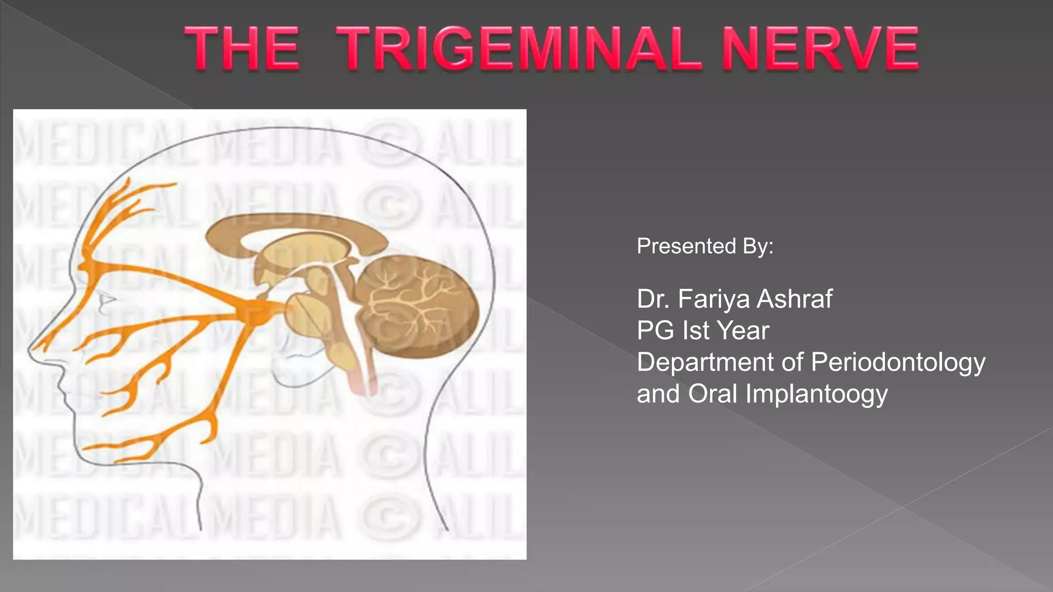
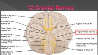
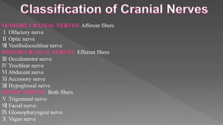
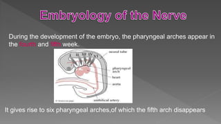
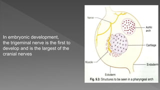
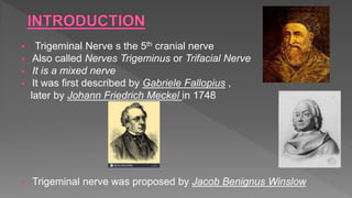
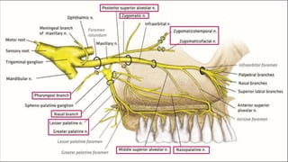
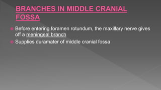
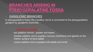
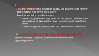
Presented By:
Dr. Fariya Ashraf
PG Ist Year
Department of Periodontology
and Oral Implantoogy
 Nerve physiology
 Embryology of pharyngeal
arches
 Introduction
 Trigeminal ganglion
 Roots
 Nuclei
 Ophthalmic nerve
 Maxillary nerve
 Mandibular nerve
 Appiled anatomy
 Examination of trigeminal
nerve
 Classification of Trigeminal
nerve injuries
 Mechanism of injury to
trigeminal nerve
 Clinical considerations
 Periodontal complications
 Treatment approaches
 Outcomes of injuries and
surgical intervention
 Conclusion
 References
Nerve :
A bundle of fibers that uses chemical and electrical signals to transmit sensory and motor
information from one part of the body to the another.
-There are twelve pairs of cranial nerves and their defining feature is that
they exit the cranial cavity through foramina or fissures.
-All cranial nerves innervate structures in the head or neck.
SENSORY CRANIAL NERVES: Afferent fibers
Ⅰ Olfactory nerve
Ⅱ Optic nerve
Ⅷ Vestibulocochlear nerve
MOTOR CRANIAL NERVES: Efferent fibers
Ⅲ Occulomotor nerve
Ⅳ Trochlear nerve
Ⅵ Abducent nerve
Ⅺ Accessory nerve
Ⅻ Hypoglossal nerve
MIXED NERVES: Both fibers
Ⅴ Trigeminal nerve
Ⅶ Facial nerve
Ⅸ Glossopharyngeal nerve
Ⅹ Vagus nerve
During the development of the embryo, the pharyngeal arches appear in
the fourth and fifth week.
It gives rise to six pharyngeal arches,of which the fifth arch disappears
In embryonic development,
the trigeminal nerve is the first to
develop and is the largest of the
cranial nerves
 Trigeminal Nerve s the 5th cranial nerve
 Also called Nerves Trigeminus or Trifacial Nerve
 It is a mixed nerve
 It was first described by Gabriele Fallopius ,
later by Johann Friedrich Meckel in 1748
 Trigeminal nerve was proposed by Jacob Benignus Winslow
Relations :
a) Lateral – middle meningeal artery
b) Medial – internal carotid artery,
posterior part of cavernous sinus
c) Inferior – foramen lacerum,
greater petrosal nerve, motor root
of trigeminal nerve
d) Superior – parahippocampal gyrus
SENSORY NUCLEUS MOTOR
NUCLEU
S
MESENCEPHALIC
NUCLEUS
SUPERIOR
SENSORY
NUCLEUS
SPINAL
NUCLEUS
• Also known as Gasserion ganglion or Semilunar ganglion.
• Lies at the apex of petrous temporal bone in a
dural cave, the Meckel’s cave
• It is somewhat crescentric or semilunarian
shape, with its convexity directed
anteromedially.
• The three divisions of the trigeminal nerve
emerges from this convexity
• The central processes of the ganglion cells forms the large sensory root of the Trigeminal
nerve, which is attached to pons at its junction with the middle cerebellar peduncle.
• The periheral processes form the three divisions of the trigeminal nerve.
Trigeminal Nerve
Opthalmic
(Sensory)
Maxillary
(Sensory)
Mandibular
(Mixed)
 1st branch
 purely sensory
 Arises from anteromedial part of trigeminal ganglion
 Crosses cavernous sinus b/w occulomotor and trochlear
nerves
 3 branches :
a. Lacrimal
b. Frontal
c. Nasociliary
ALL THESE
BRANCHES
CROSS THE
SUPERIOR
ORBITAL
FISSURE
OPHTHALMIC NERVE
ARISES FROM TRIGEMINAL
GANGLION
CROSSES CAVERNOUS SINUS
LACRIMAL
NERVE
FRONTA
L NERVE
NASOCILIARY
NERVE
Supratrochlear
nerve
Posterior ethmoidal
nerve
Anterior ethmoidal
nerve
Infratrochlear nerve
Long ciliary nerve
Supraorbital
nerve
1. LACRIMAL NERVE
 smallest branch
 connected with zygomaticotemporal branch of maxillary nerve
secretomotor fibres to lacrimal gland
 supplies : lacrimal gland and skin of upper eyelid and conjunctiva
2. FRONTAL NERVE
 largest branch
 runs b/w levator palpebrae superioris and periosteum lining the roof
of orbit
 divides into :
i. Supraorbital nerve : supplies skin of forehead and scalp upto the vertex
ii. Supratrochlear nerve : supplies upper eyelid and conjunctiva
 3. NASOCILIARY NERVE
 3rd branch
 crosses optic nerve, cavernous sinus and supraorbital
fissure
 divides near anterior ethmoidal foramen :
i. Sensory root of ciliary ganglion : carries sensory fibres that
begin in the cornea, the iris and the choroid
ii. Long ciliary nerve : innervates iris, cornea, dilator pupillae and
ciliary muscles. Also gives sensory roots to ciliary ganglia.
iii. Posterior ethmoidal nerve : supplies ethmoidal and sphenoidal
air sinuses
iv. Anterior ethmoidal nerve : supplies nasal cavity
v. Infratrochlear nerve : supplies skin of upper and lower eyelids
and over the upper part of the nose
 Lies in posterior part of orbital cavity
 Situation :
i. Lateral side of optic nerve
ii. Medial to rectus lateralis muscle
 3 roots :
i. Sensory
ii. Sympathetic
iii. Parasympathetic
 Purely sensory
 Arises from middle of the distal edge of trigeminal ganglion
 Passes through foramen rotundum
Branches from the Maxillary Nerve can be divided into the following
groups :
1. Branches in middle cranial fossa
i. Meningeal branch
2. Branches arising in pterygopalatine fossa
i. Ganglionic branches
ii. Zygomatic nerve
iii. Posterior superior alveolar nerve
3. Branches arising in infraorbital groove and canal
i. Middle superior alveolar nerve
ii. Anterior superior alveolar nerve
4. Branches in the face
i. Inferior palpebral
ii. Lateral nasal
iii. Superior labial
 Before entering foramen rotundum, the maxillary nerve gives
off a meningeal branch
 Supplies duramater of middle cranial fossa
A. GANGLIONIC BRANCHES
In pterygopalatine fossa, the maxillary nerve is connected to the pterygopalatine
ganglion by ganglionic branches
i. Branches to the palate :
 two palatine nerves : greater and lesser
 Greater palatine nerve supplies mucous membrane and glands on the
inferior surface of hard palate
 Lesser palatine nerve supplies soft palate and tonsil
ii. Branches to the nose :
 Posterior inferior nasal branches supply the posterior and inferior
part of lateral wall of the nasal cavity
 Posterior superior nasal branches :
a) Medial – supply posterior parts of the roof and septum. One nerve of this
group is large, i.e. Nasopalatine nerve – supplies anterior part of hard
palate.
b) Lateral – supply the posterosuperior part of lateral wall of nasal cavity
iii. Other branches from pterygopalatine ganglion :
a) orbital branches : supply the periosteum and orbitalis muscle
b) pharyngeal nerve
B. ZYGOMATIC NERVE
It enters the orbit through inferior orbital fissure and runs
forward along its lateral wall.
Divides into 2 branches :
i. Zygomaticotemporal nerve :
 emerges from the temporal surface of the bone
 supplies the skin over temple region
ii. Zygomaticofacial nerve :
 emerges from the bone through the zygomaticofacial foramen
present on the lateral surface of the bone
 Supplies skin of cheek
C. POSTERIOR SUPERIOR ALVEOLAR NERVE
arises from the maxillary nerve in pterygopalatine fossa
Runs down on the posterior surface of maxilla
Lies in the wall of maxillary sinus which it supplies
Divides into branches that form a plexus over the
roots of the molar teeth
1. Middle superior alveolar nerve
Arises from infraorbital nerve (nerve lies in infraorbital groove)
Entering the foramen in the floor of the groove, reaches the maxillary sinus and joins
the PSA nerve
Forms a plexus above the roots of premolars
2. Anterior Superior Alveolar Nerve
Arises from infraorbital nerve (nerve lies in infraorbital canal)
Enters bony canal through the maxillary bone (canalis
sinuosis)
Supplies incisors and canines
INFRAORBITAL NERVE
INFRAORBITAL
FORAMEN
INFERIOR
PALPEBRAL
Supply lower eyelid
LATERAL NASAL
Supply the skin on
lateral side of nose
SUPERIOR LABIAL
Supply skin of upper
lip and part of the
cheek
 Formed by union of two roots
 Sensory root arises from lateral part of trigeminal ganglion –
leaves the skull through foramen ovale
 Motor root passes through foramen ovale and unites with the
sensory root just below the foramen
 Nerve then enters the infratemporal fossa
 After a short downward course, it then divides into a smaller
anterior division and a large posterior division
SENSORY SUPPLY OF V3
1. Dura
2. Skin
3. Mucous membrane of lower lip, cheek and chin
4. External ear
5. Parotid gland
6. TMJ
7. Scalp over the temporal bone
8. Lower teeth and gingiva
9. Anterior 2/3rd of tongue
MOTOR SUPPLY OF V3
1. Muscles of mastication
2. Mylohyoid
3. Anterior belly of digastric
4. Tensor tympani
5. Tensor veli palatini
1. SENSORY FUNCTION
2. MOTOR FUNCTION
3. CORNEAL REFLEX
4. JAW JERK TEST
• Initially test the sensory branches by lightly
touching the face with a piece of cotton wool
followed by a blunt pin in three places on each
side of the face:
a. On the forehead
b. On the cheek
c. Around the jawline
Inspect for wasting of the temporal and masseter muscles.
Ask patient to clench their teeth and palpate for contraction of the temporal and masseter
muscles.
Ask the patient then to open their mouth against resistance.
 Ask the patient to look up and away, touch the cornea
 Reflex blinking of both eyes is a normal response.
 Ask the patient to open the mouth fully, and close halfway,
place index finger on the chin and tap with a patella hammer.
 When it is normal, tapping the mandible produces a brisk
contraction.
1. Local anaesthetic injection
2. Third molar surgery
3. Maxillofacial trauma
4. Orthognathic surgery
5. Maxillofacial pathology
6. Endodontic and chemical injury
(Oral & Maxillofacial Trauma, 4th edition – Fonseca)
 Trigeminal neuralgia
 Herpes Zoster Ophthalmicus
 Trigeminal neuropathy
 Wallenberg syndrome
 Also known as Fothergill’s disease,Tic
douloureux (painful jerking)
It is defined as sudden, usually, unilateral,
severe, brief, stabbing, lancinating, recurring
pain in the distribution of one or more branches
of trigeminal nerve.
Mean age: 50 years onwards
 Female predominance
male : female = 1:2 ~2.3
 It is usually idiopathic.
The probable etiologic factors are:-
1) Intra cranial tumors: Traumatic compression of the
trigeminal nerve by neoplastic(cerebellopontine
angle tumor) or vascular anomalies eg.
Arteriovenous malformations.
2) Infections: granulomatous and non granulomatous
infections involving 5th cranial nerve.
3) Petrous Ridge Compression
4) Intracranial Vascular Abnormalities
5) Post Herpetic Neuralgia
6) Demyelinating Conditions
7) Multiple Sclerosis
GENERAL CHARACTERISTICS
 Incidence : seen in about 4 in 100000 individuals
 Age : 5th or 6th decade
 Sex predilection : female predisposition (58%)
 Side involved more frequently : right side (60%)
 Trigeminal nerve involvement : mandibular > maxillary > opthalmic
 Sudden
 Unilateral
 Intermittent Paroxysmal
 Sharp shooting
 Lancinating shock like pain elicited by slight
touching
 Pain rarely crosses the midline
 Pain is of short duration and lasts for few seconds
to minutes
 In extreme cases paient has a motionless face
called the frozen or mask like face
Provocated by obvious stimuli like:
 Touching face at particular site
 Chewing
 Speaking
 Brushing
 Shaving
 Washing the face
TRIGGER POINTS:
Trigger points are most commonly located on the
cheek,lip,nose or buccal mucosa.
Diagnosis:
 Clinical examination with history is
mandatory.
 Response to treatment with tablet of
carbamazepine is universal.
 Injections of local anaesthetic agents into
patients trigger zone gives temporary relief
from pain
 CT scan – poor resolution in posterior fossa
 MRI – imaging modality of choice; reveal
MS plaques and pontine gliomas
Treatment Modalities:
o Medical treatment
o Surgical treatment:
1) Peripheral injections
2) Peripheral neurectomy
3) Cryotherapy
4) Peripheral radiofrequency
5) Neurolysis (thermocoagulation)
6) Gasserion ganglion procedures
Medicinal Treatment :
1. Peripheral nerve injections
a) Long acting anesthetic agents without adrenaline (bupivacaine)
b) Alcohol block - 0.5 -2ml of 95% absolute alcohol
2. Peripheral neurectomy
 oldest and most effective
Performed most commonly in infraorbital,inferior alveolar-metal and rarely
lingual nerves
Infraorbital neurectomy
i. Intraoral conventional approach
ii. Braun’s transantral approach
Inferiorior Alveolar Neuractomy
i. The extra oral approach
ii. Intra oral approach (via Dr Ginwalla’s incision
3) Cryotherapy or Cryoneurolysis
 Direct applications of cryotherapy probe at
temperatures colder than -60 degrees Celsius .
 In this the nerve is not sectioned but destroyed
4) Peripheral radiofrequency neurolysis
(thermocoagulation)
 Gregg and Small (1986)
 Radiofrequency electrode has the capacity to
destroy the pain fibres
5. Gasserian ganglion procedures
i. Glycerol injection
ii. thermocoagulation
iii. Balloon compression
6. Open procedures (intracranial procedures)
 microvascular decompression of sensory root
 1967-1976 by Jannetta
 most commonly performed intracranial open procedure
 open craniotomy approach is used to gain access to the
trigeminal root entry zone and adjacent brain stem
 Compressing branch of superior cerebellar artery is
carefully separated from the nerve
7. Trigeminal root section
a) Extradural sensory root section (Frazier’s approach)
b) Intradural root section by Wilkins
c) Trigeminal tractotomy (Medullary Tractotomy)
• Caused by Varicella Zoster
• Predilection for nasociliary branch of opthalmic
division of trigeminal nerve.
• Diagnosis:
History of pain
Unilateral in nature
Segmental distribution of lesions
Vesicles are presnt
Clinical Features:
1) Cutaneous lesions :
 Rash
 Vesicle
 Pustule crust permanent scar
2) Ocular lesions :
 periorbital pain
 Edema
 Conjunctivitis
 scleritis
 Corneal scarring
 Impaired vision / transient
blindness
Treatment :
 Acyclovir 800mg, 5 times/day for a week, within 4 days of onset
of rash
 Valacyclovir 1,000mg, 3times/day for a week
 Analgesic
 Antibiotic ointments
 Systemic steroids 60mg/day
 Corneal grafting
INFECTION TREATMENT
Shingles Acyclovir(Zovirax), 800 mg orally five times daily for 7-10 days
Skin Palliative with cool compresses, mechanical cleansing
Blepharitis/conjunctivitis Palliative, with cool compresses and topical lubrication
Topical broad-spectrum antibiotics for secondary bacterial infection
Epithelial keratitis Debridement or none
Stromal keratitis Topical steroids
Neurotrophic keratitis Topical lubrication, topical antibiotics for secondary infection, Tissue
adhesives and protective contact lens prevent corneal perforation
Uvetitis Topical steroids, Oral steroids, oral Acyclovir
Acute retinal necrosis/progressive outer
retinal necrosis
i.V acyclovir for 7-10 days followed by oral acyclovir for 14 weeks
Laser/surgical intervetion
 Characterized by numbness in the skin or mucosal
membranes in the distribution of the trigeminal nerve
 Neuropathic weakness in the muscles of mastication.
 TNO should not be confused with trigeminal neuralgia (TNA)
 Brief attacks of lancinating pain but without sensory
impairment or motor weakness.
 In TNO, pain may dominate the clinical picture
 As disorder progresses and neurons are destroyed,
numbness and weakness usually appear.
 In untreated idiopathic TNA, neurons are preserved,
although their myelin sheaths may be destroyed and there
is ultimately gain of function.
 In TNO as the condition advances, loss of function in the
affected nerve branches becomes evident
• A stroke which causes loss of pain/temperature sensation from one
side of the face and the other side of the body.
ETIOLOGY:-
In the medulla, the Ascending Spinothalamic Tract (which carries
pain/temperature information from the opposite side of the body) is
adjacent to the Descending Spinal Tract of the fifth nerve (which carries
pain/temperature information from the same side of the face)
 A stroke cuts off the blood supply to this area
 Destroys both tracts simultaneously.
 Results in loss of pain/temperature sensation in a
unique “checkerboard” pattern (ipsilateral face,
contralateral body)
Symptoms of Wallenberg's Syndrome
The most common symptom people with Wallenberg's syndrome have is difficulty swallowing.
• Hoarseness
• Dizziness, nausea, and vomiting
• Rapid involuntary eye movements
• Difficulty with balance and gait coordination
• Problems with body temperature sensation
• Lack of pain and temperature sensation on one side of the face, or different symptoms on each side of
the body
• Uncontrollable hiccups
• Loss of taste on one side of the tongue
• Decreased sweating
• Changes in heart rate and blood pressure
Additionally, some people with Wallenberg's syndrome have difficulty balancing while walking because
they feel like the world is tilting.
TREATMENT
 Treatment involves focusing on relief of symptoms and active
rehabilitation
 Speech Therapy - common form of rehabilitation
 In more severe cases, a feeding tube maybe inserted through the
mouth or a gastrostomy may be necessary if swallowing is impaired.
 Medication may be used to reduce or eliminate residual pain - anti-
epileptics such as gabapentin.
 Antiplatelets like aspirin or clopidogrel and statin regimen. Warfarin is
used if atrial fibrillation is present.
Other complications causing nerve damage:
 Nerve blocks
 Flap retraction during periodontal surgery
 Implant surgery
 Maxillary sinus surgery
Dental Implant Surgery
Maxillary sinus surgery
 Trigeminal nerve, its anatomic course and branches are very important from a
dentist point of view as inadvertant surgical procedure may lead to trigeminal nerve
injury.
 Disorders of Trigeminal nerve are not rare ,knowing about it will help in formulating
appropriate diagnosis and treatment thus achieving the best possible recovery of
Trigeminal nerve function.
 Nerve blocks given for carrying various dental procedures involves the various
branches of Trigeminal nerve, hence to avoid any complications ,one needs to have
a knowledge about the course and branches of the nerve .
 Anatomy for dental students – Inderbir Singh
 Grant’s Atlas of Anatomy – 10th edition
 Anand’s Human Anatomy for dental students 2nd edition
 Textbook of oral and maxillofacial surgery – Balaji 2007 edition
 Essentials of Human Anatomy – A.K. Datta
 BD Charausia , 6th edition
 Burket’s oral medicine and diagnosis – 9th edition
 Oral and Maxillofacial Trauma – Fonseca – 4th Edition
 Textbook of oral and maxillofacial surgery, Neelima Malik, 3rd
edition
Trigeminal nerve ppt

Trigeminal nerve ppt

  • 1.
    Presented By: Dr. FariyaAshraf PG Ist Year Department of Periodontology and Oral Implantoogy
  • 2.
     Nerve physiology Embryology of pharyngeal arches  Introduction  Trigeminal ganglion  Roots  Nuclei  Ophthalmic nerve  Maxillary nerve  Mandibular nerve  Appiled anatomy  Examination of trigeminal nerve  Classification of Trigeminal nerve injuries  Mechanism of injury to trigeminal nerve  Clinical considerations  Periodontal complications  Treatment approaches  Outcomes of injuries and surgical intervention  Conclusion  References
  • 3.
    Nerve : A bundleof fibers that uses chemical and electrical signals to transmit sensory and motor information from one part of the body to the another.
  • 4.
    -There are twelvepairs of cranial nerves and their defining feature is that they exit the cranial cavity through foramina or fissures. -All cranial nerves innervate structures in the head or neck.
  • 6.
    SENSORY CRANIAL NERVES:Afferent fibers Ⅰ Olfactory nerve Ⅱ Optic nerve Ⅷ Vestibulocochlear nerve MOTOR CRANIAL NERVES: Efferent fibers Ⅲ Occulomotor nerve Ⅳ Trochlear nerve Ⅵ Abducent nerve Ⅺ Accessory nerve Ⅻ Hypoglossal nerve MIXED NERVES: Both fibers Ⅴ Trigeminal nerve Ⅶ Facial nerve Ⅸ Glossopharyngeal nerve Ⅹ Vagus nerve
  • 7.
    During the developmentof the embryo, the pharyngeal arches appear in the fourth and fifth week. It gives rise to six pharyngeal arches,of which the fifth arch disappears
  • 8.
    In embryonic development, thetrigeminal nerve is the first to develop and is the largest of the cranial nerves
  • 9.
     Trigeminal Nerves the 5th cranial nerve  Also called Nerves Trigeminus or Trifacial Nerve  It is a mixed nerve  It was first described by Gabriele Fallopius , later by Johann Friedrich Meckel in 1748  Trigeminal nerve was proposed by Jacob Benignus Winslow
  • 10.
    Relations : a) Lateral– middle meningeal artery b) Medial – internal carotid artery, posterior part of cavernous sinus c) Inferior – foramen lacerum, greater petrosal nerve, motor root of trigeminal nerve d) Superior – parahippocampal gyrus
  • 11.
  • 12.
    • Also knownas Gasserion ganglion or Semilunar ganglion. • Lies at the apex of petrous temporal bone in a dural cave, the Meckel’s cave • It is somewhat crescentric or semilunarian shape, with its convexity directed anteromedially. • The three divisions of the trigeminal nerve emerges from this convexity
  • 13.
    • The centralprocesses of the ganglion cells forms the large sensory root of the Trigeminal nerve, which is attached to pons at its junction with the middle cerebellar peduncle. • The periheral processes form the three divisions of the trigeminal nerve.
  • 15.
  • 16.
     1st branch purely sensory  Arises from anteromedial part of trigeminal ganglion  Crosses cavernous sinus b/w occulomotor and trochlear nerves  3 branches : a. Lacrimal b. Frontal c. Nasociliary ALL THESE BRANCHES CROSS THE SUPERIOR ORBITAL FISSURE
  • 18.
    OPHTHALMIC NERVE ARISES FROMTRIGEMINAL GANGLION CROSSES CAVERNOUS SINUS LACRIMAL NERVE FRONTA L NERVE NASOCILIARY NERVE Supratrochlear nerve Posterior ethmoidal nerve Anterior ethmoidal nerve Infratrochlear nerve Long ciliary nerve Supraorbital nerve
  • 19.
    1. LACRIMAL NERVE smallest branch  connected with zygomaticotemporal branch of maxillary nerve secretomotor fibres to lacrimal gland  supplies : lacrimal gland and skin of upper eyelid and conjunctiva 2. FRONTAL NERVE  largest branch  runs b/w levator palpebrae superioris and periosteum lining the roof of orbit  divides into : i. Supraorbital nerve : supplies skin of forehead and scalp upto the vertex ii. Supratrochlear nerve : supplies upper eyelid and conjunctiva
  • 20.
     3. NASOCILIARYNERVE  3rd branch  crosses optic nerve, cavernous sinus and supraorbital fissure  divides near anterior ethmoidal foramen : i. Sensory root of ciliary ganglion : carries sensory fibres that begin in the cornea, the iris and the choroid ii. Long ciliary nerve : innervates iris, cornea, dilator pupillae and ciliary muscles. Also gives sensory roots to ciliary ganglia. iii. Posterior ethmoidal nerve : supplies ethmoidal and sphenoidal air sinuses iv. Anterior ethmoidal nerve : supplies nasal cavity v. Infratrochlear nerve : supplies skin of upper and lower eyelids and over the upper part of the nose
  • 22.
     Lies inposterior part of orbital cavity  Situation : i. Lateral side of optic nerve ii. Medial to rectus lateralis muscle  3 roots : i. Sensory ii. Sympathetic iii. Parasympathetic
  • 23.
     Purely sensory Arises from middle of the distal edge of trigeminal ganglion  Passes through foramen rotundum
  • 24.
    Branches from theMaxillary Nerve can be divided into the following groups : 1. Branches in middle cranial fossa i. Meningeal branch 2. Branches arising in pterygopalatine fossa i. Ganglionic branches ii. Zygomatic nerve iii. Posterior superior alveolar nerve 3. Branches arising in infraorbital groove and canal i. Middle superior alveolar nerve ii. Anterior superior alveolar nerve 4. Branches in the face i. Inferior palpebral ii. Lateral nasal iii. Superior labial
  • 26.
     Before enteringforamen rotundum, the maxillary nerve gives off a meningeal branch  Supplies duramater of middle cranial fossa
  • 27.
    A. GANGLIONIC BRANCHES Inpterygopalatine fossa, the maxillary nerve is connected to the pterygopalatine ganglion by ganglionic branches i. Branches to the palate :  two palatine nerves : greater and lesser  Greater palatine nerve supplies mucous membrane and glands on the inferior surface of hard palate  Lesser palatine nerve supplies soft palate and tonsil
  • 28.
    ii. Branches tothe nose :  Posterior inferior nasal branches supply the posterior and inferior part of lateral wall of the nasal cavity  Posterior superior nasal branches : a) Medial – supply posterior parts of the roof and septum. One nerve of this group is large, i.e. Nasopalatine nerve – supplies anterior part of hard palate. b) Lateral – supply the posterosuperior part of lateral wall of nasal cavity iii. Other branches from pterygopalatine ganglion : a) orbital branches : supply the periosteum and orbitalis muscle b) pharyngeal nerve
  • 29.
    B. ZYGOMATIC NERVE Itenters the orbit through inferior orbital fissure and runs forward along its lateral wall. Divides into 2 branches : i. Zygomaticotemporal nerve :  emerges from the temporal surface of the bone  supplies the skin over temple region ii. Zygomaticofacial nerve :  emerges from the bone through the zygomaticofacial foramen present on the lateral surface of the bone  Supplies skin of cheek
  • 30.
    C. POSTERIOR SUPERIORALVEOLAR NERVE arises from the maxillary nerve in pterygopalatine fossa Runs down on the posterior surface of maxilla Lies in the wall of maxillary sinus which it supplies Divides into branches that form a plexus over the roots of the molar teeth
  • 31.
    1. Middle superioralveolar nerve Arises from infraorbital nerve (nerve lies in infraorbital groove) Entering the foramen in the floor of the groove, reaches the maxillary sinus and joins the PSA nerve Forms a plexus above the roots of premolars
  • 32.
    2. Anterior SuperiorAlveolar Nerve Arises from infraorbital nerve (nerve lies in infraorbital canal) Enters bony canal through the maxillary bone (canalis sinuosis) Supplies incisors and canines
  • 33.
    INFRAORBITAL NERVE INFRAORBITAL FORAMEN INFERIOR PALPEBRAL Supply lowereyelid LATERAL NASAL Supply the skin on lateral side of nose SUPERIOR LABIAL Supply skin of upper lip and part of the cheek
  • 34.
     Formed byunion of two roots  Sensory root arises from lateral part of trigeminal ganglion – leaves the skull through foramen ovale  Motor root passes through foramen ovale and unites with the sensory root just below the foramen  Nerve then enters the infratemporal fossa  After a short downward course, it then divides into a smaller anterior division and a large posterior division
  • 38.
    SENSORY SUPPLY OFV3 1. Dura 2. Skin 3. Mucous membrane of lower lip, cheek and chin 4. External ear 5. Parotid gland 6. TMJ 7. Scalp over the temporal bone 8. Lower teeth and gingiva 9. Anterior 2/3rd of tongue
  • 39.
    MOTOR SUPPLY OFV3 1. Muscles of mastication 2. Mylohyoid 3. Anterior belly of digastric 4. Tensor tympani 5. Tensor veli palatini
  • 40.
    1. SENSORY FUNCTION 2.MOTOR FUNCTION 3. CORNEAL REFLEX 4. JAW JERK TEST
  • 41.
    • Initially testthe sensory branches by lightly touching the face with a piece of cotton wool followed by a blunt pin in three places on each side of the face: a. On the forehead b. On the cheek c. Around the jawline
  • 42.
    Inspect for wastingof the temporal and masseter muscles. Ask patient to clench their teeth and palpate for contraction of the temporal and masseter muscles. Ask the patient then to open their mouth against resistance.
  • 43.
     Ask thepatient to look up and away, touch the cornea  Reflex blinking of both eyes is a normal response.
  • 44.
     Ask thepatient to open the mouth fully, and close halfway, place index finger on the chin and tap with a patella hammer.  When it is normal, tapping the mandible produces a brisk contraction.
  • 45.
    1. Local anaestheticinjection 2. Third molar surgery 3. Maxillofacial trauma 4. Orthognathic surgery 5. Maxillofacial pathology 6. Endodontic and chemical injury (Oral & Maxillofacial Trauma, 4th edition – Fonseca)
  • 46.
     Trigeminal neuralgia Herpes Zoster Ophthalmicus  Trigeminal neuropathy  Wallenberg syndrome
  • 47.
     Also knownas Fothergill’s disease,Tic douloureux (painful jerking) It is defined as sudden, usually, unilateral, severe, brief, stabbing, lancinating, recurring pain in the distribution of one or more branches of trigeminal nerve. Mean age: 50 years onwards  Female predominance male : female = 1:2 ~2.3
  • 48.
     It isusually idiopathic. The probable etiologic factors are:- 1) Intra cranial tumors: Traumatic compression of the trigeminal nerve by neoplastic(cerebellopontine angle tumor) or vascular anomalies eg. Arteriovenous malformations. 2) Infections: granulomatous and non granulomatous infections involving 5th cranial nerve.
  • 49.
    3) Petrous RidgeCompression 4) Intracranial Vascular Abnormalities 5) Post Herpetic Neuralgia 6) Demyelinating Conditions 7) Multiple Sclerosis
  • 50.
    GENERAL CHARACTERISTICS  Incidence: seen in about 4 in 100000 individuals  Age : 5th or 6th decade  Sex predilection : female predisposition (58%)  Side involved more frequently : right side (60%)  Trigeminal nerve involvement : mandibular > maxillary > opthalmic
  • 51.
     Sudden  Unilateral Intermittent Paroxysmal  Sharp shooting  Lancinating shock like pain elicited by slight touching  Pain rarely crosses the midline  Pain is of short duration and lasts for few seconds to minutes  In extreme cases paient has a motionless face called the frozen or mask like face
  • 52.
    Provocated by obviousstimuli like:  Touching face at particular site  Chewing  Speaking  Brushing  Shaving  Washing the face
  • 53.
    TRIGGER POINTS: Trigger pointsare most commonly located on the cheek,lip,nose or buccal mucosa.
  • 54.
    Diagnosis:  Clinical examinationwith history is mandatory.  Response to treatment with tablet of carbamazepine is universal.  Injections of local anaesthetic agents into patients trigger zone gives temporary relief from pain  CT scan – poor resolution in posterior fossa  MRI – imaging modality of choice; reveal MS plaques and pontine gliomas
  • 55.
    Treatment Modalities: o Medicaltreatment o Surgical treatment: 1) Peripheral injections 2) Peripheral neurectomy 3) Cryotherapy 4) Peripheral radiofrequency 5) Neurolysis (thermocoagulation) 6) Gasserion ganglion procedures
  • 57.
  • 58.
    1. Peripheral nerveinjections a) Long acting anesthetic agents without adrenaline (bupivacaine) b) Alcohol block - 0.5 -2ml of 95% absolute alcohol 2. Peripheral neurectomy  oldest and most effective Performed most commonly in infraorbital,inferior alveolar-metal and rarely lingual nerves
  • 59.
    Infraorbital neurectomy i. Intraoralconventional approach ii. Braun’s transantral approach Inferiorior Alveolar Neuractomy i. The extra oral approach ii. Intra oral approach (via Dr Ginwalla’s incision
  • 61.
    3) Cryotherapy orCryoneurolysis  Direct applications of cryotherapy probe at temperatures colder than -60 degrees Celsius .  In this the nerve is not sectioned but destroyed
  • 62.
    4) Peripheral radiofrequencyneurolysis (thermocoagulation)  Gregg and Small (1986)  Radiofrequency electrode has the capacity to destroy the pain fibres 5. Gasserian ganglion procedures i. Glycerol injection ii. thermocoagulation iii. Balloon compression
  • 63.
    6. Open procedures(intracranial procedures)  microvascular decompression of sensory root  1967-1976 by Jannetta  most commonly performed intracranial open procedure  open craniotomy approach is used to gain access to the trigeminal root entry zone and adjacent brain stem  Compressing branch of superior cerebellar artery is carefully separated from the nerve 7. Trigeminal root section a) Extradural sensory root section (Frazier’s approach) b) Intradural root section by Wilkins c) Trigeminal tractotomy (Medullary Tractotomy)
  • 64.
    • Caused byVaricella Zoster • Predilection for nasociliary branch of opthalmic division of trigeminal nerve. • Diagnosis: History of pain Unilateral in nature Segmental distribution of lesions Vesicles are presnt
  • 65.
    Clinical Features: 1) Cutaneouslesions :  Rash  Vesicle  Pustule crust permanent scar 2) Ocular lesions :  periorbital pain  Edema  Conjunctivitis  scleritis  Corneal scarring  Impaired vision / transient blindness
  • 66.
    Treatment :  Acyclovir800mg, 5 times/day for a week, within 4 days of onset of rash  Valacyclovir 1,000mg, 3times/day for a week  Analgesic  Antibiotic ointments  Systemic steroids 60mg/day  Corneal grafting
  • 67.
    INFECTION TREATMENT Shingles Acyclovir(Zovirax),800 mg orally five times daily for 7-10 days Skin Palliative with cool compresses, mechanical cleansing Blepharitis/conjunctivitis Palliative, with cool compresses and topical lubrication Topical broad-spectrum antibiotics for secondary bacterial infection Epithelial keratitis Debridement or none Stromal keratitis Topical steroids Neurotrophic keratitis Topical lubrication, topical antibiotics for secondary infection, Tissue adhesives and protective contact lens prevent corneal perforation Uvetitis Topical steroids, Oral steroids, oral Acyclovir Acute retinal necrosis/progressive outer retinal necrosis i.V acyclovir for 7-10 days followed by oral acyclovir for 14 weeks Laser/surgical intervetion
  • 68.
     Characterized bynumbness in the skin or mucosal membranes in the distribution of the trigeminal nerve  Neuropathic weakness in the muscles of mastication.  TNO should not be confused with trigeminal neuralgia (TNA)  Brief attacks of lancinating pain but without sensory impairment or motor weakness.
  • 69.
     In TNO,pain may dominate the clinical picture  As disorder progresses and neurons are destroyed, numbness and weakness usually appear.  In untreated idiopathic TNA, neurons are preserved, although their myelin sheaths may be destroyed and there is ultimately gain of function.  In TNO as the condition advances, loss of function in the affected nerve branches becomes evident
  • 71.
    • A strokewhich causes loss of pain/temperature sensation from one side of the face and the other side of the body. ETIOLOGY:- In the medulla, the Ascending Spinothalamic Tract (which carries pain/temperature information from the opposite side of the body) is adjacent to the Descending Spinal Tract of the fifth nerve (which carries pain/temperature information from the same side of the face)
  • 72.
     A strokecuts off the blood supply to this area  Destroys both tracts simultaneously.  Results in loss of pain/temperature sensation in a unique “checkerboard” pattern (ipsilateral face, contralateral body)
  • 73.
    Symptoms of Wallenberg'sSyndrome The most common symptom people with Wallenberg's syndrome have is difficulty swallowing. • Hoarseness • Dizziness, nausea, and vomiting • Rapid involuntary eye movements • Difficulty with balance and gait coordination • Problems with body temperature sensation • Lack of pain and temperature sensation on one side of the face, or different symptoms on each side of the body • Uncontrollable hiccups • Loss of taste on one side of the tongue • Decreased sweating • Changes in heart rate and blood pressure Additionally, some people with Wallenberg's syndrome have difficulty balancing while walking because they feel like the world is tilting.
  • 75.
    TREATMENT  Treatment involvesfocusing on relief of symptoms and active rehabilitation  Speech Therapy - common form of rehabilitation  In more severe cases, a feeding tube maybe inserted through the mouth or a gastrostomy may be necessary if swallowing is impaired.  Medication may be used to reduce or eliminate residual pain - anti- epileptics such as gabapentin.  Antiplatelets like aspirin or clopidogrel and statin regimen. Warfarin is used if atrial fibrillation is present.
  • 76.
    Other complications causingnerve damage:  Nerve blocks  Flap retraction during periodontal surgery  Implant surgery  Maxillary sinus surgery
  • 78.
  • 80.
  • 82.
     Trigeminal nerve,its anatomic course and branches are very important from a dentist point of view as inadvertant surgical procedure may lead to trigeminal nerve injury.  Disorders of Trigeminal nerve are not rare ,knowing about it will help in formulating appropriate diagnosis and treatment thus achieving the best possible recovery of Trigeminal nerve function.  Nerve blocks given for carrying various dental procedures involves the various branches of Trigeminal nerve, hence to avoid any complications ,one needs to have a knowledge about the course and branches of the nerve .
  • 83.
     Anatomy fordental students – Inderbir Singh  Grant’s Atlas of Anatomy – 10th edition  Anand’s Human Anatomy for dental students 2nd edition  Textbook of oral and maxillofacial surgery – Balaji 2007 edition  Essentials of Human Anatomy – A.K. Datta  BD Charausia , 6th edition  Burket’s oral medicine and diagnosis – 9th edition  Oral and Maxillofacial Trauma – Fonseca – 4th Edition  Textbook of oral and maxillofacial surgery, Neelima Malik, 3rd edition