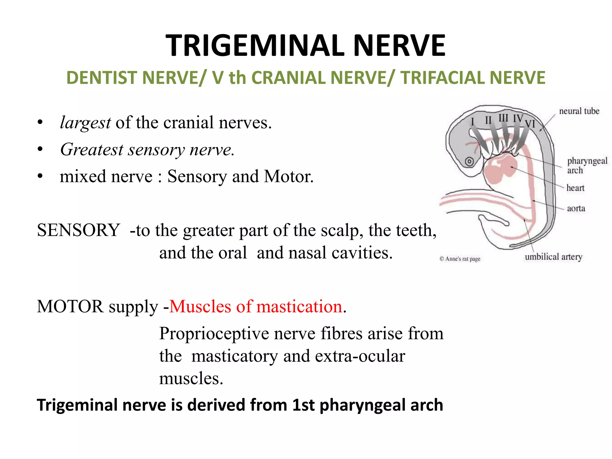
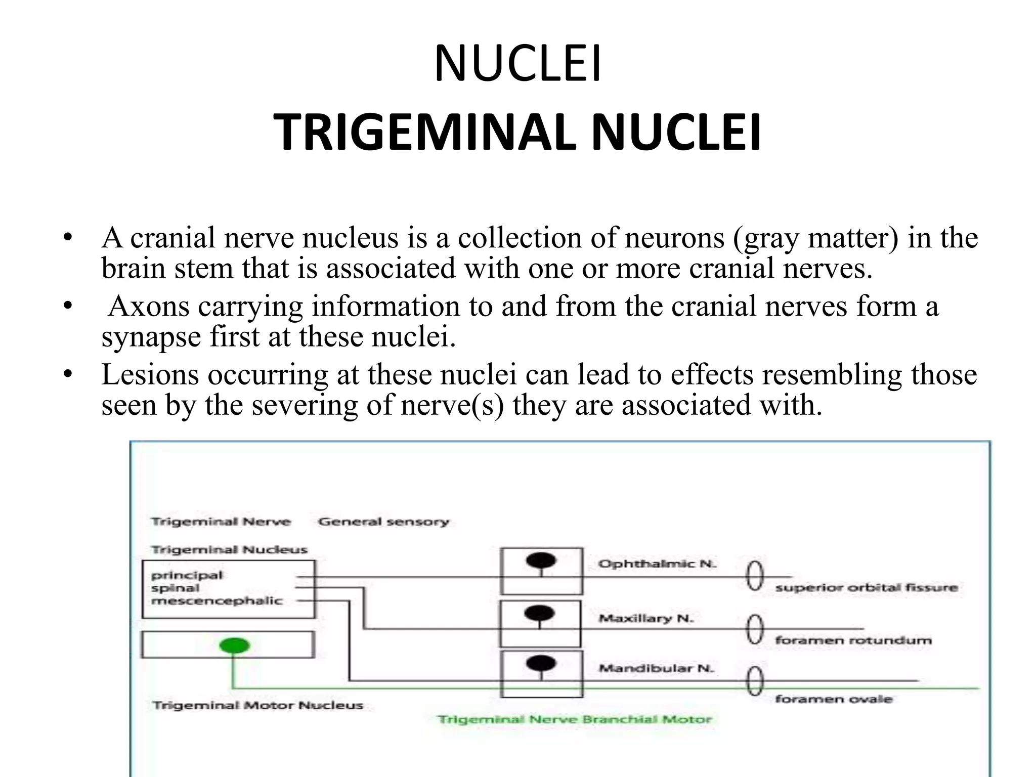
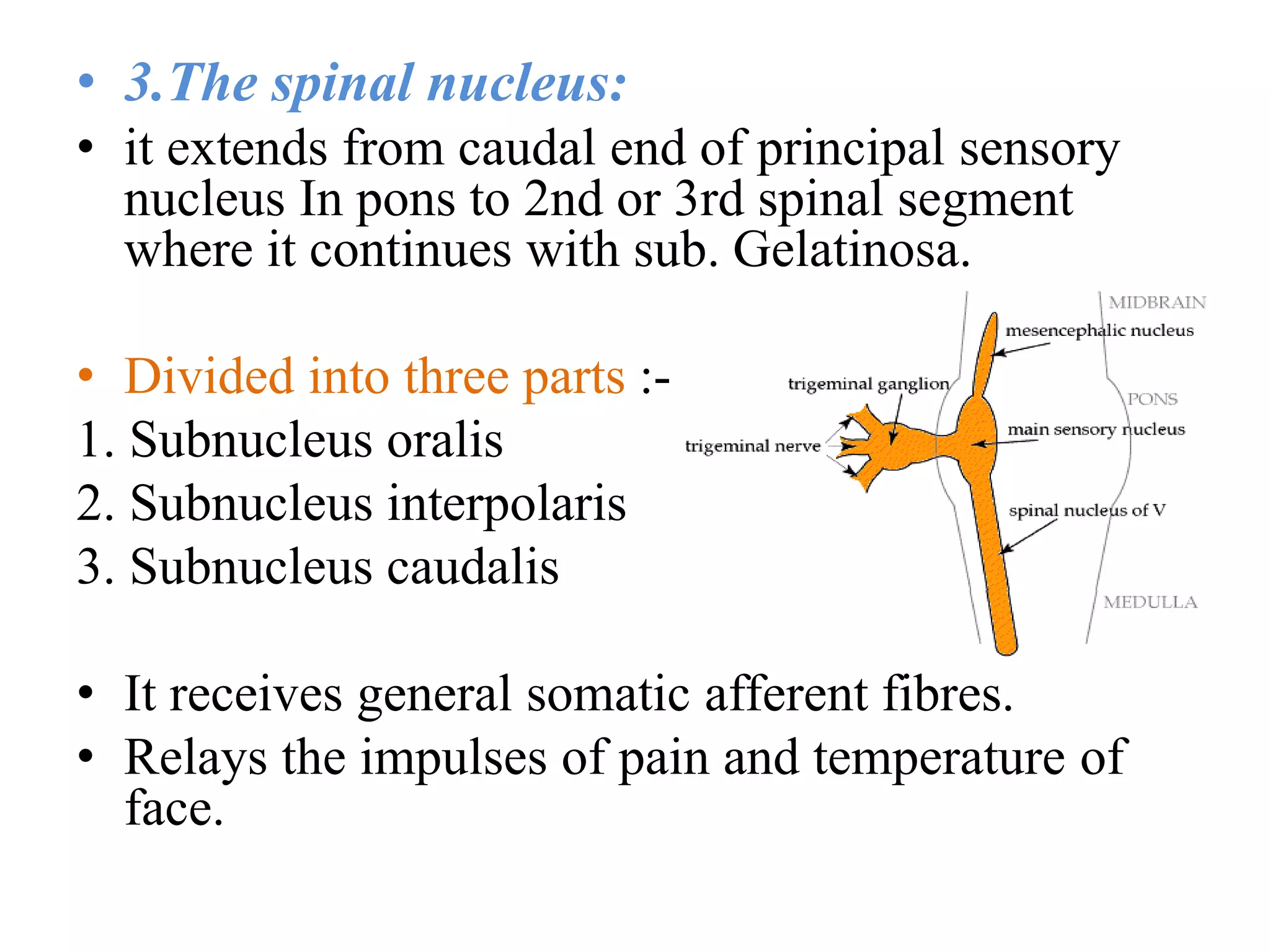
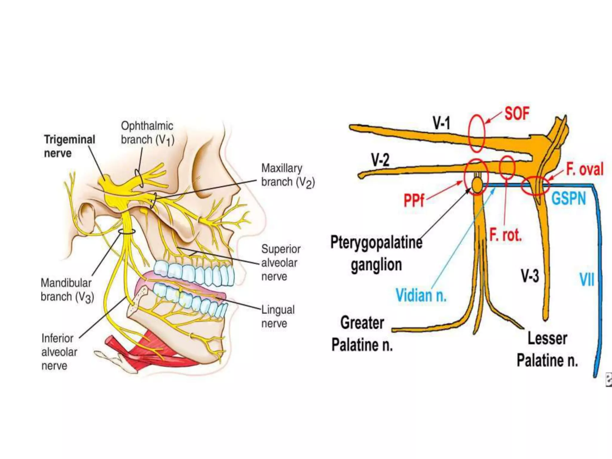
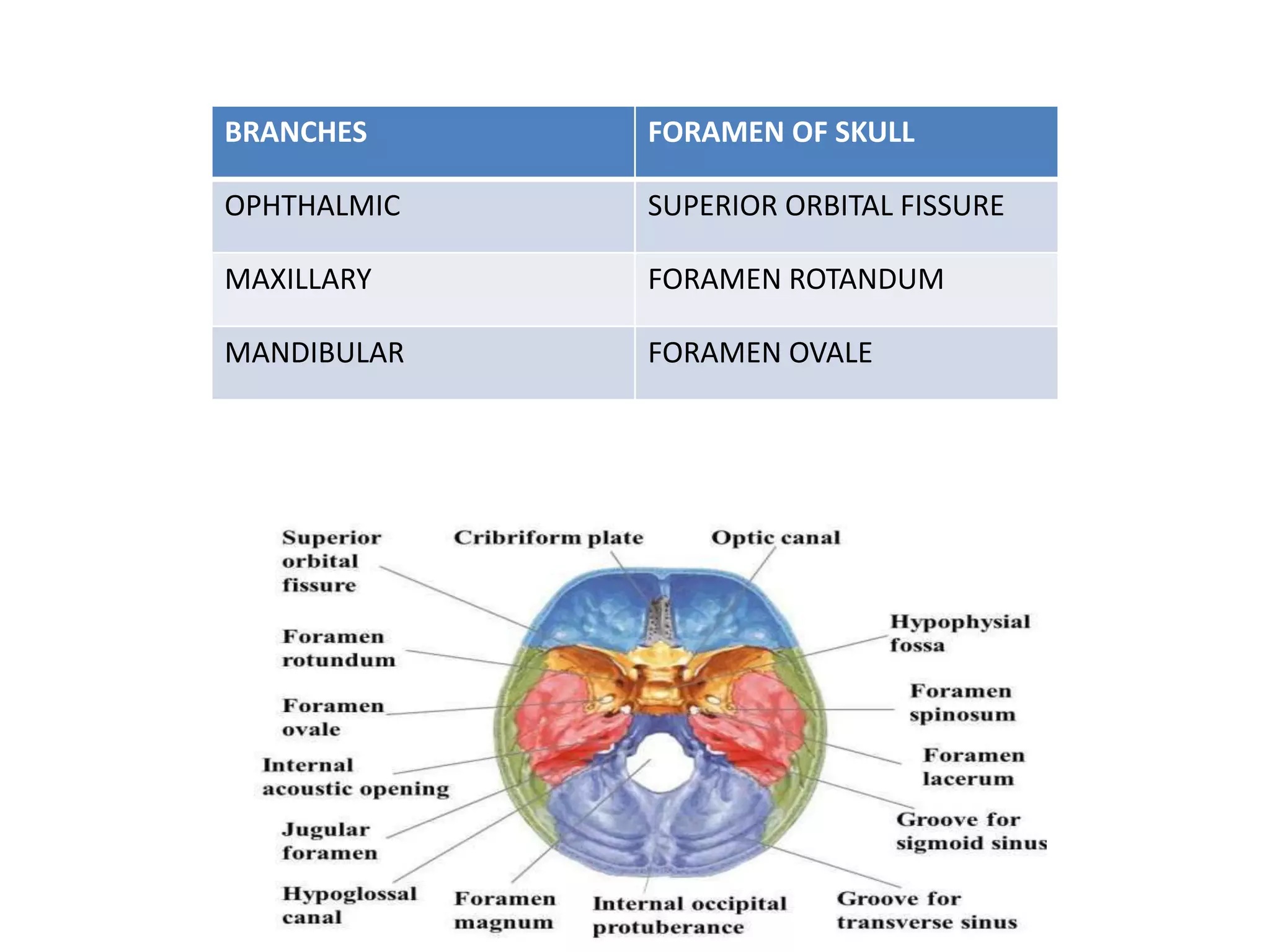
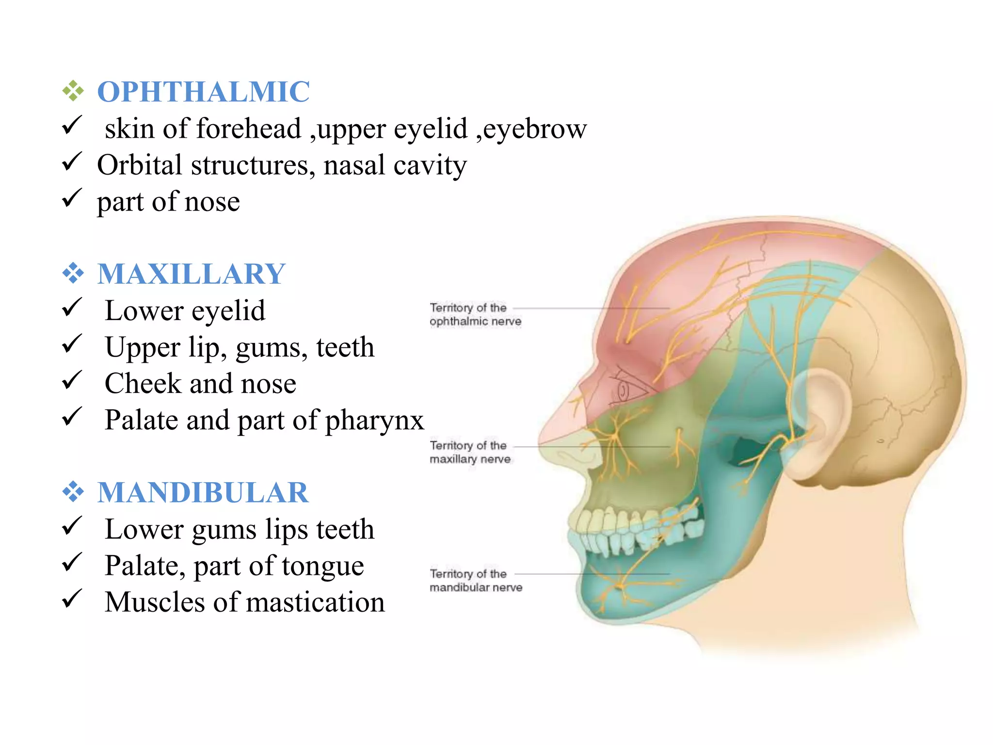
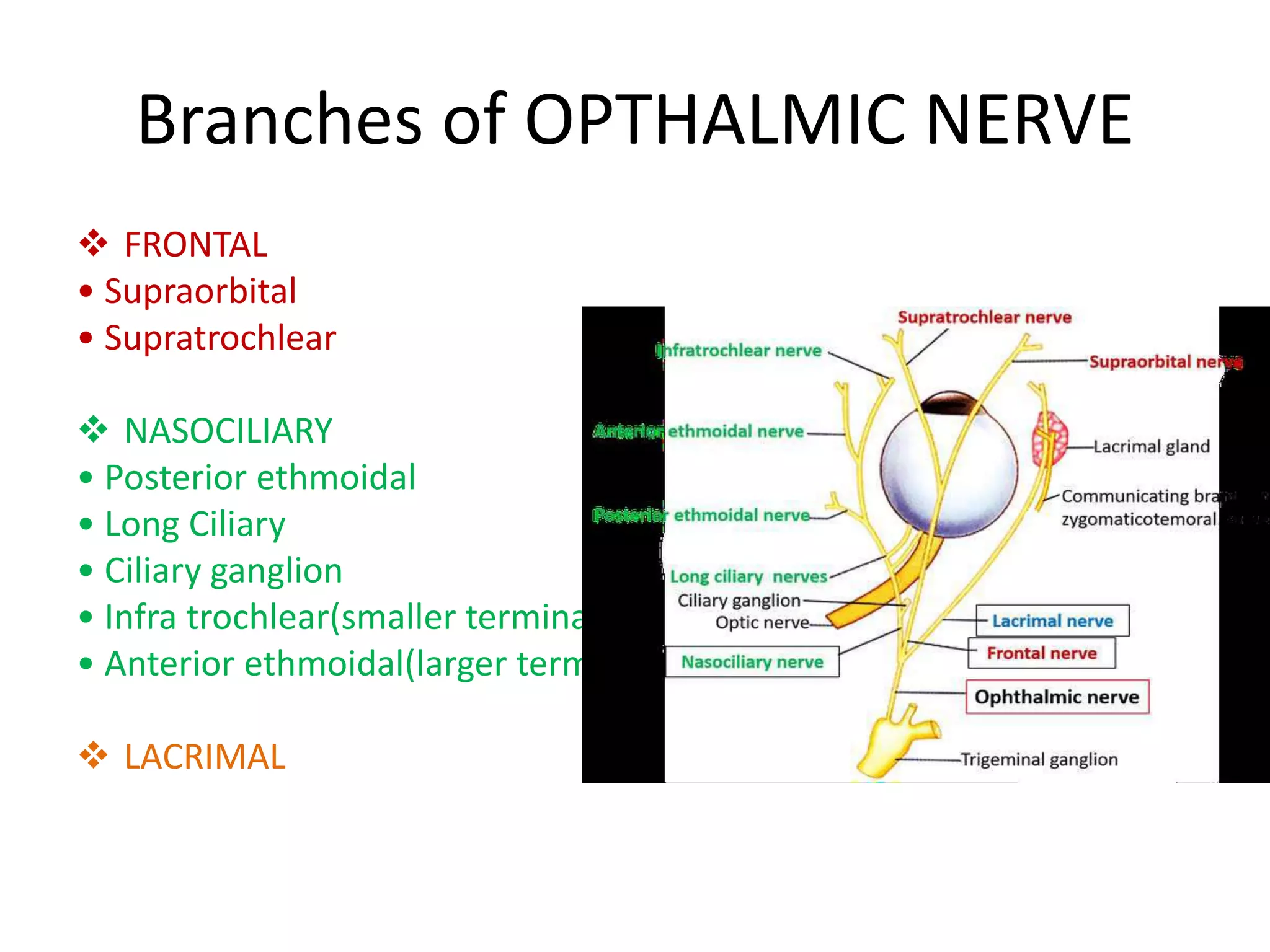
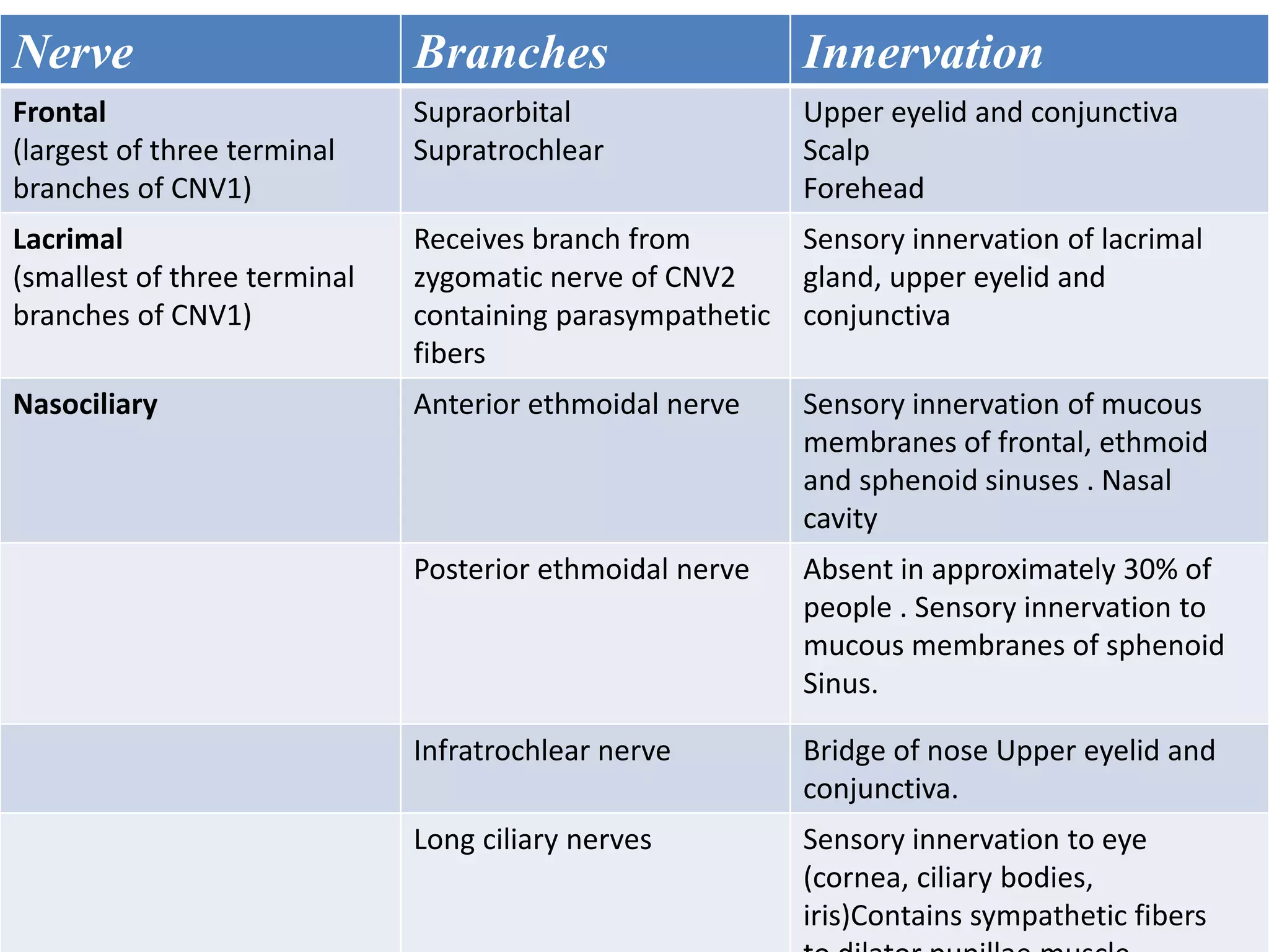
TRIGEMINAL NERVE AND ITS
APPLIED ANATOMY(PART 1)
DR KHADEEJA KULOOD M
FIRST YEAR PG
DEPT. OF PAEDODONTICS &PREVENTIVE
DENTISTRY
B. Bordoni,E.Zanier. Cranial nerves XIII and XIV: nerves in the shadows.
Journal of Multidisciplinary Healthcare 2013:6 87–91
Cranial nerves
• Cranial Nerve I - Olfactory
• Cranial Nerve II - Optic
• Cranial Nerve III - Occulomotor
• Cranial Nerve IV - Trochlear
• Cranial Nerve V - Trigeminal
• Cranial Nerve VI - Abducent
• Cranial Nerve VII - Facial
• Cranial Nerve VIII- Vestibulocochlear
• Cranial Nerve IX - Glossopharyngeal
• Cranial Nerve X - Vagus
• Cranial Nerve XI - Spinal Accessory
• Cranial Nerve XII - Hypoglossal
• Cranial nerve XIII is also known as the “zero nerve” or
“nerve N”.
(First discovered in 1870 in sharks and other types of fish, it
was initially referred to as the nerve of pinkus)
• Cranial nerve XIV was first identified in 1563, but it was
not until 1777 that it was mentioned in a textbook as the
nerve of Wrisberg.
[In modern textbooks, it is referred to as the nervus
intermedius or “intermediary nerve”. Its name is
consistent with its intermediary location between the
facial nerve (cranial nerve VII) and the superior section of
the vestibulocochlear nerve(cranial nerve VIII)]
B. Bordoni,E.Zanier. Cranial nerves XIII and XIV: nerves in the shadows.
Journal of Multidisciplinary Healthcare 2013:6 87–91
TRIGEMINAL NERVE
DENTIST NERVE/ V th CRANIAL NERVE/ TRIFACIAL NERVE
• largest of the cranial nerves.
• Greatest sensory nerve.
• mixed nerve : Sensory and Motor.
SENSORY -to the greater part of the scalp, the teeth,
and the oral and nasal cavities.
MOTOR supply -Muscles of mastication.
Proprioceptive nerve fibres arise from
the masticatory and extra-ocular
muscles.
Trigeminal nerve is derived from 1st pharyngeal arch
• Mesenchyme from the 1st arch contributes the
dermis of the face, hence sensory supply to the
skin of the face is provided by ophthalmic,
maxillary and mandibular branches of the
trigeminal nerve.
NUCLEI
TRIGEMINAL NUCLEI
• A cranial nerve nucleus is a collection of neurons (gray matter) in the
brain stem that is associated with one or more cranial nerves.
• Axons carrying information to and from the cranial nerves form a
synapse first at these nuclei.
• Lesions occurring at these nuclei can lead to effects resembling those
seen by the severing of nerve(s) they are associated with.
SENSORY NUCLEI :
The sensory nucleus is divided into three parts
 spinal
 principal sensory
 Mesencephalic
 There is also a distinct trigeminal motor
nucleus that is medial to the chief sensory
nucleus.
• Sensory trigeminal nuclei is the largest of all
nerve nuclei
1.Mesencephalic nucleus :
• Cell body of Pseudounipolar neuron
• Relay proprioception from muscles of
mastication,Extra ocular Muscles, Facial
muscles.
• Situated in Midbrain.
2.Principal sensory nucleus
• Situated in upper part of pons lateral to motor
nucleus.
• Recieves general somatic afferent fibres.
• Relays impulses of touch and pressure from
skin and mucous membrane of facial region.
• 3.The spinal nucleus:
• it extends from caudal end of principal sensory
nucleus In pons to 2nd or 3rd spinal segment
where it continues with sub. Gelatinosa.
• Divided into three parts :-
1. Subnucleus oralis
2. Subnucleus interpolaris
3. Subnucleus caudalis
• It receives general somatic afferent fibres.
• Relays the impulses of pain and temperature of
face.
• 4.THE MOTOR NUCLEUS :-
• It is situated in upper pons medial to principal
sensory nucleus.
• Contains efferent fibres.
• Innervates muscles of mastication and tensor
tympani and tensor palatini.
FUNCTIONAL COMPONENTS
• SENSORY ROOT
• General somatic afferents
face, scalp, teeth, gingiva,oral and nasal cavities
conjunctiva, PNS and cornea.
• Special visceral efferent(SVE)
innervate the muscles of mastication via the
mandibular (V3) division.
Pain,temp,light touch touch, pressure proprioception
Trigeminal ganglion bypasses trigeminal ganglion
Sensory root
Descending fibres ascending fibres
Spinal nucleus principal sensory nucleus mesencephalic
Trigeminal leminiscus
Thalamus
Post central gyrus cerebral cortex(area 3,2,1)
MOTOR ROOT
CNS
MOTOR ROOT
MOTOR NUCLEUS
MANDIBULAR NERVE
Tensor palatini
Tensor tympani
Muscles of mastication
Masseter
Lateral & Medial Pterygoids
Temporalis
TRIGEMINAL GANGLION
• Also known as Gasserian ganglion/ semilunar
ganglion,
• sensory ganglion of the trigeminal nerve occupies
a cavity (Meckel's cave) in the dura mater,
covering the trigeminal impression near the apex
of the petrous part of the temporal bone.
• crescentic or semilunar in shape, with its
convexity directed anteriomedially.
• The three divisions of the trigeminal nerve
emerges from this convexity.
• ARTERIAL SUPPLY:
Ganglionic branches of Internal Carotid Artery,
middle meningeal artery and accessory
meningeal artery.
RELATIONS
 SUPERIORLY: *superior petrosal sinus
*free margin of tentorium cerebelli
 INFERIORLY: *motor root
*greater petrosal nerve
*petrous apex
*foramen lacerum
 MEDIALLY: *posterior part of lateral wall of cavernous
sinus
*Internal Carotid Artery with its sympathetic
plexus
 LATERALLY: *uncus of temporal lobe
*middle meningeal artery and vein
*nervous spinosum
Branches of trigeminal nerve
The three major branches of
the trigeminal nerve—
• the Ophthalmic nerve (V1)
• the Maxillary nerve (V2)
• the Mandibular nerve (V3)
converge on the trigeminal
ganglion
BRANCHES FORAMEN OF SKULL
OPHTHALMIC SUPERIOR ORBITAL FISSURE
MAXILLARY FORAMEN ROTANDUM
MANDIBULAR FORAMEN OVALE
The areas of cutaneous distribution
(dermatomes) of the three branches of the
trigeminal nerve have with relatively little
overlap (unlike dermatomes in the rest of the
body, which have considerable overlap)
 OPHTHALMIC
 skin of forehead ,upper eyelid ,eyebrow
 Orbital structures, nasal cavity
 part of nose
 MAXILLARY
 Lower eyelid
 Upper lip, gums, teeth
 Cheek and nose
 Palate and part of pharynx
 MANDIBULAR
 Lower gums lips teeth
 Palate, part of tongue
 Muscles of mastication
The Ophthalmic division:-
• Superior and smallest division.
• sensory.
• Arises from the anteriomedial end of
trigeminal ganglion.
• Passes forward in the lateral wall of the
cavernous sinus, below the occulomotor and
trochlear nerves.
• Nerve is joined by the filaments from the
internal carotid sympathetic plexus.
• It communicates with the oculomotor,trochlear
• and abducent nerve.
• The latter communication may be the route by
which proprioceptive fibres from extraocular
muscles enter the trigeminal nuclear complex.
OPHTHALMIC
• Smallest of three divisions of trigeminal
nerve
• Arises from upper part of trigeminal
ganglion
• 2.5 cm long
• Passes along the lateral wall of
cavernous sinus
• gives rise to the recurrent tentorial
• branch (which supplies the tentorium
• cerebelli)
• Below the occulomotor just before
entering orbit
• Through superior orbital fissure
• Divides into three parts
LACRIMAL FRONTAL NASOCILIARY
Branches of OPTHALMIC NERVE
 FRONTAL
• Supraorbital
• Supratrochlear
 NASOCILIARY
• Posterior ethmoidal
• Long Ciliary
• Ciliary ganglion
• Infra trochlear(smaller terminal
• Anterior ethmoidal(larger terminal branch
 LACRIMAL
1.Lacrimal nerve:
• It is the smallest.
• It supplies the lacrimal gland & the
conjuntiva. It pierces the orbital septum and
ends in the skin of the upper eyelid.
2) Frontal nerve:
• It is the largest branch & appears to be the direct continuation
of the ophthalmic division.
• It enters the orbit through the SOF divides into 2 branches.
i. The supra orbital branch: It is larger & more laterally placed.
It supplies the skin of the forehead &
scalp as far back as the vertex.
It also supplies the mucous membrane
of the frontal sinus & pericranium
ii. The supra trochlear branch: It is smaller & more medially placed. It
curves upward on the forehead , close to
the bone.
It supplies the skin of the upper eyelid &
lower part of theforehead.
3.Nasociliary
• A branch of the ophthalmic
nerve
• Enters the orbit through the
superior orbital fissure (inside
the common tendinous ring)
• Crosses above the optic nerve
from lateral to medial
• Runs along the medial wall of
the orbit, between the superior
oblique and medial rectus
muscles
• Branches into Anterior
ethmoidal nerve and
Infratrochlear nerve
i. Branches in the Orbit
ii. Branches in the Nasal cavity
iii. Branches on the face
• (I) Branches in the Orbit:
i. Long root of the cilliary ganglion: It is sensory & passes
through the ganglion without synapsing and supplies the
eyeball.
ii. Long ciliary nerve: Supplies the Iris & Cornea.
iii. Posterior ethmoidal nerve: It enters the post ethmoidal
canal & supplies to the mucous membrane lining of the
Post. Ethmoidal & Sphenoidal paranasal aircells.
iv. Anterior ethmoidal nerve: It supplies to the Ant.ethmoidal
& frontal paranasal air cells. In the upper part of the nasal
cavity, it further divides into:
1) Internal nasal branches
2) External nasal branches
2) Branches in the nasal cavity:
The branches arising here supply the mucous
membrane of the nasal cavity.
3) Terminal branches on the face:
They supply sensory nerves to the skin of the
medial parts of the both eyelids, the lacrimal
sac. They also supply skin on the bridge of the
nose.
Nerve Branches Innervation
Frontal
(largest of three terminal
branches of CNV1)
Supraorbital
Supratrochlear
Upper eyelid and conjunctiva
Scalp
Forehead
Lacrimal
(smallest of three terminal
branches of CNV1)
Receives branch from
zygomatic nerve of CNV2
containing parasympathetic
fibers
Sensory innervation of lacrimal
gland, upper eyelid and
conjunctiva
Nasociliary Anterior ethmoidal nerve Sensory innervation of mucous
membranes of frontal, ethmoid
and sphenoid sinuses . Nasal
cavity
Posterior ethmoidal nerve Absent in approximately 30% of
people . Sensory innervation to
mucous membranes of sphenoid
Sinus.
Infratrochlear nerve Bridge of nose Upper eyelid and
conjunctiva.
Long ciliary nerves Sensory innervation to eye
(cornea, ciliary bodies,
iris)Contains sympathetic fibers
MAXILLARY NERVE
• second & intermediate division of the
trigeminal nerve.
• Pure sensory nerve.
• Leaves the cranial cavity through the foramen
rotundum pterygopalatine fossa
• Passes through inferior orbital fissure to continue
as infraorbital nerve
• The infraorbital n. passes through infra-orbital
groove and terminates on the face
Branches of maxillary nerve
Maxillary nerve
Cranial cavity
Pterygopalatine
fossa
infraorbital face
• The branches of the maxillary nerve can be
divided into the following 4 groups:
1) In the cranium: Meningeal
2) In the pterygopalatine fossa: Ganglionic,
Zygomatic, Post.superior alveolar
3) In the infra orbital canal: Middle sup. alveolar,
Anterior superior/ Greater alveolar
4) On the face: Palpebral, nasal, superior labial
I. Branch given off on the cranium
1. Meningeal branch:
It is given off near the foramen rotundum. It
supplies the duramater of the anterior &
middle cranial fossae.
II. Branches in the pterygopalatine fossa.
1. The ganglionic branches:
They connect the maxillary nerve to the pterygopalatine
ganglion.They contain secretomotor fibres to the lacrimal gland. They
provide sensory fibres to the orbital periosteum & mucous membrane of
the nose, palate & pharynx.
2. The zygomatic nerve:
It arises in the pterygopalatine fossa from the maxillary nerve and
travels anteriorly , entering through the inferior orbital fissure where it
divides into 2 branches.
The Zygomaticofacial nerve perforates the facial surfaces & supplies the skin
over the zygomatic bone..
The Zygomaticotemporal nerve perforates the temporal surface of
• the zygomatic bone , pierces the temporalis fascia, & supplies the
• skin over the anterior temporal fossa region.
• 3. Posterior superior alveolar nerve:
It begins in the pterygopalatine fossa but divides into 3 branches
which emerge through the pterygomaxillary fissure.
2 branches enter the posterior wall of the maxilla above the
tuberosity & supply the 3 molar teeth (except the mesiobuccal root
of first molar).
The third branch pierces the buccinator & supplies the adjoining
part of the gingiva & cheek along the buccal side of the upper
molar teeth.
III. Branches in the Infraorbital canal
( Infraorbital nerve)
1. Middle superior alveolar nerve:
 It arises from the Infra orbital nerve & runs downwards & forwardsalong
the infraorbital groove along the lateral wall of the maxillary sinus.
 It divides into branches which supply the maxillary premolars &
mesiobuccal root of the first molar teeth.
2. Anterior superior alveolar nerve:
 It also arises in the infraorbital canal near the mid point. It runs in
theanterior wall of the maxillary antrum. It runs inferiorly & divides into
the branches, which supply the canine & incisors.
 A nasal branch from this nerve, given off from the superior dental plexus
supplies the mucous membrane of the anterior part of the lateral wall &
floor of the nasal cavity. It ends in the nasal septum.
• IV. Branches given on the face:
• 1. The palpebral branches:
They arise deep to the orbicularis oculi & pierce the muscle, supplying the skin over the
lower eyelid& lateral angle of the eye along with the Zygomaticofacial & Facial
nerves.
2. The nasal branches:
They supply the skin of the nose & tip of the nasal septum & join the External nasal
branch of the anterior ethmoidal nerve.
3. The superior labial branches:
These are large & numerous. They supply the skin over the anterior part of the cheek
& upper lip including the mucous membrane & labial glands. They are joined by
the facial nerve & form the infraorbital plexus.
The branches of the Pterygopalatine ganglion are:
I. Orbital branches: periosteum of the orbit.Posterior ethmoidal &
sphenoidal air cells.
II. Palatine branches:
1.Anterior/greater palatine : mucosa of the hard palate & palatal
gingiva.
2.Middle palatine : mucous membrane of the soft palate
3.Posterior palatine : mucous membrane of the Tonsillar area.
III. Nasal branches:
1.Posterior superior lateral : posterior part of the nasal conchae
2.Nasopalatine/Sphenopalatine : mucous membrane of the premaxilla.
IV. Pharyngeal branch: mucous membrane of the nasopharynx
MANDIBULAR NERVE
• The largest division of the trigeminal nerve
• It is a mixed nerve has a sensory root and a motor root
• It leaves the skull through the
Foramen Ovale
• Below foramen ovale, the 2 roots
unite to form the trunk of the nerve
• Then , it divides into anterior &
posterior divisions
• The anterior is mainly motor
• The posterior is mainly sensory
Branches of the Mandibular nerve
I. Branches of the undivided nerve.
i. Meningeal branch/nervus spinosus.
ii. Nerve to the medial pterygoid
II. Branches of the divided nerve:
(A) Anterior division (B) Posterior division:
1.Buccal nerve 1. Auriculotemporal nerve
2.Massetric nerve 2.Lingual nerve
3.Deep temporal nerve 3.Inferior alveolar nerve
4.Nerve to the lateral pterygoid
BRANCHES OF THE UNDIVIDED NERVE
1.Meningeal nerve:
• enters the skull through the foramen spinosum along
with MMA.
• anterior & posterior divisions that supply the dura of
the middle & anterior cranial fossae.
2. Nerve to the medial pterygoid:
• It is a slender branch that supplies to the deep surface
of
• the muscle.
• • It also gives supplies tensor tympani & the
• tensor veli palati muscles
BRANCHES OF THE DIVIDED
NERVE
• I. Anterior division
• 1.The buccal nerve:
• It passes between the 2 heads of the lateral pterygoid &
descends beneath or through the temporalis. It emergesfrom
under cover of the ramus & ant. border of the masseter &
unites with the buccal branches of he facial nerve.
It supplies the skin over the ant. part of the buccinator &
mucous membrane lining the buccal surface of the gum.
• 2.The massetric nerve:
• • Passes laterally above the lateral pterygoid in front of the TMJ &
behind the tendon of temporalis.
• It passes through the mandibular notch to sink into the masseter
muscle.
• It also gives a branch to the TMJ.
3.The deep temporal nerves:
• They pass above the upper head of the lateral pterygoid, turn above
the infra temporal crest & sink into the deep part of the temporalis
muscle.
4.The nerve to the lateral pterygoid
• supplying each muscle head.
• II.Posterior Division
1.The Auriculotemporal nerve:
• Branches of the Auriculotemporal nerve:
1. Parotid branches: secretomotor, vasomotor.
2. Articular branches: to the TMJ.
3. Auricular branches: to the skin of the helix & tragus.
4. Meatal branches: Meatus of the tymphanic
membrane
5. Terminal branches: Scalp over the temporal region
Lingual nerve
• It lies between the ramus of the mandible & the muscle
in the pterygomandibular space.
• It gives off sensory fibres to the tonsil & the mucous
membrane of the posterior part of the oral cavity.
Branches of lingual nerve and its communications:
1.Chorda tympani
2.Communications with submandibular ganglion
3.Hypoglossal nerve
• Inferior alveolar nerve
• It is the largest terminal branch of the posterior
division of the mandibular nerve.
Mental nerve
Inferior alveolar
nerve
• Branches of the nerve :
1. Mental nerve: it supplies to the skin of the chin & the mucous membrane
as well as the skin of the lower lip.
2. Incisive branch: continues anteriorly from the mental nerve in the body of
the mandible to form the incisive plexus & supplies the canine & incisors.
3. Mylohyoid nerve:
it is given of before the nerve enters the canal & contains both sensory &
motor fibres.It pierces the sphenomandibular ligament, descends in a
groove in the medial side of the ramus & passes beneath the mylohyoid line
supplying the mylohyoid muscle as well as the anterior belly of the
digastric.
Ganglia Associated With The
Trigeminal Nerve
1.Cilliary Ganglion: connected with nasocilliary nerve by ganglionic
branches in orbit, sensory for orbit
2.Pterygopalatine Ganglion: connected to maxillary nerve in
infratemporal fossa sensory to orbital septum, orbicularis and nasal
cavity, max sinus, palate, nasopharynx.
3. Otic Ganglion: between trunk of mandibular n and tensor palatini,
nerve to med pterygoid passes thru but does not synapse in the
ganglion.
4.Submandibular Ganglion: related to lingual n, rests on hypoglossus
supplies post gang. Parasympathetic secretomotor fibres to sub
mandibular and sublingual gland.
ASSOCIATED
REFLEXES
Corneal Reflex
Photic sneeze reflex/Autosomal Compelling
Heliophthalmic Outburst(ACHOO)
•
• Optic-trigeminal summation
• Stimulation of the ophthalmic branch of the trigeminal
nerve may enhance the irritability of the maxillary branch,
resulting in an increased probability of sneezing
• This is similar to the mechanism by which photophobia
develops by persistent light exposure relaying signals
through the optic nerve and trigeminal nerve to produce
increased sensitivity in the ophthalmic branch.
• If this increased sensitivity occurred in the maxillary branch
instead of the ophthalmic branch, a sneeze could result
instead of photophobia
Test jaw jerk/masseter reflex
REFERENCES
 BDC Human Anatomy - Head, Neck & Brain (Volume 3)
 Gray's Anatomy for Students 3rd Ed
 Lippincott’s Concise Illustrated Anatomy - Head & Neck-Volume 3
- Ben Pansky, Thomas R. Gest – 1st Edition(2014)
 Malamed's HandBook of Local Anasthesia, 6ed
 Oral_and_Maxillofacial_Surgery_3rdEd_Neelima_Malik
 B. Bordoni,E.Zanier. Cranial nerves XIII and XIV: nerves in the
shadows.,Journal of Multidisciplinary Healthcare 2013:6 87–91

Trigeminal nerve and its applied anatomy

  • 1.
    TRIGEMINAL NERVE ANDITS APPLIED ANATOMY(PART 1) DR KHADEEJA KULOOD M FIRST YEAR PG DEPT. OF PAEDODONTICS &PREVENTIVE DENTISTRY
  • 2.
    B. Bordoni,E.Zanier. Cranialnerves XIII and XIV: nerves in the shadows. Journal of Multidisciplinary Healthcare 2013:6 87–91
  • 3.
    Cranial nerves • CranialNerve I - Olfactory • Cranial Nerve II - Optic • Cranial Nerve III - Occulomotor • Cranial Nerve IV - Trochlear • Cranial Nerve V - Trigeminal • Cranial Nerve VI - Abducent • Cranial Nerve VII - Facial • Cranial Nerve VIII- Vestibulocochlear • Cranial Nerve IX - Glossopharyngeal • Cranial Nerve X - Vagus • Cranial Nerve XI - Spinal Accessory • Cranial Nerve XII - Hypoglossal
  • 4.
    • Cranial nerveXIII is also known as the “zero nerve” or “nerve N”. (First discovered in 1870 in sharks and other types of fish, it was initially referred to as the nerve of pinkus) • Cranial nerve XIV was first identified in 1563, but it was not until 1777 that it was mentioned in a textbook as the nerve of Wrisberg. [In modern textbooks, it is referred to as the nervus intermedius or “intermediary nerve”. Its name is consistent with its intermediary location between the facial nerve (cranial nerve VII) and the superior section of the vestibulocochlear nerve(cranial nerve VIII)] B. Bordoni,E.Zanier. Cranial nerves XIII and XIV: nerves in the shadows. Journal of Multidisciplinary Healthcare 2013:6 87–91
  • 5.
    TRIGEMINAL NERVE DENTIST NERVE/V th CRANIAL NERVE/ TRIFACIAL NERVE • largest of the cranial nerves. • Greatest sensory nerve. • mixed nerve : Sensory and Motor. SENSORY -to the greater part of the scalp, the teeth, and the oral and nasal cavities. MOTOR supply -Muscles of mastication. Proprioceptive nerve fibres arise from the masticatory and extra-ocular muscles. Trigeminal nerve is derived from 1st pharyngeal arch
  • 6.
    • Mesenchyme fromthe 1st arch contributes the dermis of the face, hence sensory supply to the skin of the face is provided by ophthalmic, maxillary and mandibular branches of the trigeminal nerve.
  • 7.
    NUCLEI TRIGEMINAL NUCLEI • Acranial nerve nucleus is a collection of neurons (gray matter) in the brain stem that is associated with one or more cranial nerves. • Axons carrying information to and from the cranial nerves form a synapse first at these nuclei. • Lesions occurring at these nuclei can lead to effects resembling those seen by the severing of nerve(s) they are associated with.
  • 8.
    SENSORY NUCLEI : Thesensory nucleus is divided into three parts  spinal  principal sensory  Mesencephalic  There is also a distinct trigeminal motor nucleus that is medial to the chief sensory nucleus. • Sensory trigeminal nuclei is the largest of all nerve nuclei 1.Mesencephalic nucleus : • Cell body of Pseudounipolar neuron • Relay proprioception from muscles of mastication,Extra ocular Muscles, Facial muscles. • Situated in Midbrain.
  • 9.
    2.Principal sensory nucleus •Situated in upper part of pons lateral to motor nucleus. • Recieves general somatic afferent fibres. • Relays impulses of touch and pressure from skin and mucous membrane of facial region.
  • 10.
    • 3.The spinalnucleus: • it extends from caudal end of principal sensory nucleus In pons to 2nd or 3rd spinal segment where it continues with sub. Gelatinosa. • Divided into three parts :- 1. Subnucleus oralis 2. Subnucleus interpolaris 3. Subnucleus caudalis • It receives general somatic afferent fibres. • Relays the impulses of pain and temperature of face.
  • 11.
    • 4.THE MOTORNUCLEUS :- • It is situated in upper pons medial to principal sensory nucleus. • Contains efferent fibres. • Innervates muscles of mastication and tensor tympani and tensor palatini.
  • 13.
    FUNCTIONAL COMPONENTS • SENSORYROOT • General somatic afferents face, scalp, teeth, gingiva,oral and nasal cavities conjunctiva, PNS and cornea. • Special visceral efferent(SVE) innervate the muscles of mastication via the mandibular (V3) division.
  • 14.
    Pain,temp,light touch touch,pressure proprioception Trigeminal ganglion bypasses trigeminal ganglion Sensory root Descending fibres ascending fibres Spinal nucleus principal sensory nucleus mesencephalic Trigeminal leminiscus Thalamus Post central gyrus cerebral cortex(area 3,2,1)
  • 15.
    MOTOR ROOT CNS MOTOR ROOT MOTORNUCLEUS MANDIBULAR NERVE Tensor palatini Tensor tympani Muscles of mastication Masseter Lateral & Medial Pterygoids Temporalis
  • 16.
    TRIGEMINAL GANGLION • Alsoknown as Gasserian ganglion/ semilunar ganglion, • sensory ganglion of the trigeminal nerve occupies a cavity (Meckel's cave) in the dura mater, covering the trigeminal impression near the apex of the petrous part of the temporal bone. • crescentic or semilunar in shape, with its convexity directed anteriomedially. • The three divisions of the trigeminal nerve emerges from this convexity.
  • 18.
    • ARTERIAL SUPPLY: Ganglionicbranches of Internal Carotid Artery, middle meningeal artery and accessory meningeal artery.
  • 19.
    RELATIONS  SUPERIORLY: *superiorpetrosal sinus *free margin of tentorium cerebelli  INFERIORLY: *motor root *greater petrosal nerve *petrous apex *foramen lacerum  MEDIALLY: *posterior part of lateral wall of cavernous sinus *Internal Carotid Artery with its sympathetic plexus  LATERALLY: *uncus of temporal lobe *middle meningeal artery and vein *nervous spinosum
  • 23.
    Branches of trigeminalnerve The three major branches of the trigeminal nerve— • the Ophthalmic nerve (V1) • the Maxillary nerve (V2) • the Mandibular nerve (V3) converge on the trigeminal ganglion
  • 25.
    BRANCHES FORAMEN OFSKULL OPHTHALMIC SUPERIOR ORBITAL FISSURE MAXILLARY FORAMEN ROTANDUM MANDIBULAR FORAMEN OVALE
  • 26.
    The areas ofcutaneous distribution (dermatomes) of the three branches of the trigeminal nerve have with relatively little overlap (unlike dermatomes in the rest of the body, which have considerable overlap)
  • 27.
     OPHTHALMIC  skinof forehead ,upper eyelid ,eyebrow  Orbital structures, nasal cavity  part of nose  MAXILLARY  Lower eyelid  Upper lip, gums, teeth  Cheek and nose  Palate and part of pharynx  MANDIBULAR  Lower gums lips teeth  Palate, part of tongue  Muscles of mastication
  • 28.
    The Ophthalmic division:- •Superior and smallest division. • sensory. • Arises from the anteriomedial end of trigeminal ganglion. • Passes forward in the lateral wall of the cavernous sinus, below the occulomotor and trochlear nerves.
  • 29.
    • Nerve isjoined by the filaments from the internal carotid sympathetic plexus. • It communicates with the oculomotor,trochlear • and abducent nerve. • The latter communication may be the route by which proprioceptive fibres from extraocular muscles enter the trigeminal nuclear complex.
  • 30.
    OPHTHALMIC • Smallest ofthree divisions of trigeminal nerve • Arises from upper part of trigeminal ganglion • 2.5 cm long • Passes along the lateral wall of cavernous sinus • gives rise to the recurrent tentorial • branch (which supplies the tentorium • cerebelli) • Below the occulomotor just before entering orbit • Through superior orbital fissure • Divides into three parts LACRIMAL FRONTAL NASOCILIARY
  • 31.
    Branches of OPTHALMICNERVE  FRONTAL • Supraorbital • Supratrochlear  NASOCILIARY • Posterior ethmoidal • Long Ciliary • Ciliary ganglion • Infra trochlear(smaller terminal • Anterior ethmoidal(larger terminal branch  LACRIMAL
  • 32.
    1.Lacrimal nerve: • Itis the smallest. • It supplies the lacrimal gland & the conjuntiva. It pierces the orbital septum and ends in the skin of the upper eyelid.
  • 33.
    2) Frontal nerve: •It is the largest branch & appears to be the direct continuation of the ophthalmic division. • It enters the orbit through the SOF divides into 2 branches. i. The supra orbital branch: It is larger & more laterally placed. It supplies the skin of the forehead & scalp as far back as the vertex. It also supplies the mucous membrane of the frontal sinus & pericranium ii. The supra trochlear branch: It is smaller & more medially placed. It curves upward on the forehead , close to the bone. It supplies the skin of the upper eyelid & lower part of theforehead.
  • 34.
    3.Nasociliary • A branchof the ophthalmic nerve • Enters the orbit through the superior orbital fissure (inside the common tendinous ring) • Crosses above the optic nerve from lateral to medial • Runs along the medial wall of the orbit, between the superior oblique and medial rectus muscles • Branches into Anterior ethmoidal nerve and Infratrochlear nerve
  • 35.
    i. Branches inthe Orbit ii. Branches in the Nasal cavity iii. Branches on the face
  • 36.
    • (I) Branchesin the Orbit: i. Long root of the cilliary ganglion: It is sensory & passes through the ganglion without synapsing and supplies the eyeball. ii. Long ciliary nerve: Supplies the Iris & Cornea. iii. Posterior ethmoidal nerve: It enters the post ethmoidal canal & supplies to the mucous membrane lining of the Post. Ethmoidal & Sphenoidal paranasal aircells. iv. Anterior ethmoidal nerve: It supplies to the Ant.ethmoidal & frontal paranasal air cells. In the upper part of the nasal cavity, it further divides into: 1) Internal nasal branches 2) External nasal branches
  • 37.
    2) Branches inthe nasal cavity: The branches arising here supply the mucous membrane of the nasal cavity. 3) Terminal branches on the face: They supply sensory nerves to the skin of the medial parts of the both eyelids, the lacrimal sac. They also supply skin on the bridge of the nose.
  • 38.
    Nerve Branches Innervation Frontal (largestof three terminal branches of CNV1) Supraorbital Supratrochlear Upper eyelid and conjunctiva Scalp Forehead Lacrimal (smallest of three terminal branches of CNV1) Receives branch from zygomatic nerve of CNV2 containing parasympathetic fibers Sensory innervation of lacrimal gland, upper eyelid and conjunctiva Nasociliary Anterior ethmoidal nerve Sensory innervation of mucous membranes of frontal, ethmoid and sphenoid sinuses . Nasal cavity Posterior ethmoidal nerve Absent in approximately 30% of people . Sensory innervation to mucous membranes of sphenoid Sinus. Infratrochlear nerve Bridge of nose Upper eyelid and conjunctiva. Long ciliary nerves Sensory innervation to eye (cornea, ciliary bodies, iris)Contains sympathetic fibers
  • 39.
    MAXILLARY NERVE • second& intermediate division of the trigeminal nerve. • Pure sensory nerve.
  • 41.
    • Leaves thecranial cavity through the foramen rotundum pterygopalatine fossa • Passes through inferior orbital fissure to continue as infraorbital nerve • The infraorbital n. passes through infra-orbital groove and terminates on the face
  • 42.
  • 44.
  • 45.
    • The branchesof the maxillary nerve can be divided into the following 4 groups: 1) In the cranium: Meningeal 2) In the pterygopalatine fossa: Ganglionic, Zygomatic, Post.superior alveolar 3) In the infra orbital canal: Middle sup. alveolar, Anterior superior/ Greater alveolar 4) On the face: Palpebral, nasal, superior labial
  • 46.
    I. Branch givenoff on the cranium 1. Meningeal branch: It is given off near the foramen rotundum. It supplies the duramater of the anterior & middle cranial fossae.
  • 47.
    II. Branches inthe pterygopalatine fossa. 1. The ganglionic branches: They connect the maxillary nerve to the pterygopalatine ganglion.They contain secretomotor fibres to the lacrimal gland. They provide sensory fibres to the orbital periosteum & mucous membrane of the nose, palate & pharynx. 2. The zygomatic nerve: It arises in the pterygopalatine fossa from the maxillary nerve and travels anteriorly , entering through the inferior orbital fissure where it divides into 2 branches. The Zygomaticofacial nerve perforates the facial surfaces & supplies the skin over the zygomatic bone.. The Zygomaticotemporal nerve perforates the temporal surface of • the zygomatic bone , pierces the temporalis fascia, & supplies the • skin over the anterior temporal fossa region.
  • 48.
    • 3. Posteriorsuperior alveolar nerve: It begins in the pterygopalatine fossa but divides into 3 branches which emerge through the pterygomaxillary fissure. 2 branches enter the posterior wall of the maxilla above the tuberosity & supply the 3 molar teeth (except the mesiobuccal root of first molar). The third branch pierces the buccinator & supplies the adjoining part of the gingiva & cheek along the buccal side of the upper molar teeth.
  • 49.
    III. Branches inthe Infraorbital canal ( Infraorbital nerve) 1. Middle superior alveolar nerve:  It arises from the Infra orbital nerve & runs downwards & forwardsalong the infraorbital groove along the lateral wall of the maxillary sinus.  It divides into branches which supply the maxillary premolars & mesiobuccal root of the first molar teeth. 2. Anterior superior alveolar nerve:  It also arises in the infraorbital canal near the mid point. It runs in theanterior wall of the maxillary antrum. It runs inferiorly & divides into the branches, which supply the canine & incisors.  A nasal branch from this nerve, given off from the superior dental plexus supplies the mucous membrane of the anterior part of the lateral wall & floor of the nasal cavity. It ends in the nasal septum.
  • 50.
    • IV. Branchesgiven on the face: • 1. The palpebral branches: They arise deep to the orbicularis oculi & pierce the muscle, supplying the skin over the lower eyelid& lateral angle of the eye along with the Zygomaticofacial & Facial nerves. 2. The nasal branches: They supply the skin of the nose & tip of the nasal septum & join the External nasal branch of the anterior ethmoidal nerve. 3. The superior labial branches: These are large & numerous. They supply the skin over the anterior part of the cheek & upper lip including the mucous membrane & labial glands. They are joined by the facial nerve & form the infraorbital plexus.
  • 52.
    The branches ofthe Pterygopalatine ganglion are: I. Orbital branches: periosteum of the orbit.Posterior ethmoidal & sphenoidal air cells. II. Palatine branches: 1.Anterior/greater palatine : mucosa of the hard palate & palatal gingiva. 2.Middle palatine : mucous membrane of the soft palate 3.Posterior palatine : mucous membrane of the Tonsillar area. III. Nasal branches: 1.Posterior superior lateral : posterior part of the nasal conchae 2.Nasopalatine/Sphenopalatine : mucous membrane of the premaxilla. IV. Pharyngeal branch: mucous membrane of the nasopharynx
  • 53.
    MANDIBULAR NERVE • Thelargest division of the trigeminal nerve • It is a mixed nerve has a sensory root and a motor root • It leaves the skull through the Foramen Ovale • Below foramen ovale, the 2 roots unite to form the trunk of the nerve • Then , it divides into anterior & posterior divisions • The anterior is mainly motor • The posterior is mainly sensory
  • 58.
    Branches of theMandibular nerve I. Branches of the undivided nerve. i. Meningeal branch/nervus spinosus. ii. Nerve to the medial pterygoid II. Branches of the divided nerve: (A) Anterior division (B) Posterior division: 1.Buccal nerve 1. Auriculotemporal nerve 2.Massetric nerve 2.Lingual nerve 3.Deep temporal nerve 3.Inferior alveolar nerve 4.Nerve to the lateral pterygoid
  • 60.
    BRANCHES OF THEUNDIVIDED NERVE 1.Meningeal nerve: • enters the skull through the foramen spinosum along with MMA. • anterior & posterior divisions that supply the dura of the middle & anterior cranial fossae. 2. Nerve to the medial pterygoid: • It is a slender branch that supplies to the deep surface of • the muscle. • • It also gives supplies tensor tympani & the • tensor veli palati muscles
  • 61.
    BRANCHES OF THEDIVIDED NERVE • I. Anterior division • 1.The buccal nerve: • It passes between the 2 heads of the lateral pterygoid & descends beneath or through the temporalis. It emergesfrom under cover of the ramus & ant. border of the masseter & unites with the buccal branches of he facial nerve. It supplies the skin over the ant. part of the buccinator & mucous membrane lining the buccal surface of the gum.
  • 62.
    • 2.The massetricnerve: • • Passes laterally above the lateral pterygoid in front of the TMJ & behind the tendon of temporalis. • It passes through the mandibular notch to sink into the masseter muscle. • It also gives a branch to the TMJ. 3.The deep temporal nerves: • They pass above the upper head of the lateral pterygoid, turn above the infra temporal crest & sink into the deep part of the temporalis muscle. 4.The nerve to the lateral pterygoid • supplying each muscle head.
  • 63.
    • II.Posterior Division 1.TheAuriculotemporal nerve:
  • 65.
    • Branches ofthe Auriculotemporal nerve: 1. Parotid branches: secretomotor, vasomotor. 2. Articular branches: to the TMJ. 3. Auricular branches: to the skin of the helix & tragus. 4. Meatal branches: Meatus of the tymphanic membrane 5. Terminal branches: Scalp over the temporal region
  • 66.
    Lingual nerve • Itlies between the ramus of the mandible & the muscle in the pterygomandibular space. • It gives off sensory fibres to the tonsil & the mucous membrane of the posterior part of the oral cavity. Branches of lingual nerve and its communications: 1.Chorda tympani 2.Communications with submandibular ganglion 3.Hypoglossal nerve
  • 68.
    • Inferior alveolarnerve • It is the largest terminal branch of the posterior division of the mandibular nerve.
  • 70.
  • 71.
    • Branches ofthe nerve : 1. Mental nerve: it supplies to the skin of the chin & the mucous membrane as well as the skin of the lower lip. 2. Incisive branch: continues anteriorly from the mental nerve in the body of the mandible to form the incisive plexus & supplies the canine & incisors. 3. Mylohyoid nerve: it is given of before the nerve enters the canal & contains both sensory & motor fibres.It pierces the sphenomandibular ligament, descends in a groove in the medial side of the ramus & passes beneath the mylohyoid line supplying the mylohyoid muscle as well as the anterior belly of the digastric.
  • 73.
    Ganglia Associated WithThe Trigeminal Nerve 1.Cilliary Ganglion: connected with nasocilliary nerve by ganglionic branches in orbit, sensory for orbit 2.Pterygopalatine Ganglion: connected to maxillary nerve in infratemporal fossa sensory to orbital septum, orbicularis and nasal cavity, max sinus, palate, nasopharynx. 3. Otic Ganglion: between trunk of mandibular n and tensor palatini, nerve to med pterygoid passes thru but does not synapse in the ganglion. 4.Submandibular Ganglion: related to lingual n, rests on hypoglossus supplies post gang. Parasympathetic secretomotor fibres to sub mandibular and sublingual gland.
  • 74.
  • 75.
  • 76.
    Photic sneeze reflex/AutosomalCompelling Heliophthalmic Outburst(ACHOO) •
  • 77.
    • Optic-trigeminal summation •Stimulation of the ophthalmic branch of the trigeminal nerve may enhance the irritability of the maxillary branch, resulting in an increased probability of sneezing • This is similar to the mechanism by which photophobia develops by persistent light exposure relaying signals through the optic nerve and trigeminal nerve to produce increased sensitivity in the ophthalmic branch. • If this increased sensitivity occurred in the maxillary branch instead of the ophthalmic branch, a sneeze could result instead of photophobia
  • 78.
  • 79.
    REFERENCES  BDC HumanAnatomy - Head, Neck & Brain (Volume 3)  Gray's Anatomy for Students 3rd Ed  Lippincott’s Concise Illustrated Anatomy - Head & Neck-Volume 3 - Ben Pansky, Thomas R. Gest – 1st Edition(2014)  Malamed's HandBook of Local Anasthesia, 6ed  Oral_and_Maxillofacial_Surgery_3rdEd_Neelima_Malik  B. Bordoni,E.Zanier. Cranial nerves XIII and XIV: nerves in the shadows.,Journal of Multidisciplinary Healthcare 2013:6 87–91