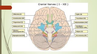
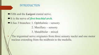
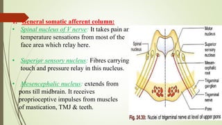
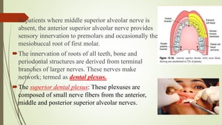
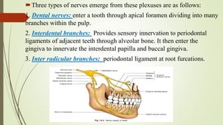
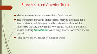
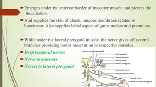
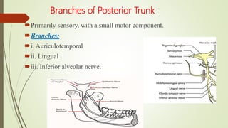
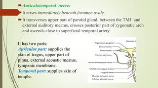
1. The trigeminal nerve is the largest of the cranial nerves and has three main divisions - the ophthalmic, maxillary, and mandibular nerves. It provides sensory innervation to most of the face and motor innervation to the muscles of mastication.
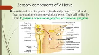
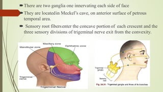
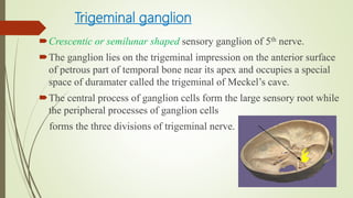
2. The trigeminal ganglion contains the cell bodies of the sensory fibers. It gives rise to the three divisions which have numerous branches that innervate the face, scalp, and oral cavity.
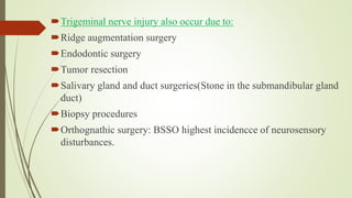
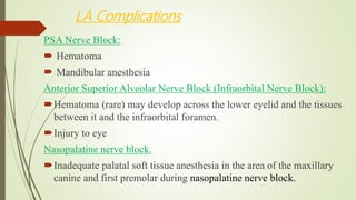
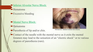
3. Clinical considerations for trigeminal nerve blocks and other procedures include risks of bleeding, infection, nerve injury, hematoma formation, and diplopia if local
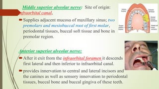
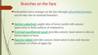
The document discusses the trigeminal nerve (CN V), which is the largest of the cranial nerves. It has both sensory and motor components and innervates the face. The trigeminal nerve consists of three major divisions - the ophthalmic, maxillary, and mandibular nerves. Each division has its own branches that supply different regions of the face, scalp and oral cavity. Trigeminal neuralgia is also mentioned. The document provides detailed information on the anatomy and branches of each division of the trigeminal nerve.
The document discusses the anatomy and branches of the maxillary nerve (V2). It begins by outlining the areas innervated and ganglia associated with the nerve. It then describes the individual branches originating from the maxillary nerve in detail, including their course, distribution and innervation. These branches include the middle meningeal nerve, zygomatic nerve, pterygopalatine branches, posterior superior alveolar nerves, and others. It concludes by noting the sphenopalatine ganglion is associated with the maxillary nerve and discusses some clinical implications like trigeminal neuralgias and facial pain.
explained with Limited matter moreover I have included all the images. if you go through a standard textbook and referred to this PPT it will help you so much I hope It helps you. ask me for the books details.
This document provides an overview of the trigeminal nerve (cranial nerve V) in 12 sections. It discusses the elementary structure of neurons, classification of cranial nerves, embryology and nuclei of the trigeminal nerve, the trigeminal ganglion, and the three divisions (ophthalmic, maxillary, mandibular) and their branches. Diagrams are included to illustrate the course and branches of the trigeminal nerve. The presentation provides detailed anatomical information about the trigeminal nerve and related structures.
The trigeminal nerve is the 5th cranial nerve and is a mixed nerve responsible for sensation in the face and motor function of the muscles of mastication. It has 3 major divisions - the ophthalmic, maxillary, and mandibular nerves. The ophthalmic nerve provides sensory innervation to the upper face and eye region. The maxillary nerve provides sensory innervation to the mid face region including the maxillary teeth. The mandibular nerve provides sensory innervation to the lower face and motor innervation to the muscles of mastication.
this presentation describes the detail anatomy of Temporo-mandibular joint with respect to its articulating surfaces, ligaments, muscles and blood and nerve supply.
This document provides an overview of the trigeminal nerve (CN V), including its nuclei, functional components, course and distribution, the trigeminal ganglion, and the three divisions of the trigeminal nerve - ophthalmic, maxillary, and mandibular nerves. It describes the sensory and motor nuclei of the trigeminal nerve in the brainstem and discusses the sensory and motor roots. It also outlines the anatomy and branches of the three divisions of the trigeminal nerve.
This document provides an overview of the trigeminal nerve (CN V) in 12 sections. It discusses the structure of neurons and nerves, lists and classifies the 12 cranial nerves, describes the embryological development and nuclei of the trigeminal nerve, details the trigeminal ganglion and course of the trigeminal nerve, and outlines its three main branches (ophthalmic, maxillary, mandibular) and their distributions. The document provides a comprehensive anatomical description of the trigeminal nerve in under 3 sentences.
The maxillary artery arises from the external carotid artery and divides into three parts - the mandibular, pterygoid, and pterygopalatine parts. It supplies structures in the face like the maxilla and mandible. The maxillary artery and its branches anastomose with other vessels and are clinically significant for conditions like nosebleeds, epidural hematomas, and complications during procedures like Le Fort I osteotomies when the branches can be injured. Precise surgical techniques are important to avoid damaging branches like the descending palatine artery.
The document provides an overview of the temporomandibular joint (TMJ), including its anatomy, components, development, function, and age-related changes. Key points include:
- The TMJ is a synovial joint that permits hinge and gliding movements of the mandible and involves the condyle of the mandible articulating with the temporal bone.
- Its main components are the mandibular condyle, glenoid fossa, articular disc, articular capsule, synovial membrane, and ligaments.
- It develops from Meckel's cartilage and functions in speech, mastication, and deglutition.
- Age-related changes include fl
The mandibular nerve is the largest of the three divisions of the trigeminal nerve. It is made up of both sensory and motor roots. It supplies sensation to the lower face, teeth, gums, lower lip, chin, and anterior two-thirds of the tongue. It also innervates the muscles of mastication. The mandibular nerve divides into anterior and posterior branches which further divide to innervate the muscles and skin of the face and mouth.
The document discusses various topics related to wound healing, including:
- The classification, types, and factors that affect wound healing.
- The two main processes in healing are regeneration (restoration of original tissues) and repair (fibrosis and scarring).
- Healing occurs through the stages of inflammation, clearance of debris, and ingrowth of granulation tissue.
- Specific examples discussed include healing of fractures, non-surgical root canals, excisional dentoalveolar wounds, replanted teeth, and dental implants.
- Complications like infection, delayed/non-union can occur if healing is disrupted.
The document discusses the anatomy and branches of the mandibular nerve, including its course, distribution, and supply. It describes various anesthetic techniques for blocking branches of the mandibular nerve, including the inferior alveolar nerve block, lingual nerve block, buccal nerve block, and mental nerve block. It also discusses potential local complications from anesthetic techniques such as needle breakage, prolonged anesthesia, and soft tissue injury.
The maxillary sinus is the largest of the paranasal sinuses. It is located within the body of the maxilla and has a pyramidal shape. The maxillary sinus develops during fetal development from the maxillary process and reaches its maximum size by age 18. It is important for functions like voice resonance and warming inhaled air. Disease processes like sinusitis, cysts, tumors or dental infections can involve the maxillary sinus. Radiographs are important for evaluating the sinus floor and its relationship to tooth roots. Surgical procedures may be needed to treat conditions like oroantral fistulas or remove foreign bodies from the sinus.
The maxillary nerve is the second division of the trigeminal nerve. It originates in the lateral wall of the cavernous sinus and passes through the foramen rotundum into the pterygopalatine fossa. It gives off several branches in the pterygopalatine fossa and infratemporal fossa, including the posterior superior alveolar nerve, zygomatic nerve, and anterior and middle superior alveolar nerves. The maxillary nerve terminates by innervating structures in the face, providing sensation to the maxillary region. A maxillary nerve block can provide anesthesia for dental procedures involving the maxillary area, such as when a large canine space abscess is
This document discusses the properties and use of zinc phosphate cement. It is the oldest luting cement, classified in two types - fine grained for luting and medium grained for luting and filling. The powder contains zinc oxide, magnesium oxide, and silica. The liquid contains phosphoric acid, water, and aluminum phosphate. When mixed, a reaction forms a zinc aluminophosphate gel. It has high compressive strength but no chemical adhesion and can irritate the pulp. Zinc phosphate cement is used for luting restorations and bases and as a temporary restoration material.
This document provides an overview of the trigeminal nerve, which is the fifth cranial nerve. It begins with an introduction to cranial nerves and then discusses the specific nuclei, ganglia, and divisions of the trigeminal nerve. The three divisions - ophthalmic, maxillary, and mandibular nerves - are described in detail regarding their course, branches, and sensory and motor functions. Key structures discussed include the trigeminal ganglion, gasserian ganglion, pterygopalatine ganglion, and submandibular ganglion. The document concludes with a recap of the main branches of the trigeminal nerve.
The document provides an overview of the anatomy, development, and surgical anatomy of the temporomandibular joint (TMJ). It discusses the key components of the TMJ, including the mandibular condyle, articular surfaces of the temporal bone, articular disc, fibrous capsule, and ligaments. It describes the development of the TMJ from two distinct blastemas beginning in the 7th week in utero. The document highlights several unique features of the TMJ, such as its articular surface being covered by fibrocartilage instead of hyaline cartilage. It also reviews the movements, vascular supply, innervation, and age-related changes of the TMJ.
The trigeminal nerve is the largest of the cranial nerves. It has both sensory and motor components. It originates from four nuclei in the brainstem and exits the skull through three divisions - ophthalmic, maxillary, and mandibular. The ophthalmic division innervates parts of the face, eye, and nasal cavity. The maxillary division innervates parts of the face, nasal cavity, and palate. The mandibular division innervates muscles of mastication and parts of the face.
The trigeminal nerve is the largest of the cranial nerves. It has both sensory and motor components. The sensory component supplies sensation to the face while the motor component innervates the muscles of mastication. It exists the skull through three divisions - ophthalmic, maxillary, and mandibular. Each division further branches to supply specific regions of the face. The trigeminal ganglion contains the cell bodies of the sensory fibers and is located in the posterior cranial fossa.
Trigeminal nerve maxillary nerve and clinical implicationDr Ravneet Kour
The document discusses the maxillary branch of the trigeminal nerve and its clinical implications. It begins by describing the basic anatomy of neurons, nerves and cranial nerves. It then focuses on the trigeminal nerve as the fifth cranial nerve, describing its nuclei, ganglion and three main branches - the ophthalmic, maxillary and mandibular nerves. Most of the document details the anatomy and branches of the maxillary nerve, including those in the pterygopalatine fossa, orbit, infraorbital canal and face. It concludes by discussing three clinical implications - trigeminal neuralgia, herpes zoster ophthalmicus and Wallenberg syndrome.
The periodontal ligament is the soft connective tissue between the cementum and alveolar bone. It has an hourglass shape that is thinnest in the middle and widens coronally and apically. During tooth eruption, fibroblasts produce collagen fibers that develop into principal fiber groups including the transseptal, alveolar crest, horizontal, oblique, apical, and interradicular fibers. The periodontal ligament contains collagen fibers, cellular elements like fibroblasts, and ground substances such as glycosaminoglycans. It functions to support the tooth, sense pressure, and maintain attachment through Sharpey's fibers embedded in the cementum and bone.
Aging causes irreversible changes to the dental hard tissues over time. The three main tissues - enamel, dentin, and cementum - all undergo changes as part of the aging process. Enamel becomes less permeable and more discolored with age. Dentin develops more dead tracts and sclerotic dentin. Cementum may experience hypercementosis and the formation of cementicles. The alveolar bone also undergoes resorption, decreasing in height and width over time. These morphological and functional changes to the dental tissues are a natural part of the biological aging process.
The temporomandibular joint (TMJ) is a bilateral joint that allows for hinge-like and gliding motions of the mandible. It is formed between the head of the mandible and the articular fossa of the temporal bone. The TMJ is unique in that it contains an articular disc that divides the joint cavity into upper and lower compartments. Common functions of the TMJ include mastication and speech. Temporomandibular disorders (TMD) refer to a group of medical conditions involving the muscles of mastication and TMJ. Major etiological factors for TMD include occlusal condition, trauma, emotional stress, deep pain input, and parafunctional activities.
The document summarizes the blood supply to the jaws and teeth. The external carotid artery divides into the maxillary artery and superficial temporal artery near the ear. The maxillary artery then divides into important branches that supply blood to different areas of the jaws and teeth. The inferior alveolar artery supplies the mandible and mandibular teeth. The posterior superior alveolar artery supplies the maxilla, roots of maxillary molars, and maxillary bone and teeth to the first bicuspid. The palatine artery supplies the hard palate and gingiva on the palatal side of maxillary teeth.
The document discusses the complex process of tooth development from initiation to eruption. It begins with the formation of the primary epithelial bands and dental lamina between 6-7 weeks in utero, which give rise to the tooth buds. The buds progress through stages of proliferation, histodifferentiation, and morphodifferentiation to form the crown and root structures. Hertwig's epithelial root sheath is responsible for root formation and shape before teeth erupt into the oral cavity.
The document discusses the trigeminal nerve (cranial nerve V) in three sentences: It describes the trigeminal nerve as the largest cranial nerve, a mixed nerve with both motor and sensory components. It originates from the trigeminal ganglion and divides into three main branches - the ophthalmic, maxillary, and mandibular nerves - which innervate the face and associated structures. The document provides detailed information on the embryology, nuclei, course and branches of the trigeminal nerve.
This document provides an overview of the trigeminal nerve (CN V), including its origins, branches, and functions. It notes that the trigeminal nerve has three main divisions - the ophthalmic, maxillary, and mandibular nerves. The cell bodies of the trigeminal nerve's sensory neurons are located in the trigeminal ganglion. The document then describes the individual branches and functions of the ophthalmic, maxillary, and mandibular nerves. It provides details on the sensory distributions and innervations of each branch.
The trigeminal nerve is the 5th cranial nerve that has both sensory and motor functions. It has three major divisions - the ophthalmic, maxillary, and mandibular nerves. The ophthalmic and maxillary nerves are purely sensory and innervate parts of the face, while the mandibular nerve contains both sensory and motor fibers that innervate muscles of mastication. The trigeminal ganglion contains the cell bodies of the sensory fibers and relays information to various sensory nuclei in the brainstem.
The trigeminal nerve is the 5th cranial nerve and is a mixed nerve responsible for sensation in the face and motor function of muscles of mastication. It has 3 main divisions - the ophthalmic, maxillary, and mandibular nerves. The mandibular nerve is the largest division and supplies motor innervation to the muscles of mastication as well as sensory innervation to parts of the face and scalp.
The trigeminal nerve is the largest cranial nerve and contains both sensory and motor fibers. It has three major divisions - the ophthalmic, maxillary, and mandibular nerves. The ophthalmic nerve innervates parts of the face, nasal cavity, and orbits. The maxillary nerve innervates parts of the face, nasal cavity, oral cavity, and maxillary sinus. The mandibular nerve is the largest division and innervates muscles of mastication and parts of the lower face. All three divisions convey sensory information from the face and motor innervation to the muscles of mastication.
The document provides information on the trigeminal nerve (CN V), including its nuclei, origin, course, branches, and functions. It describes the three main branches - ophthalmic, maxillary, and mandibular nerves. The ophthalmic nerve innervates the eye and upper face. The maxillary nerve contains sensory fibers and innervates the midface, nasal cavity, and maxillary teeth. The mandibular nerve is mixed, containing both sensory and motor fibers, and innervates the lower face, oral cavity, external ear, and muscles of mastication.
This document summarizes the trigeminal nerve, including its embryology, anatomy, branches, functions and clinical considerations. It begins with the embryology of the pharyngeal arches and how they relate to nerve development. It then describes the trigeminal ganglion, roots and nuclei. The three divisions of the trigeminal nerve and their branches are outlined. Clinical tests for examining the trigeminal nerve and classifying injuries are summarized. Common causes of trigeminal nerve injuries and their treatment approaches are briefly discussed.
The document provides information about the trigeminal nerve (CN V), which is the largest of the cranial nerves. It has both sensory and motor components. The trigeminal nerve has three main divisions - the ophthalmic, maxillary, and mandibular nerves. It innervates most of the face and provides sensory innervation to the teeth and oral cavity. The trigeminal nerve nuclei are located in the pons and midbrain. The trigeminal ganglion contains the cell bodies of pseudounipolar neurons. The branches and distribution of the three divisions of the trigeminal nerve are described in detail.
This document provides an overview of the trigeminal nerve (CN V), including its embryology, structure, branches and clinical applications. It describes the three divisions of the trigeminal nerve - ophthalmic, maxillary and mandibular nerves. The trigeminal ganglion and its sensory and motor roots are discussed. The branches and functions of the ophthalmic, maxillary and mandibular nerves are summarized. Regional nerve blocks associated with the trigeminal nerve such as the greater palatine nerve block are also outlined. Anatomical variations of the trigeminal nerve and maxillary nerve are noted.
Indian Dental Academy: will be one of the most relevant and exciting training center with best faculty and flexible training programs for dental professionals who wish to advance in their dental practice,Offers certified courses in Dental implants,Orthodontics,Endodontics,Cosmetic Dentistry, Prosthetic Dentistry, Periodontics and General Dentistry.
The trigeminal nerve is the largest cranial nerve and has three main branches - the ophthalmic, maxillary, and mandibular nerves. It arises from the semilunar ganglion and has both sensory and motor components. The ophthalmic nerve innervates the eye and forehead. The maxillary nerve innervates the midface, upper teeth, and sinuses. The mandibular nerve has both anterior and posterior divisions which innervate the lower face, scalp, ear, and lower teeth.
The trigeminal nerve is the largest cranial nerve and has three major divisions - the ophthalmic, maxillary, and mandibular nerves. The ophthalmic nerve innervates structures in the orbit and forehead. The maxillary nerve provides sensation to the midface and innervation to the maxillary teeth. The mandibular nerve is mixed with both sensory and motor components, and supplies sensation to the lower face and motor innervation to the muscles of mastication. It gives off branches including the lingual nerve which provides sensation to the anterior two-thirds of the tongue.
Indian Dental Academy: will be one of the most relevant and exciting training center with best faculty and flexible training programs for dental professionals who wish to advance in their dental practice,Offers certified courses in Dental implants,Orthodontics,Endodontics,Cosmetic Dentistry, Prosthetic Dentistry, Periodontics and General Dentistry.
The trigeminal nerve is the largest of the cranial nerves. It has both sensory and motor functions. Sensory fibers carry general somatic afferent information from the face to the trigeminal ganglion. Motor fibers innervate the muscles of mastication. The trigeminal nerve emerges from the pons and divides into three main branches: the ophthalmic, maxillary, and mandibular nerves. These branches innervate different regions of the face and cranium, carrying sensory information and motor commands. Injuries or diseases of the trigeminal nerve can cause sensory deficits or neuropathic pain conditions like trigeminal neuralgia.
The document provides information on the trigeminal nerve (CN V), including its anatomy, branches, and distribution. Some key points:
- CN V is the largest cranial nerve, supplying sensation to the face and motor function to the muscles of mastication.
- It has three main branches - the ophthalmic, maxillary, and mandibular nerves. The ophthalmic nerve innervates the forehead and orbit. The maxillary nerve innervates the midface, and the mandibular nerve innervates the lower face and jaw.
- Each branch has smaller divisions that provide both sensory and motor function to the face, mucosa, and muscles of the head and neck
The trigeminal nerve is the fifth cranial nerve that has both motor and sensory components. It has three main divisions - the ophthalmic, maxillary, and mandibular nerves. The ophthalmic nerve innervates the eye and surrounding structures. The maxillary nerve provides sensation to the midface and upper teeth. The mandibular nerve is a mixed nerve that supplies motor innervation to the muscles of mastication and sensation to the lower face and teeth. Disorders of the trigeminal nerve include trigeminal neuralgia, which causes severe facial pain, and herpes zoster ophthalmicus, which causes shingles in the eye region.
The anatomy of the nerve supply of the head and neck has many significant applications in maxillofacial surgery. Understanding these important anatomic relations- variations enables surgeons to perform the surgical procedures safely. Knowledge of these concepts helps us to recognize the problems and complications as and when they occur and manage them accordingly.
The trigeminal nerve is the largest of the cranial nerves. It has both sensory and motor functions. It has three main divisions: the ophthalmic, maxillary, and mandibular nerves. The ophthalmic nerve innervates the eye and forehead. The maxillary nerve supplies sensation to the cheek, upper teeth, and nose. The mandibular nerve provides motor input to the muscles of mastication and sensation to the lower face, teeth, and chin.
The trigeminal nerve is a mixed nerve that is the largest of the cranial nerves. It has both sensory and motor functions. Sensory branches provide sensation to the face and motor branches innervate the muscles of mastication. The trigeminal nerve has three major divisions - ophthalmic, maxillary, and mandibular nerves. These divisions branch further to innervate specific regions of the face. The trigeminal ganglion contains the cell bodies of pseudounipolar neurons that relay sensory information from the face to the brainstem trigeminal nuclei.
हिंदी वर्णमाला पीपीटी, hindi alphabet PPT presentation, hindi varnamala PPT, Hindi Varnamala pdf, हिंदी स्वर, हिंदी व्यंजन, sikhiye hindi varnmala, dr. mulla adam ali, hindi language and literature, hindi alphabet with drawing, hindi alphabet pdf, hindi varnamala for childrens, hindi language, hindi varnamala practice for kids, https://www.drmullaadamali.com
Gender and Mental Health - Counselling and Family Therapy Applications and In...PsychoTech Services
A proprietary approach developed by bringing together the best of learning theories from Psychology, design principles from the world of visualization, and pedagogical methods from over a decade of training experience, that enables you to: Learn better, faster!
Strategies for Effective Upskilling is a presentation by Chinwendu Peace in a Your Skill Boost Masterclass organisation by the Excellence Foundation for South Sudan on 08th and 09th June 2024 from 1 PM to 3 PM on each day.
Main Java[All of the Base Concepts}.docxadhitya5119
This is part 1 of my Java Learning Journey. This Contains Custom methods, classes, constructors, packages, multithreading , try- catch block, finally block and more.
Communicating effectively and consistently with students can help them feel at ease during their learning experience and provide the instructor with a communication trail to track the course's progress. This workshop will take you through constructing an engaging course container to facilitate effective communication.
Leveraging Generative AI to Drive Nonprofit InnovationTechSoup
In this webinar, participants learned how to utilize Generative AI to streamline operations and elevate member engagement. Amazon Web Service experts provided a customer specific use cases and dived into low/no-code tools that are quick and easy to deploy through Amazon Web Service (AWS.)
Walmart Business+ and Spark Good for Nonprofits.pdfTechSoup
"Learn about all the ways Walmart supports nonprofit organizations.
You will hear from Liz Willett, the Head of Nonprofits, and hear about what Walmart is doing to help nonprofits, including Walmart Business and Spark Good. Walmart Business+ is a new offer for nonprofits that offers discounts and also streamlines nonprofits order and expense tracking, saving time and money.
The webinar may also give some examples on how nonprofits can best leverage Walmart Business+.
The event will cover the following::
Walmart Business + (https://business.walmart.com/plus) is a new shopping experience for nonprofits, schools, and local business customers that connects an exclusive online shopping experience to stores. Benefits include free delivery and shipping, a 'Spend Analytics” feature, special discounts, deals and tax-exempt shopping.
Special TechSoup offer for a free 180 days membership, and up to $150 in discounts on eligible orders.
Spark Good (walmart.com/sparkgood) is a charitable platform that enables nonprofits to receive donations directly from customers and associates.
Answers about how you can do more with Walmart!"
4. INTRODUCTION
Fifth and the Largest cranial nerve.
It is the nerve of first brachial arch.
It has 3 branches: 1. Ophthalmic – sensory
2. Maxillary – sensory
3. Mandibular – mixed
• The trigeminal nerve originates from three sensory nuclei and one motor
nucleus extending from the midbrain to the medulla.
6. 1. General somatic afferent column:
• Spinal nucleus of V nerve: It takes pain and
temperature sensations from most of the
face area which relay here.
• Superior sensory nucleus: Fibres carrying
touch and pressure relay in this nucleus.
• Mesencephalic nucleus: extends from
pons till midbrain. It receives
proprioceptive impulses from muscles
of mastication, TMJ & teeth.
7. 2= Branchial efferent column
Situated at level of upper pons .
The fibres of motor nucleus supply eight muscles
derived from first branchial arch.
8. Sensory components of V Nerve
Sensation of pain, temperature, touch and pressure from skin of
face, paranasal air sinuses travel along axons. Their cell bodies lie
in the V ganglion or semilunar ganglion or Gasserian ganglion.
9. There are two ganglia one innervating each side of face
They are locatedin Meckel’s cave, on anterior surface of petrous
temporal area.
Sensory root fibersenter the concave portion of each crescent and the
three sensory divisions of trigeminal nerve exit from the convexity.
10. Motar component
It receives impulses from right and left cerebral hemisperes, red
nucleus and mesencephalic nucleus.
Supply four muscles of mastication and four other muscles that are;
Tensor veli palatini
Tensor tympani
Mylohyoid
Anterior belly of digastric
12. Ophthalmic Nerve (V1)
FUNCTIONS : The ophthalmic nerve transmits sensory innervation
from;
• Eyeball
• Skin of upper face and anterior scalp
• The lining of upper part of nasal
cavity and air cells
• The meninges of anterior cranial fossa.
• Its branches also convey parasympathetic fibers to the ciliary and iris
muscles,pupillary constriction to the lacrimal gland.
13. Origin, Course and Branches
It originates from trigeminal ganglion in middle cranial fossa. It
passes anteriorly through lateral wall of cavernous sinus.
It divides into three branches:
1. Frontal
2. Nasociliary
3. Lacrimal.
All these branches pass
through superior orbital
fissure into the orbit.
14. FRONTAL BRANCH:
It is the largest branch; supplies frontal sinus and skin of forehead and
scalp.
It divides into two branches:
Supraorbital
Supratrochlear
Frontal
15. NASOCILIARY BRANCH:
It passes the superior orbital fissure, medially within the common
tendinous ring.
It divides into the following:
a. Short ciliary nerve.
b. Long ciliary nerve.
c. Nerve to ciliary ganglion
d. Infratrochlear
e. Anterior ethmoidal
f. Posterior ethmoidal
16. LACRIMAL NERVE:
It supplies lacrimal gland and a small area of adjacent skin and
conjunctiva.
It passes through superior orbital fissure.
It receives postganglionic parasympathetic fibers from pterygopalatine
ganglion which enters orbit with zygomatic nerve for distribution to
lacrimal gland.
20. TREATMENT:
Acyclovir 800mg 5 times /day within 4 days of onset of rash
Analgesics
Antibiotic ointments
Systemic steroids 60mg/day
Corneal grafting
22. Maxillary Nerve (V2)
It is the nerve of maxillary process, that differentiates from first
pharyngeal arch.
It is purely sensory; and is intermediate in size between ophthalmic and
mandibular nerve.
23. FUNCTIONS:
The maxillary nerve transmits sensory fibers from the skin
of face between the lower eyelid and the nasal cavity and
sinuses, from the maxillary teeth.
At its origin from the pons, it contains only sensory fibers.
some of its branches receive postganglionic
parasympathetic fibers from pterygopalatine ganglion
which pass to the lacrimal, nasal and palatine glands.
24. Origin, Course and Branches
It originates from the middle part of trigeminal ganglion in middle
cranial fossa.
It gives off meningeal branch in middle cranial fossa which is sensory.
The nerve runs forwards through lower part of lateral wall of cavernous
sinus.
directed through the foramen rotundum
As it crosses pterygopalatine fossa, it gives off its main branches.
25. Maxillary nerve innervates:
1. Skin of: Middle portion of face
Lower eyelid
Side of nose
Upper lip.
2. Mucous membrane of: Nasopharynx
Maxillary sinus
Soft palate
Tonsil
Hard palate.
Maxillary teeth and periodontal tissues.
26. BRANCHES
Withincranium In pterygopalatinefossa In infraorbitalcanal Onface
MAXILLARY NERVE
Middle meningeal
nerve
• Inferior palpebral
• Lateral nasal
• Superior labial
• MSA
(middle superior
alveolar nerve)
• ASA
(anterior superior
alveolar nerve)
Zygomatic PSA
(posterior
superior
alveolar)
Pterygopalatine
•Zygomatico
temporal
•Zygomatico
facial
•Orbital
•Nasal
•Palatine
•Pharyngeal
27. Branches Within the Cranium: Middle Meningeal
Nerve
Immediately after separating from trigeminal ganglion, the
maxillary nerve gives off a small branch, middle meningeal
nerve.
It travels with middle meningeal artery.
It provides sensory innervation to the duramater of anterior
half of middle cranial fossa.
28. Branches in the Pterygopalatine Fossa
i. Pterygopalatine nerves
ii. Zygomatic nerves
iii. Posterosuperior alveolar nerves.
29. PTERYGOPALATINE NERVES
They also serve as a communication between pterygopalatine ganglion
and maxillary nerve.
By way of ganglion, the maxillary nerve has the following branches.
1. Orbital branches: Supply periosteum of orbit.
2. Nasal branches: Supply mucous membranes superior and
middle conchae, the lining of posterior ethmoidal sinus and posterior
portion of nasal septum.
These convey postganglionic parasympathetic fibers from pterygopalatine
ganglion to nasal glands.
30. Nasopalatine nerve (long sphenopalatine nerve) :
a significant branch.
It enters sphenopalatine foramen
The nasopalatine nerve continues downwards reaching the floor of nasal
cavity and giving off its branches to anterior part of nasal septum and the
floor of nose.
31. It enters incisive canal through which
it passes into the oral cavity via the
incisive foramen. The right and left
nasopalatine nerves emerge together
through this foramen and provide
sensory innervation to palatal
mucosa in the region of premaxilla.
32. 3. Palatine branches:
a. Greater palatine nerves (anterior palatine nerves)
b. Lesser palatine nerves (middle and posterior palatine nerves).
These branches provide sensory innervation to hard and soft palate.
These branches also convey postganglionic parasympathetic fibers from
pterygopalatine ganglion to palatal glands.
Greater palatine nerves :
It emerges on hard palate through greater palatine foramen
supplying sensory innervation to palatal soft tissues and bone.
Anterior to first premolar it communicates with terminal fibers of
nasopalatine nerve.
33. Lesser palatine nerve: These are sensory to soft palate. They descend
through lesser palatine foramina in palatine bone and pass backwards to
supply mucous membrane of soft palate.
4. Pharyngeal branch: It provides sensory innervation to
nasopharynx, auditory (Eustachian) tube.
34. Clinical consideration=
complications include bleeding,infection,nerve
trauma,hypertension.The possibility of needle fracture within
the greater palatine canal. Damage to intracanal vessels and
nerves.
Failure to gain access to the target area due either to imprecise
technique or greater palatine canal constriction.
Diplopia (Double vision due to anaesthesia of the ocular
muscles via diffusion of anaesthetic solution through the
inferior orbital fissure.)
35. Clinically management
(a) The patient needs reassurance.
(b) The affected eye requires corneal protection
with an eye pad dressing for the duration of
anaesthesia.
(c) Use of an eye pad will restore normal vision,
36. ZYGOMATIC NERVES
It provides sensory innervation to skin over zygomatic
region.
These nerves also convey postganglionic parasympathetic
fibers from pterygopalatine ganglion to lacrimal nerve and
glands.
It is a terminal branch. It entering the orbit through inferior
orbital fissure .
37. The zygomatic nerve enters zygomatic bone and divides
into two branches:
Zygomaticotemporal nerve: supplies skin above
zygomatic arch, anterior part of temporal bone.
Zygomaticofacial nerve: supplies skin over the
prominence of cheek.
38. POSTEROSUPERIOR ALVEOLAR NERVES
The main trunk of the maxillary nerve in the
pterygopalatine fossa.
Pass downwards through the pterygopalatine fossa and
reach the posterior surface of maxilla.
Provide sensory innervation to:
buccal gingiva in maxillary molar region; and adjacent
facial mucosal surfaces, mucous membrane of sinus and
provides sensory innervation to alveoli, periodontal
ligaments and pulpal tissues of maxillary molars, with
the exception of (25%) mesiobuccal root of first molar.
39. Clinical consideration
When the local anesthesia was administered for
the posterior superior alveolar nerve block,
forthe RCT of 16, a swelling along with large
extraoral discoloration in the mandibular
lower region.
and a small intraoral hematoma formation
takes place immediately within minutes of
administration of anesthesia.
The patient became very apprehensive about
the unaesthetic appearance of large
discoloration on the side of the face.
40. Treatment=analgesic, avoid any heat application which might increase
the size of the hematoma due to vasodilatation, application of ice
immediately after developing hematoma helps in minimizing the size by
vasoconstriction.
Any dental treatment in the involved region should be avoided until
symptoms and signs resolve.
To minimize the risk of hematoma formation, use of short needle and
minimum number of needle penetration into tissues should be
considered.
The follow up of the case was done for 4 weeks during which
discoloration was completely disappeared within in 3 weeks.
41. Branches in the Infraorbital Canal
In the infraorbital canal, the maxillary division gives two branches:
1. Middle superior alveolar nerve.
2. Anterior superior alveolar nerve.
42. Middle superior alveolar nerve: Site of origin:
infraorbital canal,
Supplies adjacent mucosa of maxillary sinus; two
premolars and mesiobuccal root of first molar,
periodontal tissues, buccal soft tissue and bone in
premolar region.
Anterior superior alveolar nerve:
After it exit from the infraorbital foramen it descends
first lateral and then inferior to infraorbital canal.
provides innervation to central and lateral incisors and
the canines as well as sensory innervation to periodontal
tissues, buccal bone and buccal gingiva of these teeth.
43. In patients where middle superior alveolar nerve is
absent, the anterior superior alveolar nerve provides
sensory innervation to premolars and occasionally the
mesiobuccal root of first molar.
The innervation of roots of all teeth, bone and
periodontal structures are derived from terminal
branches of larger nerves. These nerves make
network; termed as dental plexus.
The superior dental plexus: These plexuses are
composed of small nerve fibers from the anterior,
middle and posterior superior alveolar nerves.
44. Three types of nerves emerge from these plexuses are as follows:
1. Dental nerves: enter a tooth through apical foramen dividing into many
branches within the pulp.
2. Interdental branches: Provides sensory innervation to periodontal
ligaments of adjacent teeth through alveolar bone. It then enter the
gingiva to innervate the interdental papilla and buccal gingiva.
3. Inter radicular branches: periodontal ligament at root furcations.
45. Branches on the Face
Infraorbital nerve emerges on the face through infraorbital foramen
and divides into its terminal branches:
Inferior palpebral: supply skin of lower eyelid with sensory
innervation to both surfaces of conjunctiva.
External nasal/lateral nasal: provides sensory innervation to skin on
lateral aspect of nose.
Superior labial: provides sensory innervation to skin and mucous
membrane of whole of upper lip
46. NaOCl accident causes
ecchymosis,numbness,damage to
facial vein,chances to facial nerve
paresthesia.
NaOCl was extruded from the apex of
a right maxillary lateral incisor and
infused into the periradicular
tissues,upper and lower eyelids and
angle of the mouth,orbit shows bruis
which affects superior and inferior
palpebral (eyelid) veins, anterior
facial vein is covered by the
zygomatic muscles.
47. a) replacing NaOCl with another irrigant; b) using a lower concentration
of NaOCl; c) placing the needle passively and avoiding wedging of the
needle into the root canal; d) irrigation needle placed 1–3 mm short of
working length; e) using a side-vented needle for root canal irrigation;
and f) avoiding the use of excessive pressure during irrigation.
acetaminophen (5mg/500mg) three times a day for 7 days
amoxicillin (500 mg) three times per day for 7 days to prevent
bacterial infection,
methylprednisolone (4 mg) three times a day for 7 days to relieve
swelling.
normal tissue appearance and
absence of sequelae were
observed after 3 weeks
48. Mandibular Nerve (V3)
Mandibular nerve is the largest branch and nerve of first
(mandibular) branchial arch.
And supplies the stuctures derived from that arch.
Otic and submandibular ganglions are associated with this.
It is a mixed nerve.
49. Origin, Course and Branches
Sensory root of the mandibular division: It originates at the inferior
part of trigeminal ganglion in the middle cranial fossa
Motor root arises in motor cells located in pons and medulla
oblongata.
The two roots emerge from the cranium, separately through the
foramen ovale, the small motor root lying medial to sensory root.
They unite just outside or at the foramen ovale, and form the main
trunk of mandibular nerve.
50. Branches from Undivided Nerve
These are two branches:
Meningeal branch
Nerve to medial pterygoid.
Meningeal branch (nervus spinosus): It passes upwards and re-enters
the cranium through foramen ovale.
Here it supplies:
I. Cartilaginous part of eustachian tube
II. Middle cranial fossa; it supplies dura mater in the posterior half,
mastoid air cells.
51. Nerve to medial pterygoid: It sinks into the deep surface of muscle.
It has a branch that passes close to otic ganglion and supplies the two
tensor muscles, tensor velli palati and tensor tympani.
52. Branches from Anterior Trunk
Motor innervations to the muscles of mastication
The trunk runs forwards under lateral pterygoid muscle for a
short distance and then reaches the external surface of that
muscle by passing between its two heads. From this point it is
known as long buccal nerve (also long buccal nerve/buccinator
nerve).
The only sensory branch of anterior trunk
53. Emerges under the anterior border of masseter muscle and pierces the
buccinators.
And supplies the skin of cheek, mucous membrane related to
buccinator. Also supplies labial aspect of gums molars and premolars.
While under the lateral pterygoid muscle, the nerve gives off several
branches providing motor innervation to respective muscles.
Deep temporal nerves
Nerve to masseter
Nerves to lateral pterygoid
54. Branches of Posterior Trunk
Primarily sensory, with a small motor component.
Branches:
i. Auriculotemporal
ii. Lingual
iii. Inferior alveolar nerve.
55. Auriculotemporal nerve:
It arises immediately beneath foramen ovale.
It transverses upper part of parotid gland, between the TMJ and
external auditory meatus, crosses posterior part of zygomatic arch
and ascends close to superficial temporal artery.
It has two parts:
Auricular part: supplies the
skin of tragus, upper part of
pinna, external acoustic meatus,
tympanic membrane.
Temporal part: supplies skin of
temple.
56. Lingual nerve:
Second branch of posterior trunk of mandibular division.
It runs anterior and medial to inferior alveolar nerve.
deep to pterygomandibular raphe and below the attachment
of superior constrictor of pharynx, to reach the side of the
base of tongue, slightly below and behind and medial to
mandibular third molar.
57. It is sensory to anterior two-third of the tongue, for both general
sensation and gustation (taste) for this region
It also provides sensory innervation to mucous membrane of floor of
mouth and gingiva on lingual side of mandible.
58. Inferior alveolar nerve
Largest branch of mandibular division.
It runs vertically downward and laterally to
medial pterygoid and to the sphenomandibular
ligament.
Then it enters mandibular canal at the level of
mandibular foramen.
Throughout its path, it is accompanied by
inferior alveolar artery (a branch of internal
maxillary artery) and inferior alveolar vein. The
artery lies just anterior to the nerve.
59. The nerve divides into terminal branches at the
mental foramen:
I. Incisive nerve: Labial aspect of gums of canine
and incisors.
II. Mental nerve: Skin of chin, skin & mucous
membrane of lower lip.
60. Mylohyoid nerve:
Branches from inferior alveolar nerve prior to its entry into
the mandibular canal.
It runs downwards and forwards in mylohyoid groove on
medial surface of ramus and along the body of mandible to
reach the mylohyoid muscle.
It contains all the motor fibres from posterior division.
Supplies to mylohyoid muscle and anterior belly of
digastric.
62. OTIC GANGLION
Peripheral
parasympathetic ganglion
which relays secretomotar
fibres to parotid gland.
it is related to
mandibular nerve
but functionally it is a part
of glossopharyngeal nerve.
Trigeminal
ganglion
Otic
ganglion
63. Trigeminal ganglion
Crescentic or semilunar shaped sensory ganglion of 5th nerve.
The ganglion lies on the trigeminal impression on the anterior surface
of petrous part of temporal bone near its apex and occupies a special
space of duramater called the trigeminal of Meckel’s cave.
The central process of ganglion cells form the large sensory root while
the peripheral processes of ganglion cells
forms the three divisions of trigeminal nerve.
64. Pterigopalatine ganglion
This is the largest parasympathetic peripheral ganglion.
Lies in the pterygopalatine fossa just below the maxillary nerve.
Topographically it is related to maxillary nerve but functionally it is
connected to the facial nerve.
Submandibular ganglion
Topographically it is related to lingual nerve but functionally it is
connected to the facial nerve.
It is the relay station for secretomotor fibres to the submandibular
and subligual gland.
65. Ciliary ganglion
Parasympathetic ganglion placed in cource of occulomotor nerve.
Lies near the apex of orbit.
It has sensory, motor and sympathetic roots.
1. The sensory root comes from nesociliary nerve & contains
sensory fibres from the eyeball.
2. The motor root arises occulomotor nerve. It carries preganglionic
fibres from Edinger-Westphal nucleus to supply the sphincter
pupillae and ciliaris mucle.
3. The sympathetic root carries postganglionic fibres of superior
cervical ganglion to supply the blood vessels of eye ball and the
dilator pupillae.
67. The patient returned to the dental office concerned
over a generalized weakness of the left side of her
face and especially her inability to close her left eye,
symptoms which appeared that same morning
expressionless appearance.
Obliteration of the nasolabial fold and drooping of
the corner of the mouth were also present.
On the attempt to smile, her mouth was drawn to the
right side
68. Treatment= acyclovir combined with
prednisone.
The patient should be referred to a neurologist
for further evaluation and a clinical follow-up
must be organized. The recovery is often total,
but slow and progressive.
The patient was unable to raise her left eyebrow or
close her left eyelid.
When she was prompted to close her eyes, the left
eyeball rolled upwards.
69. Immediately after injection, the
patient reported blurred vision in
the right eye.
After approximately 25 to 30
seconds, an unexpected response
was observed. The patient
reported complete loss of vision.
Dental treatment was stopped
immediately.
The patient was reassured and
kept in the supine position
The tooth was sensitive to vertical
percussion; however, a delayed
response to electric pulp testing
was observed.
A periapical radiograph revealed a
deep carious lesion involving the
pulp and widening of the
periodontal ligament space at the
mesial and distal root of tooth .
70. Treatment= the anesthetic solution should
be slowly injected.
This would avoid injecting the solution
under pressure.
The gauge of needle used for injection
may also play an important role in this
complication.
Smaller- gauge needles are more likely to
be deflected as they pass through tissues.
The clinician can try to minimize the
potential for such complications through
effective anxiety management .
71. The sealant used was relatively nonneurotoxic, and although it was within the
bony confines of the canal, the sealant was not within the epineurium.
Steroids, administered to reduce edema and
inflammatory response to the sealant within the
rigid confines of the inferior alveolar nerve, may
provide some relief or allow surgeons to wait a day
or two before performing surgery.
The clinician immediately should perform decompression
and débridement, irrigation and cleaning of the nerve, which
may achieve the best results.
72. LA Complications
PSA Nerve Block:
Hematoma
Mandibular anesthesia
Anterior Superior Alveolar Nerve Block (Infraorbital Nerve Block):
Hematoma (rare) may develop across the lower eyelid and the tissues
between it and the infraorbital foramen.
Injury to eye
Nasopalatine nerve block.
Inadequate palatal soft tissue anesthesia in the area of the maxillary
canine and first premolar during nasopalatine nerve block.
73. Paresthesia is a neurosensitivity disorder caused by
injury to the neural tissue. It is characterized by a
burning or twinging sensation or by partial loss of local
sensitivity.
Paresthesia related to endodontic treatment can occur
because of extravasation of filling material or the
intracanal dressing, as a consequence of periapical
surgery or because of periapical infection.
74. Apical lesion of an endodontically treated second premolar with short obturation
was observed radiographically. Retreatment was unsuccessful at reaching the
apex; instead, swelling, pain, and extension of numbness on the region
In case of treatment-related paresthesia in which apical extrusion
of endodontic materials is observed, the need for surgical
a case of
periapical
lesion-induced
paresthesia
that was
successfully
treated
endodontically
symptoms completely
resolved within a year
after the treatment
75. Inferior Alveolar Nerve Block:
Hematoma
Excessive bleeding
Mental Nerve Block:
Hematoma
Paresthesia of lip and/or chin.
Contact of the needle with the mental nerve as it exits the mental
foramen may lead to the sensation of an “electric shock” or to various
degrees of paresthesia (rare).
Hematoma: bilateral mental nerve blocks.
76. Inferior alveolar nerve:
damaged by fracture of mandible,
or during surgical removal of third molar(0.41-7.5%) resulting in
paraesthesia.
This can be assessed by testing sensation over the chin.
lingual nerve
In extraction of malposed wisdom tooth(0.06-11.5%) care must be
taken not injure lingual nerve.
If injury occurs there is loss of all sensation from anterior two-third of
tongue.
During extraction buccal nerve may get involved by LA causing
temporary numbness of the cheek.
78. Examination Of Trigeminal Nerve
1. Sensation Function
2. Motor Function
3. Corneal reflex
4. Test jaw jerk
79. Sensation function
Use sterile sharp object on forehead,
cheek, and jaw If any abnormality
present then go to test of thermal
sensation and light touch
Corneal reflex
A clean piece of cotton wool
and ask the patient to look
away ,gently touch the cornea
with the cotton wool and the
patient will blink.
80. Test jaw jerk (masserter reflex)
Is a stretch reflex used to test the status of V nerve.
Doctor finger on tip of jaw, grip patellar hammer halfway
up shaft and tap finger lightly,
Normally patient as nothing is happens, or just a slight
closure.
Brisk in upper motor neuron lesions.
81. Motor examination:
The mandibular nerve is tested clinically by asking the patient to
clench her/his teeth and then filling for the contracting masseter &
temporalis muscles on the two sides.
If one masseter is paralysed, the jaw deviates to the paralysed side,
on opening mouth by the action of normal lateral pterygoid of
opposite side.
The activity of pterygoid is tested by asking the patient to move the
chin from side to side.
82. TRIGEMINAL NEURALGIA
Definition=
Sudden, usually unilateral, severe brief stabbing
pain in the distribution of one of more branches
of the V nerve.
Other names
Tic douloureux – Nicholaus Andre
Fothergill’s disease – John Fothergill
83. CLASSIFICATION:
International Headache Society (IHS);
A]Classical /idiopathic/typical B]Symptomatic
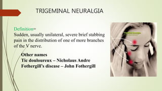
Classical trigeminal neuralgia=
1. is a unilateral disorder characterized by brief
electric, shocklike pains.
2. Abrupt in onset and termination
3. limited to the distribution of one or more
divisions of the trigeminal nerve
Symptomatic trigeminal neuralgia =
1. Pain is similar to classical type But it is caused by a
demonstrable structural lesion other than vascular
compression.
84. clinical feature
Provocated by obvious stimuli like
Touching face at particular site
Chewing
Speaking
Brushing
Shaving
Washing the face
The characteristic of the disorder being that the attacks do not occur
during sleep.
85. Rare-Middle age-Female predilection (60%)-Maxillary
division more commonly involved than opthalmic
Pain lasts for few seconds to minutes =
1. Unilateral (predominantly right side)
2. Precipitated by trigger zones
86. IHS diagnostic criteria;
A. Paroxysmal attacks of facial or frontal pain that last a few seconds to
less than 2 minutes, affecting one or more divisions of the trigeminal
nerve and criteria B and C.
B. Pain has at least one of the following characteristics:
1.Intense, sharp, superficial or stabbing
2. Precipitated from trigger areas or by trigger factors
3. The patient is entirely asymptomatic between paroxysms.
C. Attacks are stereotyped in the individual patient.
D. There is no clinically evident neurological deficit.Not attributed
to another disorder.
88. Sweet diagnostic 5 major criteria:
1. The pain is paroxysmal
2. The pain may be provoked by light touch to the face (trigger
zones)
3. The pain is confined to trigeminal distribution
4. The pain is unilateral
5. The clinical sensory examination is normal
89. Surgical management
Surgical options have been reserved for those patients
1. who have a clearly defined secondary cause for the
trigeminal neuralgia,
2. who are unresponsive,
3. who have severe, unremitting pain that limits their
ability to eat,
4. for whom multiple medications are intolerable or
contraindicated
91. Carbamazepine and phenytoin are the traditional anticonvulsants.
initially dose should be kept small to minimum especially in elderly
patients to avoid nausea, vomiting and gastric irritation.
Once the pain remission has being achieved the drug dose should be
kept at maintainance level or withdrawn and restarted if symptoms
reappear
When carbamazepine is contraindicated clonazepam can be given
Co-administration of phenytoin or baclofen is also advocated.
Medical treatment
92. Surgical treatment
Peripheral Nerve Injections:
Long acting anesthetic agents: without adrenaline bupivacaine
with or without corticosteroids.
The selective nerve blocks can be given as an emergency measure,
where the patient is suffering quite a lot.
Alcohol injections:
The intraoral injection of 95% absolute alcohol in small quantities
(0.5 to 2 ml).
Repeated alcohol injections should be avoided, as it causes local
tissue toxicity, inflammation and fibrosis.
93. The results are variable. Sometimes it provides relief for a period of 6
to 12 months or sometimes patient comes back with pain immediately
within short time span.
94. Gamma knife radiosurgery
The Gamma Knife is a focused 201 intercepting beams of
gamma radiation, produced by separate cobalt sources.
1. The dose is 70–90 Gy.
2. Pain relief is usually not immediate.
3. The mean time to pain relief in two series was
approximately one month.
95. Peripheral neurectomy (nerve Avulsion) :
Oldest and the most effective procedure
Simple
Relatively reliable
Indicated in patients in whom craniotomy is contraindicated due to
age, debility, limited life expectancy.
Acts by interrupting the flow of a significant number of afferent
impulses to central trigeminal apparatus.
Performed mostly on infraorbital, inferior alveolar, mental and
rarely lingual nerve.
It has a disadvantage of producing full anesthesia or deep
hypoesthesia related dysfunction.
96. Cryotherapy:
Direct application of cryotherapy probe (nitrous oxide probe)
Temperature colder than -60 ºC for 2-3 minutes
Repeated three times
97. Peripheral radiofrequency neurolysis thermocoagulation:
Radiofrequency electrode that has the capacity to destroy the pain
fibres is used in this procedure.
Temperature being 65 to 75 ºC for 1 to 2 minutes.
Shown to induce pain remissions in 80% of cases.
Advantages: Low morbidity in high risk/ elderly patients.
99. GYCEROL INJECTIONS:
Absolute alcohol or phenol-glycerol mixture can be used as the
neurolytic agents.
Agent is injected into meckel’s cave or in the ganglion.
It induces pain relief in 80% of the cases.
Also spares the ophthalmic division and the motor root. .
100. THERMOCOAGULATION:
A radiofrequency electrode that has the capacity to destroy pain fibres
is used.
Alternating currents of high frequency is passed through the
electrode.
It produces ionization in biological tissues leads to coagulation of
tissues.
101. BALLON COMPRESSION:
A Fogarty catheter 1 to 2cm is advanced within the meckel’s cave
through foramen ovale.
Inflated upto 0.75ml at the ventral aspect of the ganglion root for 1
minute.
It destroys the root fibres.
102. Trigeminal Neuralgia Diet
1. Low fat, high protein, high calorie
2. Easy chewable foods
Trigeminal Neuralgia Association recommend avoidance of:
avoiding things such as caffeine, citrus fruits and bananas.
extreme hot/cold, hot sauce,chili, spicy salsa, mints, black
pepper,cinnamon, ginger, nutmeg
103. The diagnosis and management of nerve injury
during endodontic treatment
Mechanical nerve injuries during endodontic treatment may be
classified Neuropraxia (cases in which there is a brief altered
sensation); Axonotmesis (partial damage to the nerve fibers that
may fully recover within 2– 12 months); and Neurotmesis (a
nerve that has been entirely cut, with poor prognosis for recovery)
Diagnosis of endodontic treatment-related nerve injury=
Periapical radiography, treatment planning, and for the
management of endodontic complications, computed tomography
scans.
104.
105. Diagnosis and management of suspected nerve injury following an endodontic
treatment.
(1)Non-routine sequel: any event that is not part of the routine and/or expected
sequel during or following the endodontic procedure.
(2) Suspected altered sensation: any event that occurred during or following
the endodontic procedure, was reported by the patient, or is suspected based on
clinical and/or radiographic evaluation, which may suggest the presence of
altered sensation following the endodontic procedure.
(3) Clinical evaluation: evaluation performed to determine the presence, nature,
and extent of the sensory disturbance.
106. (4) Definitive/non-definitive diagnosis: the practitioner is
able/unable (respectively) to determine the presence, nature, and
extent of the suspected altered sensation; OR is able/unable
(respectively) to determine the required treatment and/or to
monitor recovery.
(5) Early intervention: an intervention performed even prior to
a definitive diagnosis, aimed at preventing permanent nerve
damage and enabling a better clinical and medico-legal response
107. Para trigeminal or Readers syndrome:
characterized by severe, unilateral facial pain and headache
in the distribution of the ophthalmic division of the
trigeminal nerve
in combination with ipsilateral oculosympathetic palsy or
Horner syndrome.
UNIlateral pain of head without vasomotor disturbances.
108. Frey syndrome / Gustatory sweating:
First described by frey.
It is localised gustatory sweating in the area supplied by
auriculotemporal nerve.
It arries as a result of damage to auriculotemporal nerve and subsequent
innervation of sweat glands by parasympathetic salivary fibres.
The patient typically exhibits flushing and
sweating of the involves side of the face,
chiefly in temporal area, during eating.
109. It can be commonly seen in:
Penetrating wound of the parotid region
Parotid surgery
Mandibular & Zygomatic bone fractures
TMJ surgery
110. Treatment:
Botox injection: reduce sweating
Topical anti- perspirant (20% aluminium chloride solution)
Application of an ointment containing an anti-cholinergic drug such
as 3% scopolamine
Blockage of parasympathetic outflow by way of alcohol injection or
2%
lignocaine injections at various sites such as the otic ganglion &
the auriculo-temporal nerve.
Partial parotidectomy.
111. Trotter syndrome:
In nasopharyngeal carcinoma, the tumor may extend laterally and
involve the sinus of Morgagni involving the mandibular nerve.
This produces a triad of symptoms known as Trotter's Triad.
These symptoms are:
1) Conductive deafness (due to eustachian tube involvement)
2) Ipsilateral immobility of the soft palate
3) Trigeminal neuralgia
112. In injury to:
Opthalmic nerve: loss of corneal blink reflex.
Maxillary nerve: loss of sneeze reflex. As it is a afferent path for
sneeze reflex.
Mandibular nerve: loss of jaw jerk reflex.
A lesion of foramen ovale leads to paraesthesia along mandible,
tongue, temporal region and paraesthesia of mucles of mastication.
113. Trigeminal nerve injury also occur due to:
Ridge augmentation surgery
Endodontic surgery
Tumor resection
Salivary gland and duct surgeries(Stone in the submandibular gland
duct)
Biopsy procedures
Orthognathic surgery: BSSO highest incidencce of neurosensory
disturbances.
114. Maxillofacial trauma:
Fracture of mandibular body and ramus
LeFort fractures
Fracture of condylar segment medially
Mandibular angle, body and symphysis fracture
Pathologic lesions:
Surgical removal
Use of Carnoy’s solution
115. Conclusion
Since Trigeminal nerve is mixed nerve, supplies mainly head and neck
region.
Hence as a dentist one should know throughly about intracranial and
extracranial course and distribution of Trigeminal nerve,
Diagnose the pathologies associated with Trigeminal nerve and for
appropriate treatment.
116. REFERENCES
Human anatomy,head neck face chaurasia
Textbook Oral and maxillofacial surgery Neelima malik
Local anaesthesia- malamed
Damage to the inferior alveolar nerve as the result of root canal therapyM.
Anthony Pogrel, DDS, MD, FRCS, FACS
Severe Facial Ischemia After Endodontic Treatment Peter Lindgren, MD, DDS,*
Karl-Fredrik Eriksson, MD, PhD,† and Anita Ringberg, MD, PhD‡
Endodontic-related Paresthesia: A Case Report and Literature Review Maria
Ahonen, DDS,* and Leo Tj€aderhane, DDS, PhD*†
Amaurosis, an Unusual Complication Secondary to Inferior Alveolar Nerve
Anesthesia: A Case Report and Literature Review Rahul Pandey, MDS,*
Nivedita Dixit, MDS,† Kuldeep K*
117. Permanent mimic musculature and nerve damage caused by sodium
hypochlorite: a case report Matthias Pelka, DMD,a and Anselm
Petschelt, DMD,b Erlangen, Germany
Hematoma - A Complication of Posterior Superior Alveolar Nerve
Block Nidhi Gupta1, Kunwarjeet Singh2* and Sidarth Sharma3
Endodontic-related inferior alveolar nerve injuries: A review and a
therapeutic flow chart R. Castro c,b,*, M. Guivarc’h b, J.M. Foletti
a, J.H. Catherine a,b, C. Chossegros c, L. Guyot a
8 muscle=temporalis, masseter,medial pterygoid,lateral pterygoid,ant belly of diagastric,post belly of dia’tensor tympani ,tensor palatine
Supraorbital: It is the larger and lateral branch.
It crosses supraorbital margin in the notch or foramen, turns upwards to supply skin of forehead and scalp as far posterior as the vertex.
2. Supratrochlear: It is the smaller and medial branch
It supplies upper eyelid
landmark=Mucobuccal fold,zygomatic process of maxilla,infratemporal surface of maxilla,anterior border and coronoid process of ramus of mandible,maxillary tuberosity.
A 45 years old female patient reported to our dental centre with chief complaint of severe pain in the maxillary right posterior region which aggravates during sleeping and on intake of the hot food. The clinical and radiographic evaluation reveals carious exposed maxillary right first molar (The intraoral photograph was taken after removal of carious lesion and temporary restoration). The root canal treatment followed by fabrication of the crown was planned for the same.
These is clinical case in which pt visited to clinic with complaining pain in upper anterior tooth.after investigation dentist advise a rct treatment.dentist was use sod.hypo as irrigation agent .these all happens due to incorrect procedure of use of irrigation technique.
For avoide these we can use .0.5 to 5.25
The main trunk remains undivided only for a short distance of 2 to 4 mm and then it divides into a “cat of nine tails.”
Insert needle into depression or imagionary line between pterygoid raphe and coronoid process above mandi occlusal plane
20-year-old female patient visited her local dentist for a routine restorative procedure, carried out on the lower left first molar.
A left inferior alveolar block anesthesia was administered, using a cartridge dental syringe with a needle of 25 gauge and 34 mm length.
The local anesthetic used was articaine hydrochloride 4% with adrenaline 1 ∶ 100.000.
A total amount of 1.7 mL (one cartridge) was delivered.
More specifically, if the injection is administered too far posteriorly, the anesthetic solution could be injected into the parotid substance, whose deep lobe extends around the posterior ramus of the mandible and projects forward on the medial surface of the ramus. Most often, the gland envelopes the facial nerve, thus leading to the direct anesthesia of the latter.1
These is another case report pt visite clinic for pain in lower rt back tooth.
Also, because of the smaller lumen, the anesthetic may be injected under greater pressure. A 25-G needle is preferred for injections in which the risk of positive aspiration is high.
The clinician can try to minimize the potential for such complications through effective anxiety management .
mechanical trauma from overinstrumentation into the inferior alveolar canal; pressure phenomenon from the presence of the endodontic point or sealant within the inferior alveolar canal neurotoxic effect from the medicaments used to clean the canal . Endodontic treatment of mandibular molar teeth has the potential to damage the inferior alveolar nerve via direct trauma, pressure or neurotoxicity. Ph 9.2 to 11.7 alkaline.
Paresthesia was present on the left side of the lower lip extending from the mandibular midline to the second premolar both extraorally and intraorally in the area of mental nerve stem.
Normal=slight closure of jaw
Abnormal=brisk complete closure.
International classification of headache disorder ,STREOTYPE-PERSON TO PERSON
50 unit of clostridium botulinm,type a neurotoxin,0.25 mg albumin,0.45 sodium chloride.
Carnoys=100% ethanol,chloroform,glacial acetic acid,ferric chloride,
Use= for tissue fixsation