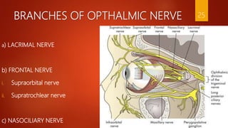
1
TRIGEMINAL NERVE
DEPARTMENT OF PUBLIC HEALTH DENTISTRY
2
GUIDED BY
DR.(PROF) VEERANNA
RAMESH
PRESENTED BY
DR.ANANT KUMAR
PG FIRST YEAR
B.I.D.S.H
CONTENTS:-
 INTRODUCTION
 TRIGEMINAL NUCLEI
 FUNCTIONAL COMPONENTS
 COURSE AND DISTRIBUTION
 TRIGEMINAL GANGLION
 DIVISION OF TRIGEMINAL NERVE
 CLINICAL EXAMINATION
 APPLIED ANATOMY
 CONCLUSION
 REFERENCES
3
INTRODUCTION
 Trigeminal nerve is the largest cranial nerve and resembles a
spinal nerve, its origin by two roots(sensory and motor).
 It is also known as Trifacial and Trigeminus nerve.
 It arises by two roots,a posterior larger(sensor)root
and the anterior smaller(motor root).
 The smaller root consist of three or four bundles, in the larger,
bundles are more numerous, varying in number seventy to a
hundred .
 It is attached to the lateral part of the pons by its two roots,
sensory and motor.
4
 The two roots are seperated from one another by a few of the transverse fibers of the pons.
 The deep origin of the larger or sensory root may be traced between the transverse fibers of the pons
varolii to the lateral tract of the medulla oblongata, immediately behind the olivary body.
 The two roots of the nerve passes forwards through an oval opening in the dura mater, at the apex of
the petrous portion of the temporal bone.
 The fibers of the larger root enter a large semilunar ganglion(GASSERIAN), while the smaller root
passes beneath the ganglion without having any connection with it, and joins outside the cranium with
one of the trunks derived from it.
5
1. Sensory root :-
supply hard palate ,floor of mouth, upper lip, cheek, mucosa,
mucus membrane of the maxillary sinus.
2. Motor root :-
Receives impulses from the right and left cerebral
it supply four muscles of mastication and tensor veli palatine,
tensor tympani, mylohyoid and anterior belly of digastric.
6
NUCLEI
7
TRIGEMINAL NUCLEI
 A cranial nerve nucleus is a collection of neurons (grey matter)
in the brain stem that is associated with one or more cranial
nerve.
 Axons carrying information to and from the cranial
nerves form a synapse first at these nuclei.
 Lesions occurring at these nuclei can lead to effects
resembling those seen by the severing of nerves they are
associated with.
8
SENSORY NUCLEUS
1. MESENCEPHALIC NUCLEUS :-
 The mesencephalic nucleus is a paired structure located at the
mesopontine junction, which projects cephalad through the mid
brain.
 It is situated along the anterolateral aspect of the grey matterand
lies ventral to the inferior colliculi at the level of midbrain.
 It lies superior to the main sensory nucleus of the trigeminal
nerve with which it is continuous within the pons.
 It is also the nucleus of masseteric reflex.
9
1. PRINCPLE/ SUPERIOR SENSORY NUCLEUS :-
 It lies in pons continuous with spinal nucleus.
 Relys for the sensation of touch and and pressure
from face and scalp.
3. INFERIOR SENSORY/ SPINAL NUCLEUS :-
 It lies in medulla oblongata.
 It also relys for Pain and Temperature from face
and scalp.
 It is also a nucleus for the ordinary sensation of the
areas supplied by VII,IX and X cranial nerves.
10
MOTOR NUCLEUS
 It is located in pons medial to principal
sensory nucleus.
 Derived from first brancheal arch.
 It supply to all muscles derived from 1st
branchial arch.
 Connected with corticonuclear fibres of the
pyramidal tract of both sides.
11
FUNCTIONAL COMPONENTS
12
FUNCTIONAL COMPONENTS
1. SENSORY COMPONENTS :-
 Sensation of pain, temperature, touch and pressure from skin
of face, mucous membrane of nose, most of the tongue,
paranasal air sinuses travel along axons.
 The cell bodies lies in the trigeminal ganglion or semilunar
ganglion or Gasserian ganglion.
 It lies at the apex of petrous temporal bone in adural cave,
the Meckel’s cave.
 The central processes of trigeminal ganglion form sensory
root. some fibres ascend and other descend.
13
 Ascending fibres end in superior sensory nucleus.
 Descending fibers end in the spinal nucleus of V nerve.
2. MOTOR COMPONENTS :-
 The motor nucleus receives impulses from the right and
left cerebral hemispheres, red nucleus and mesencephalic
nucleus.
 Fibres of motor root supply four muscles of mastication
and four other muscles are tensor veli palatini ,tensor
tympani, mylohyoid and anterior belly of digastric.
14
COURSE AND DISTRIBUTION
15
COURSE AND DISTRIBUTION
 Both motor and sensory root are attached ventrally to
junction of pons and middle cerebellar peduncle with
motor root lying ventromedially to the sensory root.
 It Passes anteriorly in middle cranial fossa to lie below
tentorium cerebelli in cavum trigeminale , here motor
root lies inferior to sensory root.
 Its Sensory root connected to posteromedial concave
border of the trigeminal ganglion.
16
 Convex anterolateral margin of the ganglion gives
attachment to the 3rd division of the trigeminal nerve.
 Motor root turns further inferior with sensory component of
3rd division emerge out of foramen ovale as Mandibular
nerve.
 Opthalmic and maxillary division emerges through superior
orbital fissure and foramen rotundum .
17
GANGLION
18
THE TRIGEMINAL GANGLION
 SEMILUNAR OR GASSERIAN GANGLION:-
It is Crecentric in shape with convexity anterolaterally.
Contains cell bodies of pseudounipolar neurons.
 LOCATION :
It Lies in a bony fossa at apex of the petrous temporal
bone on floor of middle cranial fossa, just lateral to
posterior part of lateral wall of cavernous sinus.
19
 BLOOD SUPPLY :-
The Ganglion is supplied by
(a)Internal carotid artery
(b)Middle meningeal artery
(c)Accessory meningeal arteries.
(d)The meningeal branch of ascending
pharyngeal artery.
20
RELATIONS:-
 SUPERIORLY: superior petrosal sinus,
free margin of tentorium cerebelli.
 INFERIORLY: motor root,
greater petrosal nerve,
petrous apex,
foramen lacerum.
 MEDIALLY: posterior part of lateral wall of cavernous sinus,
internal carotid artery with its sympathetic
plexus.
 LATERALLY: uncus of temporal lobe , nervous spinosum,
middle meningeal artery and vein.
21
DIVISIONS OF TRIGEMINAL NERVE
22
DIVISIONS OF TRIGEMINAL NERVE
1. OPTHALMIC BRANCH (V1)
2. MAXILLARY BRANCH (V2)
3. MANDIBULAR BRANCH (V3)
23
OPTHALMIC NERVE
 It is the smallest of the three branches of the semilunar ganglion.(CNV1)
 It leaves the anterior medial part of the ganglion and passes forward in the the lateral wall of the
cavernous sinus.
 Its fibers are sensory, or afferent from the scalp ,the skin of the forehead, the upper eyelid lining, the
frontal sinus, the conjunctiva of the eyeball, the lacrimal gland and the skin of the lateral angle of the
eye.
 It also transmits sensory impulses from the sclera of the eyeball and the lining of the ethmoid cells.
24
BRANCHES OF OPTHALMIC NERVE
a) LACRIMAL NERVE
b) FRONTAL NERVE
i. Supraorbital nerve
ii. Supratrochlear nerve
c) NASOCILIARY NERVE
25
A) LACRIMAL NERVE
 It is the smallest of the all three branches of the
ophthalmic nerve.
 It passes into the orbit at the lateral angle of the
superior orbital fissure.
 It supplies sensory fibres to the gland and adjacent
conjunctiva.
26
B) FRONTAL NERVE
 It is the largest of the all three branches of ophthalmic nerve.
 It appears to be a direct continuation of the ophthalmic division.
 It enters the orbit by way of the superior orbital fissure.
Frontal nerve further divides into two branches:-
a)Supraorbital nerve
b)Supratrochlear nerve
27
a) Supraorbital nerve:-
 it is the largest branch of the frontal nerve.
 It passes forward and leaves the orbit through the supraorbital foramen to supply
skin of the upper eyelid ,the forehead and the anterior scalp region to the vertex of
the skull.
b) Supratrochlear nerve:-
 It is the smallest branch of the frontal nerve.
 It passes towards the upper medial angle of the orbit.
 It supply the skin of the upper eyelid and lower medial portion of the forehead.
28
C) NASOCILIAY NERVE
 It is the third main branch of the ophthalmic division.
 It enters the orbit through the superior orbital fissure.
 Branches of the nasociliary divided into those arising in the orbit, in the nasal cavity
and on the face.
BRANCHES in the orbit:-
1.Long root of the ciliary ganglion
2.Long ciliary nerve
3.Posterior ethmoidal nerve
4.Anterior ethmoidal nerve
29
1. LONG ROOT OF CILIARY GANGLION:-
 It arises from the nasociliary nerve.
 It contains sensory fibres ,which passes through
the ganglion without synapsing and continue
on to the eyeball by means of the short ciliary
nerves.
2. LONG CILIARYN NERVE :-
 There are usually two or three long ciliary
branching from the nasociliary nerve.
 They are distributed to the iris and cornea.
30
3. POSTERIOR ETHMOIDAL NERVE :-
It enters the posterior ethmoidal canal to
be distributed to the mucous membrane lining
the posterior ethmoidal cells and the sphenoid
sinus.
4. ANTERIOR ETHMOID NERVE :-
The anterior ethmoidal nerve gives of
filaments that supply the mucous membrane
of the anterior ethmoidal cells and frontal
sinus.
31
MAXILLARY DIVISION (V2)
32
MAXILLARY NERVE
 Maxillary nerve is one of the branches of the
trigeminal nerve and is the nerve of 1st branchial
arch.it is a sensory nerve.
 It takes origin from the sensory root of trigeminal
nerve from the trigeminal ganglia.
COURSE :-
A. INTRACRANIAL COURSE :- from the trigeminal
ganglia , the maxillary division of the trigeminal nerve
becomes the maxillary nerve and passes in the middle
cranial fossa through the lateral wall of the cavernous
sinus into the foramen rotundum. It comes out into the
pterygopalatine fossa.
33
B. EXTRACRANIAL COURSE :-
In the pterygopalatine fossa
a) It passes above the pterygopalatine ganglia and gives a
branch, <.e. sensory root of the trigeminal ganglia
which passes through the ganglia without relaying in it.
b) The sensory root passes through the pterygopalatine
ganglia and gives the 3 branches.
BRANCHES AND DISTRIBUTION :
1. Branches In the middle cranial fossa
2. Branches in the pterygopalatine fossa
3. Other branches
34
1. Branches In the middle cranial fossa :-
I. MENINGEAL BRANCHES- supply the dura mater of
middle cranial fossa.
2 . Branches in the pterygopalatine fossa :-
It gives two sensory roots through the pterygopalatine
ganglia , which pass through it without relay and give the
following branches.
1) NASAL BRANCHES – They pass through the spheno-
palatine foramen into the nasal cavity so they are known
sphenopalatine nerves.
A. LATERAL POSTERIOR SUPERIOR NASAL NERVES –
Supply
35
a) Posterosuperior part or quadrant of nasal cavity
b) Superior nasal concha
c) Middle nasal concha.
B. MEDIAL POSTERIOR SUPERIOR NASAL NERVES – Supply
a)Posterior part of roof of nasal cavity.
b)Nasal septum
c. NASOPALATINE NERVE /LONG SPHENOPALATINE NERVE -
Which is the largest of this group. it passes downwards and
forwards on the nasal septum. It descends through the incisive
foramen into the hard palate and supplies the anterior part of the
hard palate.
36
2) PALATINI BRANCHES –
1. GREATER (anterior) PALATINE :- it passes through the greater
palatine canal and descends through the greterpalatine foramen on the
under surface of hard palate. It supplies:
a) Hard palate (except its anterior part which is supplied by
nasopalatine nerve).
b) Lateral wall of the nasal cavity of inferior nasal concha and adjoining
meatuses.
2 . LESSER OR (MIDDLE AND POSTERIOR) PALATINE NERVE :-
it emerges through lesser palatine foramen and passes backwards to
supply.
a) Soft palate for ordinary sensation as well as taste sensation. Taste
fibers come along the greater superficial petrosal nerves which pass
through the sphenopalatine ganglia.
37
b) Tonsil for ordinary sensation.
3. PHARYNGEAL BRANCH :- it passes through the palatino-
vaginal canal and supplies Nasopharynx behind the
pharyngotympanic tube.
4. ORBITAL BRANCH :- they pass through the inferior orbital
fissure into the orbit and supply:
a) periosteum of the orbit.
b) Orbitalis muscles by the sympathetic nerve fibres, which come
along the sphenopalatine ganglia.
38
3. OTHER BRANCHES -
5. ZYGOMATIC NERVE :-
a) It divides into zygomaticofacial and zygomaticotemporal
nerves. They supply the skin of the face and scalp.
b) It also carries the postganglionic parasympathetic
fibres arising from the pterygopalatine ganglia for
the supply of the lacrimal gland.
6. POSTERIOR SUPERIOR ALVEOLAR NERVE :-
it enters the posterior surface of body of maxilla and supplies
and supplies the upper three molar teeth and adjoining parts
of the gum.
39
7. INFRAORBITAL NERVE :-
it passes through the infraorbital canal and then comes out through
the infraorbital foramen into the face.
A) In the infraorbital canal, it divides into -
a) Middle superior alveolar nerve, which supplies the premolar teeth
and maxillary sinus.
b) Anterior superior alveolar nerve , which supplies canine and
teeth and maxillary sinus.
40
BRANCHES ON THE FACE
The infraorbital nerve emerges through the infraorbital foramen onto the face
it divide into its terminal branches.
a. INFERIOR PALPEBRAL :-
supply the skin and conjunctiva of lower eyelid.
b. EXTERNAL NASAL :-
supply the skin of the side of the nose and of the septum.
c. SUPERIAL LABIAL :-
it is the largest and most numerous, Distributed to the skin of the upper
lip and also supply to the mucous membrane of mouth, and labial
glands.
they are joined immediately beneath the orbit by filaments from the
facial nerve ,forming with them the infraorbital plexus.
41
MANDIBULAR DIVISION (V3)
42
MANDIBULAR
NERVE The mandibular division of the trigeminal nerve is the largest of the
three division, it is formed by the union of the large sensory bundle
of fibers and a small motor bundle of fibers.
 The sensory root fibers are peripheral extensions of unipolar cells
located located in the semilunar ganglion.
 The motor root fibers are derived from motor cells located in the
medulla oblongata.
 The large sensory root arises from the semilunar ganglion , its fibers
are distributed to the dura, skin and the mucous membrane of the
chin, cheek and lower lip.
 The motor root is located in the middle cranial fossa ,it joins the
sensory root after the latter leaves the semilunar ganglion.
43
BRANCHES AND DISTRIBUTION
44
1. BRANCHES FROM NERVE TRUNK :-
A. NERVOUS SPINOSUS - It is a sensory nerve and ascends up.
It passes through the foramen spinosum along with middle
meningeal artery , branches of maxillary artery. Both the
nerve and artery supply the middle meningeal artery.
B. NERVE TO MEDIAL PTERYGOID – it supplies
a)Medial pterygoid from its deep surface. It gives a motor
root to otic ganglia but do not relay in it. then it supplies
the Tensor palate , Tensor tympani.
3. BRANCHES FROM ANTERIOR DIVISION :-
45Below the level of the undivided part of the mandibular division,
the trunk separate into two parts :
(A) ANTERIOR DIVISION – The anterior division is smaller then
the posterior division. It receives sensory and motor fiber that
supply the muscles of mastication, the skin and mucous
membrane of the cheek, and the buccal gingivae and lower
molars. It passes downward and forward ,where it divides
into four branches.
1. NERVE TO LATERAL PTERYGOID
2. DEEP TEMPORAL NERVE
3. MASSETERIC NERVE
4. BUCCAL NERVE
1. PTERYGOID NERVE :- it supplies the lateral pterygoid
from its deep surface.
2. NERVES TO THE TEMPORAL MUSCLE :- two in
number and supply the temporalis from its deep
surface.
a. ANTERIOR DEEP TEMPORAL NERVE
b. POSTERIOR DEEP TEMPORAL NERVE
3. MASSETER NERVE :- this nerve passes above the
lateral pterygoid through the masseteric notch. The
nerve passes along with masseteric vesssels , supply the
masseter from its deep surface.
46
4. BUCCAL NERVE :-
 It is a sensory nerve . It passes between the two heads of
origin of lateral pterygoid muscle and supplies the buccal
region of the face .
a) Mucous membrane of the buccal region deep to
buccinators.
b) Skin of face superficial to buccinator .
c) Lateral surface (labial surface) of the molar and
teeth.
47
3. BRANCHES FROM POSTERIOR DIVISION-
 It gives three sensory branches , one of which also carries
motor fibres.
1. AURICULO-TEMPORAL NERVE :- it is a sensory nerve and
ascends upwards between the neck of the mandible and
sphenomandibular ligaments. It lies lateral to spine of the
sphenoid.
o It gives a auricular branch to lateral surface of the upper part of
the upper part of the auricle and tragus of the auricle and
outer surface of the tympanic membrane.
o It supplies the temporomandibular joint.
o It supplies the skin of the face , scalp and temporal region.
48
2.LINGUAL NERVE :-
It takes origin from the posterior division of the mandibular nerve
1cm below the scalp.
Courses : it passes between-
a)Tensor palatii and lateral pterygoid
b)Medial pterygoid and lateral pterygoid
c)It is joined by the chorda tympani nerve 2 cm below the
skull.
d)below the lower border of the lateral pterygoid ,it runs
downwards and forwards between the ramus of medial
pterygoid.
49
e) Now , it comes in direct relation with the mucous
membrane covering the cusp of 3rd molar tooth of
mandible , the origins of superior constrictor and
myelohyoid .
f) Then, it runs over the hyoglossus deep the myelohyoid
g) Lastly it lies on the surface of genioglossus dep to
myelohyoid . Here, it winds round the submandibular
duct and terminates by giving its branches .
TERMINATION :- It terminates in sub – mandibular
region and gives branches.
BRANCHES AND DISTRIBUTION :-
Its sensory fibres supplies.
50
a) Anterior 2/3rd of the tongue for ordinary sensation.
b) Floor of the mouth, lingual surface of gums of teeth of mandible.
3. INFERIOR ALVEOLAR NERVE :- it is a sensory nerve for the teeth of
the lower jaw, but it also carries some motor fibres to supply the
mylohyoid and anterior belly of digastric.
ORIGIN : It takes origin from the posterior division of the mandibular
nerve.
COURSE :
a) inferior alveolar nerve passes downward and forward and comes to lie
on the inner surface of ramus mandible lateral to the spheno-mandibular
ligament.
b) It enters through the mandibular foramen ,passes in the mandibular
canal, and lastly comes out in the face as the mental nerve.
51
c) Its motor fibres separate and give a branch known as nerve to
mylohyoid vessels and pierces the spheno-mandibular ligament
and then come into mylohyoid groove of inner surface of body
of mandible.
AUTONOMIC GANGLIA ASSOSIATED WITH THE MANDIBULAR
DIVISION :- two ganglia are associated with the mandibular division
submandibular and otic ganglia.
A. Submandibular (submaxillary) ganglion :-
the submandibular ganglion is a small ovoid body that is suspended
from the lingual nerve .these parasympathetic fibers are preganglionic ,
having their origins in the superior salivatory nucleus in the medulla.
They course within the intermediate nerve and in the facial canal and
group together to form the chorda tympani nerve.
52
B. OTIC GANGLION :-
The otic ganglia is a flattened ovoid body located on the medial
side of the undivided mandibular division of the trigeminal nerve. It
below the foramen ovale and infront of the middle meningeal
It has two main roots.
1. PARASYMPATHETIC PREGANGLIONIC(SECRETORY) FIBERS :-
These parasympathetic fibers arise in inferior salivatory nucleus.
This group of cells lies in the floor of the fourth ventricle in the
medulla. The efferent fibres pass by way of the glossopharyngeal
nerve through the jugular canal.
53
2. SYMPATHETIC ROOT :-
 The sympathetic root is made up of post ganglionic fibres that have
originated in the superior cervical sympathetic ganglion and the plexus
of the middle meningeal artery .
 these sympathetic fibres pass interruptedly through the otic ganglion .
With the post ganglionic parasympathetic fibres , they join the
auriculotemporal nerve and with its glandular branches , continue to
parotid gland.
 affrerent or sensory fibres from the parotid gland pass by way of the
auriculotemporal nerve.
54
CLINICAL EXAMINATION
55
CLINICAL EXAMINATION
1. Testing for the integrity of the trigeminal nerve :-
a) For its motor function , ask the patient to clench his teeth
firmly then feel the contraction of masseter muscle. And
observe for deviation of the the mandible as the jaws are
opened. if there is 7th nereve weakness this makes it easier to
perceive deviation is to the weak side.
b) For its sensory function for touch ,touch the different
areas of the skin of the face with a wisp of cotton wool. For
its sensory function of pain, give a gentle pressure with a pin
of the face.
56
APPLIED ANATOMY
57
APPLIED ANATOMY
1.TRIGEMINAL NEURALGIA
2.HERPES ZOSTER OPTHALMICUS
3.WALLENBERG SYNDROME.
1.TRIGEMINAL NEURALGIA :-
 It is a paroxysmal , intermittent, excruciating pain confined to one of
the branches of the trigeminal nerve.
 Characterised by unilateral affliction, not crossing the midline ,
presence of trigger zones, cessation of pain during sleep.
 The etiology is not definitely known . Suggested cause and viral
lesions of the ganglion, demyelination of the nerves, narrowing of the
formation and idiopathic.
58
Trigeminal neuralgia affects mainly adults, especially older people. It is more
common among women.
ETIOLOGY :-
 Trigeminal neuralgia is usually caused by Compression of the trigeminal nerve at
its root by an aberrant loop of an intracranial artery (e.g. anterior inferior cerebellar artery,
ectopic basilar artery).
 Other less common causes include compression by a tumor, an arteriovenous malformation, an
aneurysm, and occasionally a multiple sclerosis plaque at the root entry zone (usually in younger
patients), but these causes are usually distinguished by accompanying sensory loss and other
deficits.
59
SYMPTOMS AND SIGNS :-
 Pain due to trigeminal neuralgia occurs along the distribution of one or more sensory
of the trigeminal nerve, most often the maxillary. The pain is paroxysmal, lasting seconds up
2 minutes, but attacks may recur rapidly-as often as 100 times a day. It is lancinating,
excruciating, and sometimes incapacitating.
 Pain is often precipitated by stimulating a facial trigger point (eg, by chewing, brushing the
teeth, or smiling). Sleeping on that side of the face is often intolerable. Usually, only one side
the face is affected.
DIAGNOSIS :-
 Clinical evaluation :- Symptoms of trigeminal neuralgia are often pathognomonic. Thus,
some other disorders that cause facial pain can be differentiated clinically.
 POSTTHEREPTIC PAIN is differentiated by its constant duration (without paroxysms), typical
antecedent rash, scarring, and predilection for the ophthalmic division.
60
 Migraine, which may cause atypical facial pain, is differentiated by pain that is more
prolonged and often throbbing.
 Sinusitis and odontogenic pain, which can usually be differentiated by their
associated findings (eg, nasal discharge, fever, positional headache, tooth sensitivity)
 Neurologic examination is normal in trigeminal neuralgia. Thus, neurologic deficits
(usually loss of facial sensation) suggest that the trigeminal neuralgia like pain is
caused by another disorder (eg, tumor, stroke, multiple sclerosis plaque, vascular
malformation, other lesions that compress the trigeminal nerve or disrupt its brain
stem pathways).
61
7 TRIGGERS ZONE OF TRIGEMINAL NEURALGIA :-
1.YAWNING
2.SNEEZING
3.BENDING OVER
4.LOUD NOISES
5.BRIGHT LIGHTS
6.BLOWING UP BALLONS
7.DRINKING THROUGH STRAW
62
TREATMENT
 Trigeminal neuralgia is treated with carbamazepine 200 mg orally 3 or 4 times a day, which is usually
effective for long periods; it is begun at 100 mg orally twice a day, increasing the dose by 100 to 200
mg/day until pain is controlled (maximum daily dose 1200 mg).
 If carbamazepine is ineffective one of the following oral drugs may be tried:
Oxcarbazepine 150 to 300 mg B.D
Baclofen 5 mg T.D.S
Lamotrigine 25 mg O.D for 2 weeks.
Gabapentin 300 mg OD on day1, 300 mg B.D on day 2,
300 mg T.D.S on day 3
Phenytoin 100 to 200 mg B.D
63
 Injection of 60 to 90% alcohol in the nerve trunk or ganglion.
 Peripheral nurectomy or cryotherapy of the peripheral trigger
zone.
 Microvascular nerve root decompression procedure . It is a
neurosurgical procedure wherein the internal vascular loops of
the superior cerebellar artery is made to compess on the
trigeminal nerve root.
64
2.HERPES ZOSTER OPTHALMICUS :-
 Herpes zoster ophthalmicus is reactivation of a varicella-zoster virus
infection (shingles) involving the eye. Symptoms and signs, which may be
severe, include dermatomal forehead rash and painful inflammation of all
the tissues of the anterior and, rarely, posterior structures of the eye.
 Diagnosis is based on the characteristic appearance of the anterior
structures of the eye plus zoster dermatitis of the first branch of the
trigeminal nerve (V1). Treatment is with oral antivirals, mydriatics , and
topical corticosteroids.
 Herpes zoster of the forehead involves the globe in three fourths of cases
when the nasociliary nerve is affected (as indicated by a lesion on the tip of
the nose) and in one third of cases not involving the tip of the nose. Overall,
the globe is involved in half of patients.
65
Symptoms and Signs :-
 A prodrome of tingling of the forehead may occur. During acute disease, in addition to the
painful forehead rash, symptoms and signs may include severe ocular pain; marked eyelid
edema; conjunctival, episcleral, and circumcorneal conjunctival hyperemia; corneal edema;
and photophobia.
Diagnosis :-
 Zoster rash on the forehead or eyelid plus eye findings
 Diagnosis is based on a typical acute herpes zoster rash on the forehead, eyelid, or both or
a characteristic history plus signs of previous zoster rash.
 Vesicular or bullous lesions in this distribution that do not yet involve the eye suggest
significant risk and should prompt an ophthalmologic consultation to determine whether
eye is involved.
66
COMPLICATIONS :- 67
 Keratitis or uveitis may be severe and followed by scarring. Late sequelae—glaucoma, cataract, chronic
or recurrent uveitis, corneal scarring, corneal neovascularization, and hypesthesia are common and
may threaten vision. Post-therpetic neuralgia may develop later.
 Patients may develop episcleritis (without increased risk of visual loss) and/or retinitis (with risk of
severe visual loss).
TREATMENT :-
 Oral antivirals (eg. acyclovir, famciclovir , valacyclovir )
 Sometimes topical corticosteroids.
PREVENTION :-
Recombinant herpes zoster vaccine is recommended for immunocompetent adults ≥ 50 years, regardless of
whether they have had herpes zoster or been given the older, live-attenuated vaccine. This recombinant vaccine
decreases the chance of getting herpes zoster by 97% for adults 50 to 69 years and 91% for adults ≥ 70 years.
Peripheral nerve block provides temporary relief.
 If pain is severe despite these measures, neuroablative treatments are considered;
however, efficacy may be temporary, and improvement may be followed by recurrent
pain that is more severe than the preceding episodes, and surgery that relieves pain
may result in facial numbness. Painful numbness (anesthesia dolorosa) may also
it occurs in 4% of patients after rhizotomy.
 In a posterior fossa craniectomy, a small pad can be placed to separate the pulsating
vascular loop from the trigeminal root (called microvascular decompression, or the
Jannetta procedure). In gamma knife radiosurgery, gamma radiation is focused on
proximal trigeminal nerve where it exits the brain stem; this procedure interrupts
signals to the brain.
 Electrolytic or chemical lesions or balloon compression of the trigeminal (gasserian)
ganglion can be made via a percutaneous stereotactically positioned needle.
Occasionally, the trigeminal nerve fibers between the gasserian ganglion and brain
stem are cut.
68
WALLENBERG SYNDROME
INTRODUCTION :-
 Wallenberg syndrome is also known as lateral medullary syndrome or the
posterior inferior cerebellar artery syndrome. Wallenberg described the first case
in 1895.
 This neurological disorder is associated with a variety of symptoms that occur as
a result of damage to the lateral segment of the medulla posterior to the inferior
olivary nucleus. It is the most typical posterior circulation ischemic stroke
syndrome in clinical practice.
ETIOLOGY :-
 Wallenberg syndrome is caused most commonly by atherothrombotic occlusion
of the vertebral artery, followed most frequently by the posterior inferior
cerebellar artery, and least often, the medullary arteries.
69
 Hypertension is the most prevalent risk factor, followed by smoking and diabetes.
Cerebral embolism is a less frequent cause of the infarction.
 The other important cause to remember is vertebral artery dissection, which may have risk factors
including neck manipulation or injury, Marfan syndrome, Ehlers Danlos syndrome, and fibromuscular
dysplasia.
 Vertebral artery dissection is the commonest cause of Wallenberg syndrome in younger patients.
DIAGNOSIS :-
 The diagnosis is usually made or suspected from a clinical exam and history of presentation. MRI
with diffusion-weighted imaging (DWI) is the best diagnostic test to confirm the infarct in the
inferior cerebellar area or lateral medulla. .
 A CT angiogram or MR angiogram is very helpful in identifying the site of vascular occlusion
 An ECG is useful in excluding any underlying atrial fibrillation. Checking the serum electrolytes is
essential.
70
DIIFERENTIAL DIAGNOSIS :-
a) Chronic pain syndrome
b) Lacunar stroke
c) Middle cerebral artery stroke
d) Migraine headache
e) Multiple sclerosis
f) Posterior reversible encephalopathy syndrome
g) Subarachnoid hemorrhage
h) Subdural hematoma
i) Systemic lupus erythematosus
j) Vertebrobasilar stroke
71
Different combinations of the following deficits may all be found in
Wallenberg syndrome :-
ON THE SIDE OF LESION -
 Vertigo with nystagmus (inferior vestibular nucleus and pathways). The nystagmus is typically
central, beating to the direction of gaze. Nausea and vomiting, and sometimes hiccups, are
associated with vertigo. Hiccups can often be intractable.
 Dysphonia, dysarthria, and dysphagia (different nuclei and fibers of the IX and X nerves), often
present with ipsilateral loss of gag reflex.
 Horner syndrome; miosis, ptosis, and anhydrosis (sympathetic fibers)
 Ipsilateral ataxia with a tendency to fall to the ipsilateral side (inferior cerebellar hemisphere,
spinocerebellar fibers, and inferior cerebellar peduncle)
 Pain and numbness with impaired facial sensation on the face (descending trigeminal tract).
72
ON THE CONTRALATERAL SIDE –
 Impaired pain and temperature sensation in the arms and legs (spinothalamic tract).
 It is important to note that there is no or only minimal weakness of the contralateral
side (corticospinal fibers are ventral in location).
73
CONCLUSION
 The range of pathologies responsible for trigeminal neuralgia is vast and the possible topographical
areas of impairment are extensive ranging from the cervical spine to the face and skull base.
 MRI investigation is based on an initial imaging protocol that can be completed by examination if
lesions are detected.
 In this context, clinical understanding of the patient prior to the MRI scan is often a precious aid in
directing the examination (essential or secondary neuralgia, affected dermatomes).
 In the case of essential neuralgia, the most commonly observed lesion is neurovascular compression.
Diagnosis is based on T2-weighted inframillimetric acquisition by visualising a vessel perpendicular to
the REZ (at 2–6 mm from the emergence of the brainstem). 3D TOF then makes it possible to
determine the arterial or venous origin of the compression.
74
REFERENCES
75
• Chaurasia’s B D human anatomy, 7th edition volume 3
• Gray’s anatomy for students, first south asia edition
• Prasad Jagannath human anatomy 1st edition volume 3
• Bennett C.Richard Monheim’s Local anesthesia and pain control
in dental practice 7th edition.
• Lui Forshing , Tadi Prasanna , C. Anil kumar Arayamparambil ,
(NCBI)National Center for Biotechnology Information Wellenberg
syndrome 6 july 2020.
• I.Roat Melvin, MD, FACS, Sidney Kimmel Medical College at
Thomas Jefferson University, may 2020.
76

Trigeminal nerve

  • 1.
  • 2.
    TRIGEMINAL NERVE DEPARTMENT OFPUBLIC HEALTH DENTISTRY 2 GUIDED BY DR.(PROF) VEERANNA RAMESH PRESENTED BY DR.ANANT KUMAR PG FIRST YEAR B.I.D.S.H
  • 3.
    CONTENTS:-  INTRODUCTION  TRIGEMINALNUCLEI  FUNCTIONAL COMPONENTS  COURSE AND DISTRIBUTION  TRIGEMINAL GANGLION  DIVISION OF TRIGEMINAL NERVE  CLINICAL EXAMINATION  APPLIED ANATOMY  CONCLUSION  REFERENCES 3
  • 4.
    INTRODUCTION  Trigeminal nerveis the largest cranial nerve and resembles a spinal nerve, its origin by two roots(sensory and motor).  It is also known as Trifacial and Trigeminus nerve.  It arises by two roots,a posterior larger(sensor)root and the anterior smaller(motor root).  The smaller root consist of three or four bundles, in the larger, bundles are more numerous, varying in number seventy to a hundred .  It is attached to the lateral part of the pons by its two roots, sensory and motor. 4
  • 5.
     The tworoots are seperated from one another by a few of the transverse fibers of the pons.  The deep origin of the larger or sensory root may be traced between the transverse fibers of the pons varolii to the lateral tract of the medulla oblongata, immediately behind the olivary body.  The two roots of the nerve passes forwards through an oval opening in the dura mater, at the apex of the petrous portion of the temporal bone.  The fibers of the larger root enter a large semilunar ganglion(GASSERIAN), while the smaller root passes beneath the ganglion without having any connection with it, and joins outside the cranium with one of the trunks derived from it. 5
  • 6.
    1. Sensory root:- supply hard palate ,floor of mouth, upper lip, cheek, mucosa, mucus membrane of the maxillary sinus. 2. Motor root :- Receives impulses from the right and left cerebral it supply four muscles of mastication and tensor veli palatine, tensor tympani, mylohyoid and anterior belly of digastric. 6
  • 7.
  • 8.
    TRIGEMINAL NUCLEI  Acranial nerve nucleus is a collection of neurons (grey matter) in the brain stem that is associated with one or more cranial nerve.  Axons carrying information to and from the cranial nerves form a synapse first at these nuclei.  Lesions occurring at these nuclei can lead to effects resembling those seen by the severing of nerves they are associated with. 8
  • 9.
    SENSORY NUCLEUS 1. MESENCEPHALICNUCLEUS :-  The mesencephalic nucleus is a paired structure located at the mesopontine junction, which projects cephalad through the mid brain.  It is situated along the anterolateral aspect of the grey matterand lies ventral to the inferior colliculi at the level of midbrain.  It lies superior to the main sensory nucleus of the trigeminal nerve with which it is continuous within the pons.  It is also the nucleus of masseteric reflex. 9
  • 10.
    1. PRINCPLE/ SUPERIORSENSORY NUCLEUS :-  It lies in pons continuous with spinal nucleus.  Relys for the sensation of touch and and pressure from face and scalp. 3. INFERIOR SENSORY/ SPINAL NUCLEUS :-  It lies in medulla oblongata.  It also relys for Pain and Temperature from face and scalp.  It is also a nucleus for the ordinary sensation of the areas supplied by VII,IX and X cranial nerves. 10
  • 11.
    MOTOR NUCLEUS  Itis located in pons medial to principal sensory nucleus.  Derived from first brancheal arch.  It supply to all muscles derived from 1st branchial arch.  Connected with corticonuclear fibres of the pyramidal tract of both sides. 11
  • 12.
  • 13.
    FUNCTIONAL COMPONENTS 1. SENSORYCOMPONENTS :-  Sensation of pain, temperature, touch and pressure from skin of face, mucous membrane of nose, most of the tongue, paranasal air sinuses travel along axons.  The cell bodies lies in the trigeminal ganglion or semilunar ganglion or Gasserian ganglion.  It lies at the apex of petrous temporal bone in adural cave, the Meckel’s cave.  The central processes of trigeminal ganglion form sensory root. some fibres ascend and other descend. 13
  • 14.
     Ascending fibresend in superior sensory nucleus.  Descending fibers end in the spinal nucleus of V nerve. 2. MOTOR COMPONENTS :-  The motor nucleus receives impulses from the right and left cerebral hemispheres, red nucleus and mesencephalic nucleus.  Fibres of motor root supply four muscles of mastication and four other muscles are tensor veli palatini ,tensor tympani, mylohyoid and anterior belly of digastric. 14
  • 15.
  • 16.
    COURSE AND DISTRIBUTION Both motor and sensory root are attached ventrally to junction of pons and middle cerebellar peduncle with motor root lying ventromedially to the sensory root.  It Passes anteriorly in middle cranial fossa to lie below tentorium cerebelli in cavum trigeminale , here motor root lies inferior to sensory root.  Its Sensory root connected to posteromedial concave border of the trigeminal ganglion. 16
  • 17.
     Convex anterolateralmargin of the ganglion gives attachment to the 3rd division of the trigeminal nerve.  Motor root turns further inferior with sensory component of 3rd division emerge out of foramen ovale as Mandibular nerve.  Opthalmic and maxillary division emerges through superior orbital fissure and foramen rotundum . 17
  • 18.
  • 19.
    THE TRIGEMINAL GANGLION SEMILUNAR OR GASSERIAN GANGLION:- It is Crecentric in shape with convexity anterolaterally. Contains cell bodies of pseudounipolar neurons.  LOCATION : It Lies in a bony fossa at apex of the petrous temporal bone on floor of middle cranial fossa, just lateral to posterior part of lateral wall of cavernous sinus. 19
  • 20.
     BLOOD SUPPLY:- The Ganglion is supplied by (a)Internal carotid artery (b)Middle meningeal artery (c)Accessory meningeal arteries. (d)The meningeal branch of ascending pharyngeal artery. 20
  • 21.
    RELATIONS:-  SUPERIORLY: superiorpetrosal sinus, free margin of tentorium cerebelli.  INFERIORLY: motor root, greater petrosal nerve, petrous apex, foramen lacerum.  MEDIALLY: posterior part of lateral wall of cavernous sinus, internal carotid artery with its sympathetic plexus.  LATERALLY: uncus of temporal lobe , nervous spinosum, middle meningeal artery and vein. 21
  • 22.
  • 23.
    DIVISIONS OF TRIGEMINALNERVE 1. OPTHALMIC BRANCH (V1) 2. MAXILLARY BRANCH (V2) 3. MANDIBULAR BRANCH (V3) 23
  • 24.
    OPTHALMIC NERVE  Itis the smallest of the three branches of the semilunar ganglion.(CNV1)  It leaves the anterior medial part of the ganglion and passes forward in the the lateral wall of the cavernous sinus.  Its fibers are sensory, or afferent from the scalp ,the skin of the forehead, the upper eyelid lining, the frontal sinus, the conjunctiva of the eyeball, the lacrimal gland and the skin of the lateral angle of the eye.  It also transmits sensory impulses from the sclera of the eyeball and the lining of the ethmoid cells. 24
  • 25.
    BRANCHES OF OPTHALMICNERVE a) LACRIMAL NERVE b) FRONTAL NERVE i. Supraorbital nerve ii. Supratrochlear nerve c) NASOCILIARY NERVE 25
  • 26.
    A) LACRIMAL NERVE It is the smallest of the all three branches of the ophthalmic nerve.  It passes into the orbit at the lateral angle of the superior orbital fissure.  It supplies sensory fibres to the gland and adjacent conjunctiva. 26
  • 27.
    B) FRONTAL NERVE It is the largest of the all three branches of ophthalmic nerve.  It appears to be a direct continuation of the ophthalmic division.  It enters the orbit by way of the superior orbital fissure. Frontal nerve further divides into two branches:- a)Supraorbital nerve b)Supratrochlear nerve 27
  • 28.
    a) Supraorbital nerve:- it is the largest branch of the frontal nerve.  It passes forward and leaves the orbit through the supraorbital foramen to supply skin of the upper eyelid ,the forehead and the anterior scalp region to the vertex of the skull. b) Supratrochlear nerve:-  It is the smallest branch of the frontal nerve.  It passes towards the upper medial angle of the orbit.  It supply the skin of the upper eyelid and lower medial portion of the forehead. 28
  • 29.
    C) NASOCILIAY NERVE It is the third main branch of the ophthalmic division.  It enters the orbit through the superior orbital fissure.  Branches of the nasociliary divided into those arising in the orbit, in the nasal cavity and on the face. BRANCHES in the orbit:- 1.Long root of the ciliary ganglion 2.Long ciliary nerve 3.Posterior ethmoidal nerve 4.Anterior ethmoidal nerve 29
  • 30.
    1. LONG ROOTOF CILIARY GANGLION:-  It arises from the nasociliary nerve.  It contains sensory fibres ,which passes through the ganglion without synapsing and continue on to the eyeball by means of the short ciliary nerves. 2. LONG CILIARYN NERVE :-  There are usually two or three long ciliary branching from the nasociliary nerve.  They are distributed to the iris and cornea. 30
  • 31.
    3. POSTERIOR ETHMOIDALNERVE :- It enters the posterior ethmoidal canal to be distributed to the mucous membrane lining the posterior ethmoidal cells and the sphenoid sinus. 4. ANTERIOR ETHMOID NERVE :- The anterior ethmoidal nerve gives of filaments that supply the mucous membrane of the anterior ethmoidal cells and frontal sinus. 31
  • 32.
  • 33.
    MAXILLARY NERVE  Maxillarynerve is one of the branches of the trigeminal nerve and is the nerve of 1st branchial arch.it is a sensory nerve.  It takes origin from the sensory root of trigeminal nerve from the trigeminal ganglia. COURSE :- A. INTRACRANIAL COURSE :- from the trigeminal ganglia , the maxillary division of the trigeminal nerve becomes the maxillary nerve and passes in the middle cranial fossa through the lateral wall of the cavernous sinus into the foramen rotundum. It comes out into the pterygopalatine fossa. 33
  • 34.
    B. EXTRACRANIAL COURSE:- In the pterygopalatine fossa a) It passes above the pterygopalatine ganglia and gives a branch, <.e. sensory root of the trigeminal ganglia which passes through the ganglia without relaying in it. b) The sensory root passes through the pterygopalatine ganglia and gives the 3 branches. BRANCHES AND DISTRIBUTION : 1. Branches In the middle cranial fossa 2. Branches in the pterygopalatine fossa 3. Other branches 34
  • 35.
    1. Branches Inthe middle cranial fossa :- I. MENINGEAL BRANCHES- supply the dura mater of middle cranial fossa. 2 . Branches in the pterygopalatine fossa :- It gives two sensory roots through the pterygopalatine ganglia , which pass through it without relay and give the following branches. 1) NASAL BRANCHES – They pass through the spheno- palatine foramen into the nasal cavity so they are known sphenopalatine nerves. A. LATERAL POSTERIOR SUPERIOR NASAL NERVES – Supply 35
  • 36.
    a) Posterosuperior partor quadrant of nasal cavity b) Superior nasal concha c) Middle nasal concha. B. MEDIAL POSTERIOR SUPERIOR NASAL NERVES – Supply a)Posterior part of roof of nasal cavity. b)Nasal septum c. NASOPALATINE NERVE /LONG SPHENOPALATINE NERVE - Which is the largest of this group. it passes downwards and forwards on the nasal septum. It descends through the incisive foramen into the hard palate and supplies the anterior part of the hard palate. 36
  • 37.
    2) PALATINI BRANCHES– 1. GREATER (anterior) PALATINE :- it passes through the greater palatine canal and descends through the greterpalatine foramen on the under surface of hard palate. It supplies: a) Hard palate (except its anterior part which is supplied by nasopalatine nerve). b) Lateral wall of the nasal cavity of inferior nasal concha and adjoining meatuses. 2 . LESSER OR (MIDDLE AND POSTERIOR) PALATINE NERVE :- it emerges through lesser palatine foramen and passes backwards to supply. a) Soft palate for ordinary sensation as well as taste sensation. Taste fibers come along the greater superficial petrosal nerves which pass through the sphenopalatine ganglia. 37
  • 38.
    b) Tonsil forordinary sensation. 3. PHARYNGEAL BRANCH :- it passes through the palatino- vaginal canal and supplies Nasopharynx behind the pharyngotympanic tube. 4. ORBITAL BRANCH :- they pass through the inferior orbital fissure into the orbit and supply: a) periosteum of the orbit. b) Orbitalis muscles by the sympathetic nerve fibres, which come along the sphenopalatine ganglia. 38
  • 39.
    3. OTHER BRANCHES- 5. ZYGOMATIC NERVE :- a) It divides into zygomaticofacial and zygomaticotemporal nerves. They supply the skin of the face and scalp. b) It also carries the postganglionic parasympathetic fibres arising from the pterygopalatine ganglia for the supply of the lacrimal gland. 6. POSTERIOR SUPERIOR ALVEOLAR NERVE :- it enters the posterior surface of body of maxilla and supplies and supplies the upper three molar teeth and adjoining parts of the gum. 39
  • 40.
    7. INFRAORBITAL NERVE:- it passes through the infraorbital canal and then comes out through the infraorbital foramen into the face. A) In the infraorbital canal, it divides into - a) Middle superior alveolar nerve, which supplies the premolar teeth and maxillary sinus. b) Anterior superior alveolar nerve , which supplies canine and teeth and maxillary sinus. 40
  • 41.
    BRANCHES ON THEFACE The infraorbital nerve emerges through the infraorbital foramen onto the face it divide into its terminal branches. a. INFERIOR PALPEBRAL :- supply the skin and conjunctiva of lower eyelid. b. EXTERNAL NASAL :- supply the skin of the side of the nose and of the septum. c. SUPERIAL LABIAL :- it is the largest and most numerous, Distributed to the skin of the upper lip and also supply to the mucous membrane of mouth, and labial glands. they are joined immediately beneath the orbit by filaments from the facial nerve ,forming with them the infraorbital plexus. 41
  • 42.
  • 43.
    MANDIBULAR NERVE The mandibulardivision of the trigeminal nerve is the largest of the three division, it is formed by the union of the large sensory bundle of fibers and a small motor bundle of fibers.  The sensory root fibers are peripheral extensions of unipolar cells located located in the semilunar ganglion.  The motor root fibers are derived from motor cells located in the medulla oblongata.  The large sensory root arises from the semilunar ganglion , its fibers are distributed to the dura, skin and the mucous membrane of the chin, cheek and lower lip.  The motor root is located in the middle cranial fossa ,it joins the sensory root after the latter leaves the semilunar ganglion. 43
  • 44.
    BRANCHES AND DISTRIBUTION 44 1.BRANCHES FROM NERVE TRUNK :- A. NERVOUS SPINOSUS - It is a sensory nerve and ascends up. It passes through the foramen spinosum along with middle meningeal artery , branches of maxillary artery. Both the nerve and artery supply the middle meningeal artery. B. NERVE TO MEDIAL PTERYGOID – it supplies a)Medial pterygoid from its deep surface. It gives a motor root to otic ganglia but do not relay in it. then it supplies the Tensor palate , Tensor tympani.
  • 45.
    3. BRANCHES FROMANTERIOR DIVISION :- 45Below the level of the undivided part of the mandibular division, the trunk separate into two parts : (A) ANTERIOR DIVISION – The anterior division is smaller then the posterior division. It receives sensory and motor fiber that supply the muscles of mastication, the skin and mucous membrane of the cheek, and the buccal gingivae and lower molars. It passes downward and forward ,where it divides into four branches. 1. NERVE TO LATERAL PTERYGOID 2. DEEP TEMPORAL NERVE 3. MASSETERIC NERVE 4. BUCCAL NERVE
  • 46.
    1. PTERYGOID NERVE:- it supplies the lateral pterygoid from its deep surface. 2. NERVES TO THE TEMPORAL MUSCLE :- two in number and supply the temporalis from its deep surface. a. ANTERIOR DEEP TEMPORAL NERVE b. POSTERIOR DEEP TEMPORAL NERVE 3. MASSETER NERVE :- this nerve passes above the lateral pterygoid through the masseteric notch. The nerve passes along with masseteric vesssels , supply the masseter from its deep surface. 46
  • 47.
    4. BUCCAL NERVE:-  It is a sensory nerve . It passes between the two heads of origin of lateral pterygoid muscle and supplies the buccal region of the face . a) Mucous membrane of the buccal region deep to buccinators. b) Skin of face superficial to buccinator . c) Lateral surface (labial surface) of the molar and teeth. 47
  • 48.
    3. BRANCHES FROMPOSTERIOR DIVISION-  It gives three sensory branches , one of which also carries motor fibres. 1. AURICULO-TEMPORAL NERVE :- it is a sensory nerve and ascends upwards between the neck of the mandible and sphenomandibular ligaments. It lies lateral to spine of the sphenoid. o It gives a auricular branch to lateral surface of the upper part of the upper part of the auricle and tragus of the auricle and outer surface of the tympanic membrane. o It supplies the temporomandibular joint. o It supplies the skin of the face , scalp and temporal region. 48
  • 49.
    2.LINGUAL NERVE :- Ittakes origin from the posterior division of the mandibular nerve 1cm below the scalp. Courses : it passes between- a)Tensor palatii and lateral pterygoid b)Medial pterygoid and lateral pterygoid c)It is joined by the chorda tympani nerve 2 cm below the skull. d)below the lower border of the lateral pterygoid ,it runs downwards and forwards between the ramus of medial pterygoid. 49
  • 50.
    e) Now ,it comes in direct relation with the mucous membrane covering the cusp of 3rd molar tooth of mandible , the origins of superior constrictor and myelohyoid . f) Then, it runs over the hyoglossus deep the myelohyoid g) Lastly it lies on the surface of genioglossus dep to myelohyoid . Here, it winds round the submandibular duct and terminates by giving its branches . TERMINATION :- It terminates in sub – mandibular region and gives branches. BRANCHES AND DISTRIBUTION :- Its sensory fibres supplies. 50
  • 51.
    a) Anterior 2/3rdof the tongue for ordinary sensation. b) Floor of the mouth, lingual surface of gums of teeth of mandible. 3. INFERIOR ALVEOLAR NERVE :- it is a sensory nerve for the teeth of the lower jaw, but it also carries some motor fibres to supply the mylohyoid and anterior belly of digastric. ORIGIN : It takes origin from the posterior division of the mandibular nerve. COURSE : a) inferior alveolar nerve passes downward and forward and comes to lie on the inner surface of ramus mandible lateral to the spheno-mandibular ligament. b) It enters through the mandibular foramen ,passes in the mandibular canal, and lastly comes out in the face as the mental nerve. 51
  • 52.
    c) Its motorfibres separate and give a branch known as nerve to mylohyoid vessels and pierces the spheno-mandibular ligament and then come into mylohyoid groove of inner surface of body of mandible. AUTONOMIC GANGLIA ASSOSIATED WITH THE MANDIBULAR DIVISION :- two ganglia are associated with the mandibular division submandibular and otic ganglia. A. Submandibular (submaxillary) ganglion :- the submandibular ganglion is a small ovoid body that is suspended from the lingual nerve .these parasympathetic fibers are preganglionic , having their origins in the superior salivatory nucleus in the medulla. They course within the intermediate nerve and in the facial canal and group together to form the chorda tympani nerve. 52
  • 53.
    B. OTIC GANGLION:- The otic ganglia is a flattened ovoid body located on the medial side of the undivided mandibular division of the trigeminal nerve. It below the foramen ovale and infront of the middle meningeal It has two main roots. 1. PARASYMPATHETIC PREGANGLIONIC(SECRETORY) FIBERS :- These parasympathetic fibers arise in inferior salivatory nucleus. This group of cells lies in the floor of the fourth ventricle in the medulla. The efferent fibres pass by way of the glossopharyngeal nerve through the jugular canal. 53
  • 54.
    2. SYMPATHETIC ROOT:-  The sympathetic root is made up of post ganglionic fibres that have originated in the superior cervical sympathetic ganglion and the plexus of the middle meningeal artery .  these sympathetic fibres pass interruptedly through the otic ganglion . With the post ganglionic parasympathetic fibres , they join the auriculotemporal nerve and with its glandular branches , continue to parotid gland.  affrerent or sensory fibres from the parotid gland pass by way of the auriculotemporal nerve. 54
  • 55.
  • 56.
    CLINICAL EXAMINATION 1. Testingfor the integrity of the trigeminal nerve :- a) For its motor function , ask the patient to clench his teeth firmly then feel the contraction of masseter muscle. And observe for deviation of the the mandible as the jaws are opened. if there is 7th nereve weakness this makes it easier to perceive deviation is to the weak side. b) For its sensory function for touch ,touch the different areas of the skin of the face with a wisp of cotton wool. For its sensory function of pain, give a gentle pressure with a pin of the face. 56
  • 57.
  • 58.
    APPLIED ANATOMY 1.TRIGEMINAL NEURALGIA 2.HERPESZOSTER OPTHALMICUS 3.WALLENBERG SYNDROME. 1.TRIGEMINAL NEURALGIA :-  It is a paroxysmal , intermittent, excruciating pain confined to one of the branches of the trigeminal nerve.  Characterised by unilateral affliction, not crossing the midline , presence of trigger zones, cessation of pain during sleep.  The etiology is not definitely known . Suggested cause and viral lesions of the ganglion, demyelination of the nerves, narrowing of the formation and idiopathic. 58
  • 59.
    Trigeminal neuralgia affectsmainly adults, especially older people. It is more common among women. ETIOLOGY :-  Trigeminal neuralgia is usually caused by Compression of the trigeminal nerve at its root by an aberrant loop of an intracranial artery (e.g. anterior inferior cerebellar artery, ectopic basilar artery).  Other less common causes include compression by a tumor, an arteriovenous malformation, an aneurysm, and occasionally a multiple sclerosis plaque at the root entry zone (usually in younger patients), but these causes are usually distinguished by accompanying sensory loss and other deficits. 59
  • 60.
    SYMPTOMS AND SIGNS:-  Pain due to trigeminal neuralgia occurs along the distribution of one or more sensory of the trigeminal nerve, most often the maxillary. The pain is paroxysmal, lasting seconds up 2 minutes, but attacks may recur rapidly-as often as 100 times a day. It is lancinating, excruciating, and sometimes incapacitating.  Pain is often precipitated by stimulating a facial trigger point (eg, by chewing, brushing the teeth, or smiling). Sleeping on that side of the face is often intolerable. Usually, only one side the face is affected. DIAGNOSIS :-  Clinical evaluation :- Symptoms of trigeminal neuralgia are often pathognomonic. Thus, some other disorders that cause facial pain can be differentiated clinically.  POSTTHEREPTIC PAIN is differentiated by its constant duration (without paroxysms), typical antecedent rash, scarring, and predilection for the ophthalmic division. 60
  • 61.
     Migraine, whichmay cause atypical facial pain, is differentiated by pain that is more prolonged and often throbbing.  Sinusitis and odontogenic pain, which can usually be differentiated by their associated findings (eg, nasal discharge, fever, positional headache, tooth sensitivity)  Neurologic examination is normal in trigeminal neuralgia. Thus, neurologic deficits (usually loss of facial sensation) suggest that the trigeminal neuralgia like pain is caused by another disorder (eg, tumor, stroke, multiple sclerosis plaque, vascular malformation, other lesions that compress the trigeminal nerve or disrupt its brain stem pathways). 61
  • 62.
    7 TRIGGERS ZONEOF TRIGEMINAL NEURALGIA :- 1.YAWNING 2.SNEEZING 3.BENDING OVER 4.LOUD NOISES 5.BRIGHT LIGHTS 6.BLOWING UP BALLONS 7.DRINKING THROUGH STRAW 62
  • 63.
    TREATMENT  Trigeminal neuralgiais treated with carbamazepine 200 mg orally 3 or 4 times a day, which is usually effective for long periods; it is begun at 100 mg orally twice a day, increasing the dose by 100 to 200 mg/day until pain is controlled (maximum daily dose 1200 mg).  If carbamazepine is ineffective one of the following oral drugs may be tried: Oxcarbazepine 150 to 300 mg B.D Baclofen 5 mg T.D.S Lamotrigine 25 mg O.D for 2 weeks. Gabapentin 300 mg OD on day1, 300 mg B.D on day 2, 300 mg T.D.S on day 3 Phenytoin 100 to 200 mg B.D 63
  • 64.
     Injection of60 to 90% alcohol in the nerve trunk or ganglion.  Peripheral nurectomy or cryotherapy of the peripheral trigger zone.  Microvascular nerve root decompression procedure . It is a neurosurgical procedure wherein the internal vascular loops of the superior cerebellar artery is made to compess on the trigeminal nerve root. 64
  • 65.
    2.HERPES ZOSTER OPTHALMICUS:-  Herpes zoster ophthalmicus is reactivation of a varicella-zoster virus infection (shingles) involving the eye. Symptoms and signs, which may be severe, include dermatomal forehead rash and painful inflammation of all the tissues of the anterior and, rarely, posterior structures of the eye.  Diagnosis is based on the characteristic appearance of the anterior structures of the eye plus zoster dermatitis of the first branch of the trigeminal nerve (V1). Treatment is with oral antivirals, mydriatics , and topical corticosteroids.  Herpes zoster of the forehead involves the globe in three fourths of cases when the nasociliary nerve is affected (as indicated by a lesion on the tip of the nose) and in one third of cases not involving the tip of the nose. Overall, the globe is involved in half of patients. 65
  • 66.
    Symptoms and Signs:-  A prodrome of tingling of the forehead may occur. During acute disease, in addition to the painful forehead rash, symptoms and signs may include severe ocular pain; marked eyelid edema; conjunctival, episcleral, and circumcorneal conjunctival hyperemia; corneal edema; and photophobia. Diagnosis :-  Zoster rash on the forehead or eyelid plus eye findings  Diagnosis is based on a typical acute herpes zoster rash on the forehead, eyelid, or both or a characteristic history plus signs of previous zoster rash.  Vesicular or bullous lesions in this distribution that do not yet involve the eye suggest significant risk and should prompt an ophthalmologic consultation to determine whether eye is involved. 66
  • 67.
    COMPLICATIONS :- 67 Keratitis or uveitis may be severe and followed by scarring. Late sequelae—glaucoma, cataract, chronic or recurrent uveitis, corneal scarring, corneal neovascularization, and hypesthesia are common and may threaten vision. Post-therpetic neuralgia may develop later.  Patients may develop episcleritis (without increased risk of visual loss) and/or retinitis (with risk of severe visual loss). TREATMENT :-  Oral antivirals (eg. acyclovir, famciclovir , valacyclovir )  Sometimes topical corticosteroids. PREVENTION :- Recombinant herpes zoster vaccine is recommended for immunocompetent adults ≥ 50 years, regardless of whether they have had herpes zoster or been given the older, live-attenuated vaccine. This recombinant vaccine decreases the chance of getting herpes zoster by 97% for adults 50 to 69 years and 91% for adults ≥ 70 years.
  • 68.
    Peripheral nerve blockprovides temporary relief.  If pain is severe despite these measures, neuroablative treatments are considered; however, efficacy may be temporary, and improvement may be followed by recurrent pain that is more severe than the preceding episodes, and surgery that relieves pain may result in facial numbness. Painful numbness (anesthesia dolorosa) may also it occurs in 4% of patients after rhizotomy.  In a posterior fossa craniectomy, a small pad can be placed to separate the pulsating vascular loop from the trigeminal root (called microvascular decompression, or the Jannetta procedure). In gamma knife radiosurgery, gamma radiation is focused on proximal trigeminal nerve where it exits the brain stem; this procedure interrupts signals to the brain.  Electrolytic or chemical lesions or balloon compression of the trigeminal (gasserian) ganglion can be made via a percutaneous stereotactically positioned needle. Occasionally, the trigeminal nerve fibers between the gasserian ganglion and brain stem are cut. 68
  • 69.
    WALLENBERG SYNDROME INTRODUCTION :- Wallenberg syndrome is also known as lateral medullary syndrome or the posterior inferior cerebellar artery syndrome. Wallenberg described the first case in 1895.  This neurological disorder is associated with a variety of symptoms that occur as a result of damage to the lateral segment of the medulla posterior to the inferior olivary nucleus. It is the most typical posterior circulation ischemic stroke syndrome in clinical practice. ETIOLOGY :-  Wallenberg syndrome is caused most commonly by atherothrombotic occlusion of the vertebral artery, followed most frequently by the posterior inferior cerebellar artery, and least often, the medullary arteries. 69
  • 70.
     Hypertension isthe most prevalent risk factor, followed by smoking and diabetes. Cerebral embolism is a less frequent cause of the infarction.  The other important cause to remember is vertebral artery dissection, which may have risk factors including neck manipulation or injury, Marfan syndrome, Ehlers Danlos syndrome, and fibromuscular dysplasia.  Vertebral artery dissection is the commonest cause of Wallenberg syndrome in younger patients. DIAGNOSIS :-  The diagnosis is usually made or suspected from a clinical exam and history of presentation. MRI with diffusion-weighted imaging (DWI) is the best diagnostic test to confirm the infarct in the inferior cerebellar area or lateral medulla. .  A CT angiogram or MR angiogram is very helpful in identifying the site of vascular occlusion  An ECG is useful in excluding any underlying atrial fibrillation. Checking the serum electrolytes is essential. 70
  • 71.
    DIIFERENTIAL DIAGNOSIS :- a)Chronic pain syndrome b) Lacunar stroke c) Middle cerebral artery stroke d) Migraine headache e) Multiple sclerosis f) Posterior reversible encephalopathy syndrome g) Subarachnoid hemorrhage h) Subdural hematoma i) Systemic lupus erythematosus j) Vertebrobasilar stroke 71
  • 72.
    Different combinations ofthe following deficits may all be found in Wallenberg syndrome :- ON THE SIDE OF LESION -  Vertigo with nystagmus (inferior vestibular nucleus and pathways). The nystagmus is typically central, beating to the direction of gaze. Nausea and vomiting, and sometimes hiccups, are associated with vertigo. Hiccups can often be intractable.  Dysphonia, dysarthria, and dysphagia (different nuclei and fibers of the IX and X nerves), often present with ipsilateral loss of gag reflex.  Horner syndrome; miosis, ptosis, and anhydrosis (sympathetic fibers)  Ipsilateral ataxia with a tendency to fall to the ipsilateral side (inferior cerebellar hemisphere, spinocerebellar fibers, and inferior cerebellar peduncle)  Pain and numbness with impaired facial sensation on the face (descending trigeminal tract). 72
  • 73.
    ON THE CONTRALATERALSIDE –  Impaired pain and temperature sensation in the arms and legs (spinothalamic tract).  It is important to note that there is no or only minimal weakness of the contralateral side (corticospinal fibers are ventral in location). 73
  • 74.
    CONCLUSION  The rangeof pathologies responsible for trigeminal neuralgia is vast and the possible topographical areas of impairment are extensive ranging from the cervical spine to the face and skull base.  MRI investigation is based on an initial imaging protocol that can be completed by examination if lesions are detected.  In this context, clinical understanding of the patient prior to the MRI scan is often a precious aid in directing the examination (essential or secondary neuralgia, affected dermatomes).  In the case of essential neuralgia, the most commonly observed lesion is neurovascular compression. Diagnosis is based on T2-weighted inframillimetric acquisition by visualising a vessel perpendicular to the REZ (at 2–6 mm from the emergence of the brainstem). 3D TOF then makes it possible to determine the arterial or venous origin of the compression. 74
  • 75.
    REFERENCES 75 • Chaurasia’s BD human anatomy, 7th edition volume 3 • Gray’s anatomy for students, first south asia edition • Prasad Jagannath human anatomy 1st edition volume 3 • Bennett C.Richard Monheim’s Local anesthesia and pain control in dental practice 7th edition. • Lui Forshing , Tadi Prasanna , C. Anil kumar Arayamparambil , (NCBI)National Center for Biotechnology Information Wellenberg syndrome 6 july 2020. • I.Roat Melvin, MD, FACS, Sidney Kimmel Medical College at Thomas Jefferson University, may 2020.
  • 76.