Nerve physiology
RestingMembrane Potential & Action Potential
Dr. Dina Hamdy Merzeban
Lecturer of physiology Fayoum university
www.facebook.com/physiology-department-fayoum-university
www.youtube.com/physiology ‫فكر‬
‫تاني‬
http://slideshare.net/merzeban
1. Leak channels or pores – always open (Resting Membrane Potential)
2. Gated channels which open and close
 Chemically (or ligand)-gated channels – open with binding of a specific
neurotransmitter (the ligand) (graded potential)
 Mechanically-gated channels – open and close in response to physical
deformation of receptors (graded potential)
 Voltage-gated channels – open and close in response to changes in the
membrane potential (action potential)
TYPES OF PLASMA MEMBRANE ION
CHANNELS
MEMBRANE
POTENTIAL
• Separation of opposite charges
across the membrane
Or
• Difference in relative number of
cations and anions in the ECF
and ICF
• Separated charges create
the ability to do work
• Membrane potential is
measured in millivolts
• 1mv = 1/1000 volts Which has the greatest membrane potential?
• Plasma membrane of all living cells has
a membrane potential (polarized
electrically)
• Due to differences in concentration and
permeability of key ions ie Na+ K+ and
large intracellular proteins in ICF
• Nerve and muscle cells
• They are Excitable cells
• Have ability to produce rapid, transient
changes in their membrane potential
when excited which serves as electric
signals
MEMBRANE POTENTIAL
– Inside is negative with respect to the outside
– This is measured using microelectrodes and
oscilloscope
– This is about -70 to -90 mV
RESTING MEMBRANE
POTENTIAL (RMP)
• Constant membrane
potential present in cells of
non excitable tissues and
excitable tissues when they
are at rest (not excited)
• The ions primarily
responsible for the
generation of resting
membrane potential are
Na+ &K+
• The concentration
difference of Na+ and K+
are maintained by the Na+
K+ pump.
TYPE OF CELL RMP
SKELETAL MUSCLE - 90 mvs
SMOOTH MUSCLE - 60mvs
CARDIAC MUSCLE - 85 to - 90 mvs
NERVE CELL - 70 mvs
GIBBS DONNAN EQUILIBRIUM
 When two solutions containing
ions are separated by
semipermeable membrane
 Electrical and chemical
energies on either side of the
membrane are equal and
opposite to each other
Key point
• Concentration gradient for K
is towards outside and for Na
is towards inside
• but the electric gradient for
both of these ions is towards
the negatively charged side
of the membrane
MORE PERMEABILITY OF K+ AS COMPARED
TO NA+ IN RESTING STATE
•The plasma membrane is more permeable to K+ in resting
state than Na+ because the membrane has got 100 times
more leak channels for K+ than for Na+
•Moreover the hydrated form of K+ is smaller than the
hydrated form of Na+
• Ions with highest permeability or conductance
at rest will make the greatest contributions to
the resting membrane potential
• K is more permeable at rest so highest
contribution by K and least by Na.
CAUSES OF GENERATION OF
RMP
1. Only 20% of the RMP is directly generated by
Na K pump
1. 80% of the RMP is caused by the passive
diffusion of Na and K down the concentration
gradient through leak channels
The potential at which no further net diffusion of ion
occurs down the concentration gradient due to equal
and opposite electrical gradient is called Nernst
potential or equilibriumpotential
NERNST POTENTIAL (EQUILIBRIUM
POTENTIAL)
Plasma membrane
ECF ICF
Concentration
gradient for K+
Electrical
gradient for K+
EK+ = –94mV
Effect
of
movement
of
K+
alone
on
RMP
(K+
equilibrium
potential)
EMF = ± 61 x log (Cin / Cout)
Where 61 is constant & is = RT / z F
Where R= Universal Gas constant
T = Absolute temperature z = ion Valence
F = Faraday, an electrical Constant.
NERNST POTENTIAL (EQUILIBRIUM
POTENTIAL)
• The potential level across the membrane that
will prevent net diffusion of an ion
• Nernst equation determines this potential
E = ± 61 x log (Cin / Cout)
Where 61 is constant & is = RT / z F
the sign of the potential is positive (+) if the ion diffusing is a
negative ion,
and it is negative (-) if the ion is positive.
EMF = ± 61 x log (Cin / Cout)
Where 61 is constant & is = RT / z F
Where R= Universal Gas constant
T = Absolute temperature z = ion Valence
F = Faraday, an electrical Constant.
Conc K+ ions inside =140 mEq/l
Conc K+ ions outside = 4 mEq/l
E = ± 61 x log (Cin / Cout)
E (mv)= - 61 log 140
4
= -61 log 35
= - 94mv
So , if potassium ions were the only
factor causing the resting potential, the
resting potential would be equal to -94
mv .
EQUILIBRIUM (NERNST) POTENTIAL FOR
K+
Plasma membrane
ECF ICF
Concentration
gradient for Na+
Electrical
gradient for Na+
ENa+ = +61 mV
Effect
of
movement
of
Na+
alone
on
RMP
(Na+
equilibrium
potential)
EMF = ± 61 x log (Cin / Cout)
Where 61 is constant & is = RT / z F
Where R= Universal Gas constant
T = Absolute temperature z = ion Valence
F = Faraday, an electrical Constant.
Conc of Na+ ions inside the cell=14 mEq/l
Conc of Na+ ions outside the cell= 142
mEq/l
E = ± 61 x log (Cin / Cout)
E = -61 log 14 = - 61 log 0.1
140
= -61X-1 = +61
mv
Therefore, if Na ions were the only factor
causing the resting potential, the resting
EQUILIBRIUM (NERNST) POTENTIAL FOR
NA+
GOLDMAN EQUATION
• When the membrane is permeable to several ions the
equilibrium potential is calculated using Goldman
Equation (or GHK Equation)
• that develops depends on
–Polarity of each ion
–Membrane permeability
–Ionic conc
• In the resting state
– K+ permeability is 50-100 times more than that ofNa+
GOLDMAN EQUATION
• In the resting state
– K+ permeability is 50-100 times more than that ofNa+
E (mv)= -61 X log CNa+
in PNa+ + CK+
in PK+ + CCl-
out PCl-
CNa+
out PNa+ + CK+
out PK+ + CCl- in PCl-
Where ‘C’=Concentration & P= Permeability
the Goldman equation gives a potential inside the
membrane of -86 mv, which is near the potassium
potential.
Nernst potential for Potassium -94mv.
Nernst potential for Sodium +61mv
RESTING MEMBRANE POTENTIAL IN NERVES IS -90 MV
SO , -86MV IS DUE TO THE EQUILIBRIUM POTENTIAL
OF SEVERAL IONS AS CALCULATED BY GOLDMAN
EQUATION WHICH IS NEARER TO K+ DIFFUSING
POTENTIAL
AND - 4MV IS PROVIDED BY NA- K PUMP
Role of Na-K Atpase in creating & maintaining
the RMP
• Two contributions of this pump
1. Direct electrogenic contribution of Na K pump by
pumping 3 Na out and 2 K in since more +ve ions move
outside so causes negativity of -4 mvs on inside (creating)
2. Indirect contribution is in maintaining the
concentration gradient for K across the cell
membrane, which is then responsible for the K
diffusion potential that derives the membrane
potential toward the K equilibrium potential.
Nerve physiology
RestingMembrane Potential & Action Potential
Dr. Dina Hamdy Merzeban
Lecturer of physiology Fayoum university
www.facebook.com/physiology-department-fayoum-university
www.youtube.com/physiology ‫فكر‬
‫تاني‬
http://slideshare.net/merzeban
EXCITABLE TISSUES
• Excitability is the ability of living cells to respond
to changes in their environment (stimulus)
• ( i.e. generation and transmission of electrochemical
impulses along the membrane)
Nerve
EXCITABLE
TISSUES
excitable Non-excitable
Red cell
GIT
•RBC
•Intestinal cells
•Fibroblasts
•Adipocytes
neuron
muscle
•Nerve
•Muscle
•Skeletal
•Cardiac
•Smooth
EXCITABILITY
•TYPES OF STIMULI:
1. ELECTRICAL
2. CHEMICAL
3. MECHANICAL
4. THERMAL
THE STIMULUS:
It is an external force or event which when applied to an excitable tissue
produces a characteristic response. Examples of various types of stimuli
are:
1)Electrical: use to produce an action potential in neurons .
2)Hormonal: hormones are released i.e. adrenaline act on heart to
increases its rate
3)Thermal: stimulation of thermal receptors in skin by hot or cold
objects.
4)Electromagnetic receptor: stimulation of rods & cone of retina by
light.
5)Chemical: stimulation of taste receptors on the tongue
6)Sound: stimulation of auditory hair cells
FACTORS AFFECTING DEGREE OF
RESPONSE TO THE STIMULUS
-STRENGTH.
-DURATION.
-RATE OF APPLICATION.
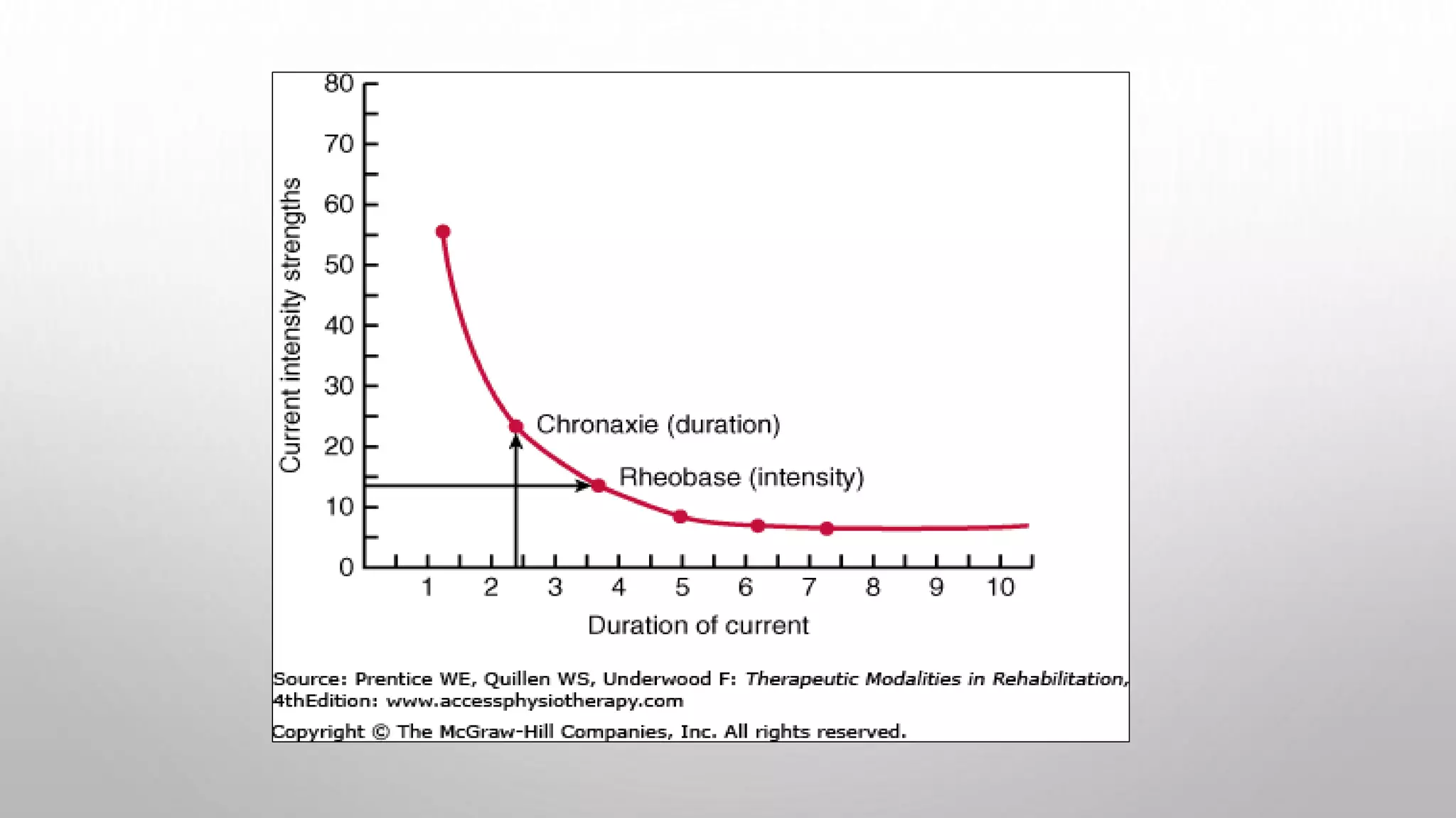
2- STRENGTH-DURATION CURVE
Utilization Time
Chronaxie
Minimal time
1- RATE OF APPLICATION
 SUDDENLY APPLIED STIMULUS OF CERTAIN INTENSITY IS
MORE EFFECTIVE THAN IF A WEAKER STIMULUS APPLIED
AND GRADUALLY INCREASED TO THE SAME INTENSITY
• SLOW CHANGES >>> ADAPTATION ( ↓ RESPONSE BY
CONTINUOUS STIMULATION)
ELECTRICAL SIGNALS ARE PRODUCED BY CHANGES IN ION
MOVEMENT ACROSS THE PLASMA MEMBRANE
• Triggering agent (stimulus)
• Change in membrane permeability
• Alter ion flow by opening and closing of gates
• Membrane potential fluctuates
•Two types of electrical signal generated ie
graded potential and action potential
ELECTRICAL SIGNALS:
GRADED POTENTIAL AND ACTION POTENTIAL
• In excitable cells changes in ion movement
in turn are brought about by changes in
membrane permeability in response to a
triggering agent or a stimuli
ION CHANNELS
• Non-gated
• Always open
• Gated
• Open or close in response to stimuli
• Chemical (ligand)
• Electrical (voltage)
• Mechanical
• When gated channels are open:
• Ions move quickly across the
membrane
• Movement is along their
electrochemical gradients
• An electrical current is created
• Voltage changes across the
membrane
Leaky channels
Gated channels
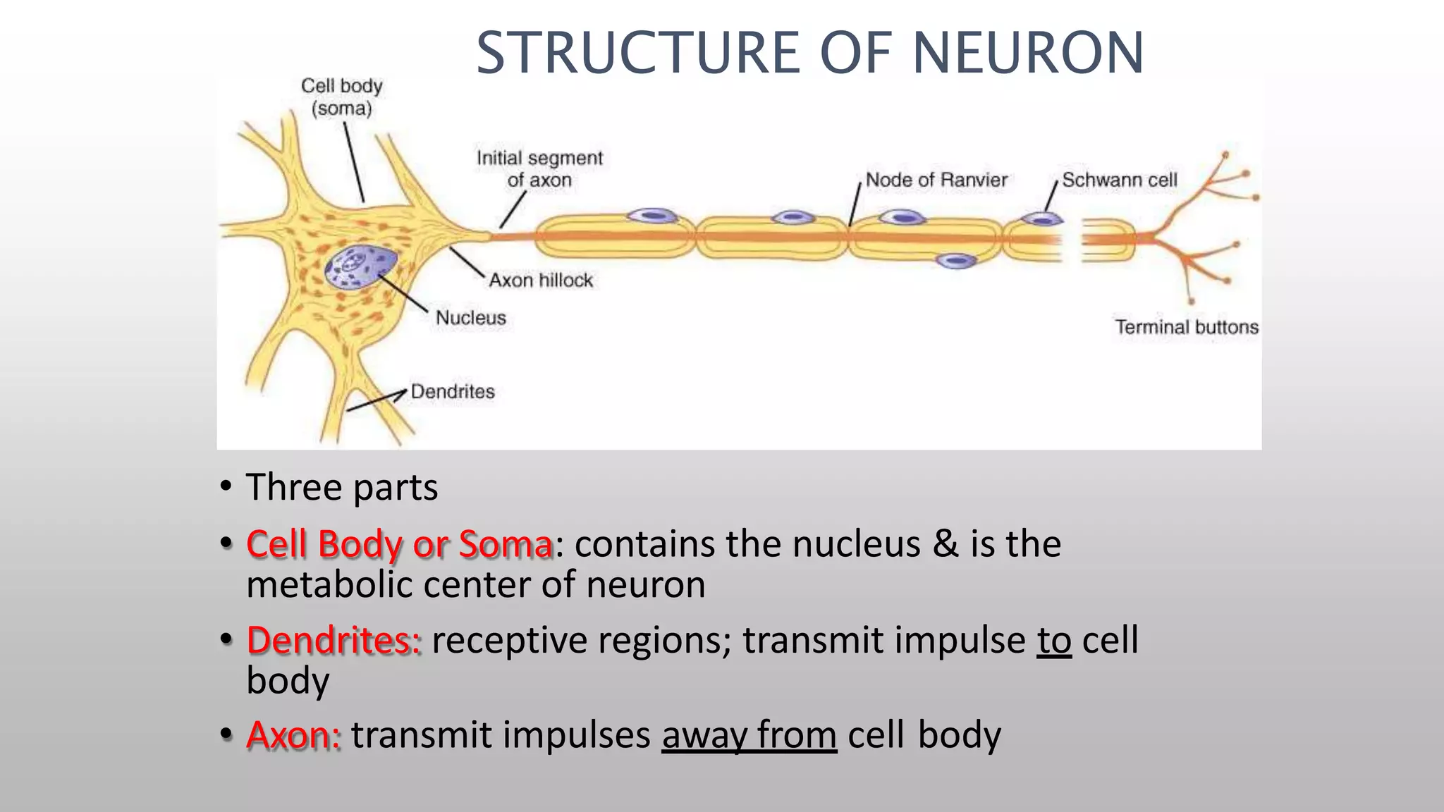
• Three parts
• Cell Body or Soma: contains the nucleus & is the
metabolic center of neuron
• Dendrites: receptive regions; transmit impulse to cell
body
• Axon: transmit impulses away from cell body
STRUCTURE OF NEURON
Terminologies Associated with Changes in Membrane Potential
• Polarization- other than 0
• Depolarization- membrane
potential less negative than RMP.
• Overshoot- when the inside of the
cell becomes +ve due to the
reversal of the membrane potential
polarity.
• Repolarization- returning to the
RMP .
• Hyperpolarization- membrane
potential more negative than RMP.
ACTION POTENTIAL
(A.P.)
• When an impulse is generated
– Inside becomes positive
– Causes depolarisation
– Nerve impulses are transmitted as AP
+30
RMP -70
Hyperpolarisation
ACTION POTENTIALS
(APS)
The AP is a brief, rapid large change
in membrane potential during which
potential reverses and the RMP
becomes +ve & then restored back to
resting state
APs do not decrease in strength with
distance so serve as long distance
signals.
Events of AP generation and
transmission are the same for skeletal
muscle cells and neurons
PARTS OF A NEURON:
AXON
• Initial Segment: Initial 50-100 um
area after axon hillock is most
excitable part; rich in Voltage
gated Na channels; site where AP
generates so called trigger zone
• Once AP generated it always
propagates towards axon
terminals
• Branches at its distal end into
many axons terminals at end of
which is an enlarged area
synaptic knob or button
•
Inside of the membrane is Negative
– During RMP
• Positive
– When an AP is generated
-70
+30
• Initially membrane is slowly depolarised
• Until the threshold level is reached
– (This may be caused by the stimulus)
+30
Threshold level
-70
• Then a sudden
change in polarisation
causes sharp
upstroke
(depolarisation) which
goes beyond the zero
level up to +35 mV -70
+30
• Then a sudden
decrease in
polarisation causes
initial sharp down
stroke (repolarisation)
-70
+30
• Spike potential
– Sharp upstroke and
downstroke
• Time duration of AP
– 1 msec
-70
+30
1 msec
PHYSIOLOGICAL BASIS OF
AP
-70
• When the threshold level is reached
– Voltage-gated Na+ channels open up
– Since Na+ conc outside is more than the inside
– Na+ influx will occur
– Positive ion coming inside increases the positivity of the
membrane potential and causes depolarisation
+30
outside
inside
Na+
Voltage-gated Na+ channel
PHYSIOLOGICAL BASIS OF
AP
– When membrane potential reaches +30, Na+
channels are inactivated
– Then Voltage-gated K+ channels open up
– K+ efflux occurs
– Positive ion leaving the inside causes more negativity inside
the membrane
– Repolarisation occurs
-70
+30
outside
inside
At +30
K+
• Depolarisation
– Change of polarity of the membrane
• from -70 to + 30 mV
• Repolarisation
– Reversal of polarity of the membrane
• from +30 to -70 mV
-70
+30
-70
+30
HYPERPOLARISATION
• When reaching the
Resting level rate
slows down
• Can go beyond the
resting level
– Hyperpolarisation
• (membrane becoming
more negative)
-70
+30
ROLE OF NA+/K+
PUMP
• Since Na+ has come in and K+ has gone out
• Membrane has become negative
• But ionic distribution has become unequal
• Na+/K+ pump restores Na+ and K+ conc slowly
– By pumping 3 Na+ ions outward and 2+ K ions
inward
VOLTAGE-GATED ION CHANNELS
Activation gate
outside
inside
Inactivation gate
• Na+ channel
– This has two gates
• Activation and inactivation gates
•
•
At rest: the activation gate is closed
At threshold level: activation gate opens
–
–
Na+ influx will occur
Na+ permeability increases to 500 fold
•
•
•
when reaching +30, inactivation gate closes
– Na influx stops
Inactivation gate will not reopen until resting membrane potential is reached
Na+ channel opens fast
outside
inside
-70
Na+
THRESHOLD
LEVEL
NA+
outside
inside
+30
Na+
m gate
outside
inside
h gate
SUMMA
RY
Plasma membrane
ECF ICF
Concentration
gradient for K+
Electrical
gradient for K+
EK+ = –94mV
Effect
of
movement
of
K+
alone
on
RMP
(K+
equilibrium
potential)
• When Na+ channel opens
• Na+ influx will occur
• Membrane depolarises
• Rising level of voltage causes many channels to open
• This will cause further Na+ influx
• Thus there a positive feedback cycle
• It does not reach Na+ equilibrium potential (+60mV)
• Because Na+ channels inactivates after opening
• Voltage-gated K+ channels open
• This will bring membrane towards K+ equilibrium
potential
INITIATION OF ACTION
POTENTIAL
•Toinitiate an AP a triggering
event causes the membrane
to depolarize from the resting
potential of -90
mvs to a
threshold of-65 to –55 mvs .
•At threshold explosive
depolarization occurs.
(positive feed back)
VOLTAGE-GATED K+ CHANNEL
• K+ channel
– This has only one gate
outside
inside
– At rest: K+ channel is closed
– At +30
• K+ channel open up slowly
• This slow activation causes K+ efflux
• This will cause membrane to become more negative
• Repolarisation occurs
outside
inside
-70
AT
+30
K+ K+
n gate
outside
inside
BASIS OF
HYPERPOLARISATION
• After reaching the resting
still slow K+ channels
may remain open:
causing further negativity
of the membrane
• This is known as
hyperpolarisation
-70
+30
outside
inside
K+
SUMMA
RY
ROLE OF THE SODIUM-
POTASSIUM PUMP IN ACTION
POTENTIAL
Repolarization restores the resting electrical conditions
of the neuron, but does not restore the resting ionic
conditions
Ionic redistribution is accomplished by the sodium-
potassium pump following repolarization
NA+/K+ PUMP
3 Na+
• Re-establishment of Na+ & K+ concentration
after action potential
– Na+/K+ Pump is responsible for this
– Energy is consumed
– Turnover rate of Na+/K+ is pump is much slower
than the Na+, K+ diffusion through channels
2 K+
ATP ADP
NA+ AND K+ CONCENTRATIONS DO NOT CHANGE DURING AN
ACTION POTENTIAL
• Although during an action potential, large changes
take place in the membrane potential as a result of
Na+entry into the cell and K+exit from the cell
• Actual Na+and K+concentrations inside and outside of
the cell generally do not change
• This is because compared to the total number of Na+
and K+ions in the intracellular and extracellular
solutions, only a small number moves across the
membrane during the action potential
Nerve physiology
Propagation of Action Potential and refractory period
Dr. Dina Hamdy Merzeban
Lecturer of physiology Fayoum university
www.facebook.com/physiology-department-fayoum-university
www.youtube.com/physiology ‫فكر‬
‫تاني‬
http://slideshare.net/merzeban
PROPAGATION OF ACTION POTENTIALS
(BASIS OF NERVE CONDUCTION)
• Two types of propagation
• Contiguous conduction
• Conduction in unmyelinated fibers
• Action potential spreads along every portion of the membrane
• Saltatory conduction
• Rapid conduction in myelinated fibers
• Impulse jumps over sections of the fiber covered with
insulating myelin
PROPAGATION OF
AP
• When one area is depolarised
• A potential difference exists between that site
and the adjacent membrane
• A current flow is initiated
• Current flow through this local circuit is
completed by extra cellular fluid
PROPAGATION OF
AP
• This local current flow will cause opening of
voltage-gated Na+ channel in the adjacent
membrane
• Na+ influx will occur
• Membrane is depolarised
PROPAGATION OF
AP
• Then the previous area become repolarised
• This process continue to work
• Resulting in propagation of AP
PROPAGATION OF
AP
PROPAGATION OF
AP
PROPAGATION OF
AP
PROPAGATION OF
AP
PROPAGATION OF
AP
PROPAGATION OF
AP
PROPAGATION OF
AP
PROPAGATION OF
AP
MYELIN
• Myelin
• Most axons are myelinated.
• Primarily composed of lipids sphingomyelin
• Formed by oligodendrocytes in CNS
• Formed by Schwann cells in PNS
• Myelin is insulating, preventing passage of ions over the
membrane as it is made up of lipids so water soluble ions
cannot permeate so current cannot leak out in the ECF
AP PROPAGATION ALONG MYELINATED NERVES
• Na+ channels are conc
around nodes
• Therefore depolarisation
mainly occurs at nodes
AP PROPAGATION ALONG MYELINATED
NERVES
•
•
Local current will flow from one node to another
Thus propagation of action potential and therefore nerve
conduction through myelinated fibres is faster than
unmyelinated fibre
– Conduction velocity of thick myelinated A alpha fibres is
about 70-100 m/s whereas in unmyelinated fibres it is about
1-2 m/s
SALTATORY
CONDUCTION
• This fast conduction through myelinated fibres
is called “saltatory conduction”
• Saltatory word means “jumping”
• This serves many purposes
– By causing depolarisation process to jump at long
intervals it increases the conduction velocity
– It conserves energy for the axon because less loss
of ions due to action potential occurring only at the
nodes
•The resistance of the membrane: Large
diameter axons provide a low resistance to current
flow within the axon and this in turn, speeds up
conduction.
•Myelin sheath: which wraps around
vertebrate axons prevents current leak out
of the cells. Acts like an insulator, for
example, plastic coating surrounding
electric wires.
•Nodes of Ranvier: are present at about 1
mm intervals along the length of axons .
High concentration of Na+ channels are
found at these nodes so AP occurs only at
nodes
2 WAYS TO INCREASE AP PROPAGATION SPEED
DISTRIBUTION OF NA+
CHANNELS
• Number of Na+ channels per
square micrometer of membrane
in mammalian neurons
50 to 75
350 – 500
< 25
2000 – 12,000
20 – 75
in the cell body
in the initial segment
on the surface of
myelin
at the nodes of
Ranvier
at the axon
terminal
PARTS OF A NEURON:
AXON
• Initial Segment: Initial 50-100 um
area after axon hillock is most
excitable part; rich in Voltage
gated Na channels; site where AP
generates so called trigger zone
• Once AP generated it always
propagates towards axon
terminals
• Branches at its distal end into
many axons terminals at end of
which is an enlarged area
synaptic knob or button
IMPORTANCE OF SALTATORY
CONDUCTION
• Increases
conduction
the
velocity
myelinated
through
nerve fiber.
• Conserves energy for
the axon
CLINICAL IMPORTANCE
• Local anaesthetics (eg. procaine) block voltage
gated sodium channels in pain nerve fibres
• Thereby pain signal transmission is blocked
CLINICAL IMPORTANCE
• Demyelinating diseases
– In certain diseases antibodies would form against myelin
and demyelination occurs
– Nerve conduction slows down drastically
• eg. Guillain-Barre Syndrome (a patient suddenly find difficult to walk,
weakness rapidly progress to upper limbs and respiratory difficulty
will also occur)
Voltage gated channels- responsible for AP
•Action potential takes place as a result of
the triggered opening and subsequent
closing of 2 specific types of channels
Voltage gated Na+ channels
Voltage gated K+ channels
VOLTAGE GATED NA+
CHANNELS
• Most important channels during AP
• It has two gates and 3 states
• Activation gates outside & inactivation
gates inside
1. At RMP activation gates are closed so
no Na+ influx at RMP thru these
channels
2. Activation gates open at threshold
3. The same increase in voltage that
open the activation gates also closes
the inactivation gates but closing of
gates is a slower process than
opening so large amount of Na+
influx has occurred
4. Inactivation gate will not reopen until
the membrane potential returns to
or near the original RMP.
Local anesthetics like lidocaine, procaine, tetracaine
block voltage gated Na channels so block the
occurrence of action potential
K+
CHANNEL
• During RMP Voltage gated K+ channels
are closed
• The same stimulus which open voltage
gated Na+ channels also open voltage
gated K+ channel
• Due to slow opening of these channels
they open just at the same time that the
Na+ channels are beginning to close
because of inactivation.
• So now decrease Na+ influx and
simultaneous increase in K+ out flux
cause membrane potential to go back to
resting state (recovery of RMP)
• These channels close when membrane
potential reaches back to RMP
Phases of action potential
•Depolarization
•Repolarization
•Hyperpolarization
PROPERTIES OF ACTION POTENTIALS
1. The All or None Principle:
Action Potentials occur in all or none fashion
depending on the strength of the stimulus
2. The Refractory Period:
Two phases:
a) Absolute refractory period
b) Relative refractory period
ALL OR NONE LAW
• Until the threshold level the potential is graded
• Once the threshold level is reached
– AP is set off and no one can stop it !
– Like a gun
ALL OR NONE LAW
• The principle that the strength by which a nerve
or muscle fiber responds to a stimulus is not
dependent on the strength of the stimulus
• If the stimulus strength is above threshold, the
nerve or muscle fiber will give a complete
response or otherwise no response at all
ALL-OR-NONE PRINCIPLE
• If any portion of the membrane is depolarized to
threshold an Action potential is initiated which will go to
its maximum height.
• A supra-threshold stimulus does not produce a large
Action potential.
• A sub-threshold stimulus does not trigger the Action
potential at all, but produce local response.
• Strength of the stimulus above the threshold is coded as the frequency
of action potentials
REFRACTORY PERIOD
(UNRESPONSIVE OR STUBBORN)
•A new action potential cannot occur in an
excitable membrane as long as the membrane is
still depolarized from the preceding action
potential.
ABSOLUTE REFRACTORY PERIOD
• Membrane cannot produce
another Action potential no
matter how great the stimulus is.
• Last for almost entire duration of
action potential.
• Cause: closure of inactivation
gates of voltage gated Na
channels in response to
depolarization. They remain
closed until the cell is repolarized
back to RMP.
REFRACTORY
PERIOD
• Absolute refractory
period
– During this period nerve
membrane cannot be
excited again
– Because of the closure
of inactivation gate
-70
+30
outside
inside