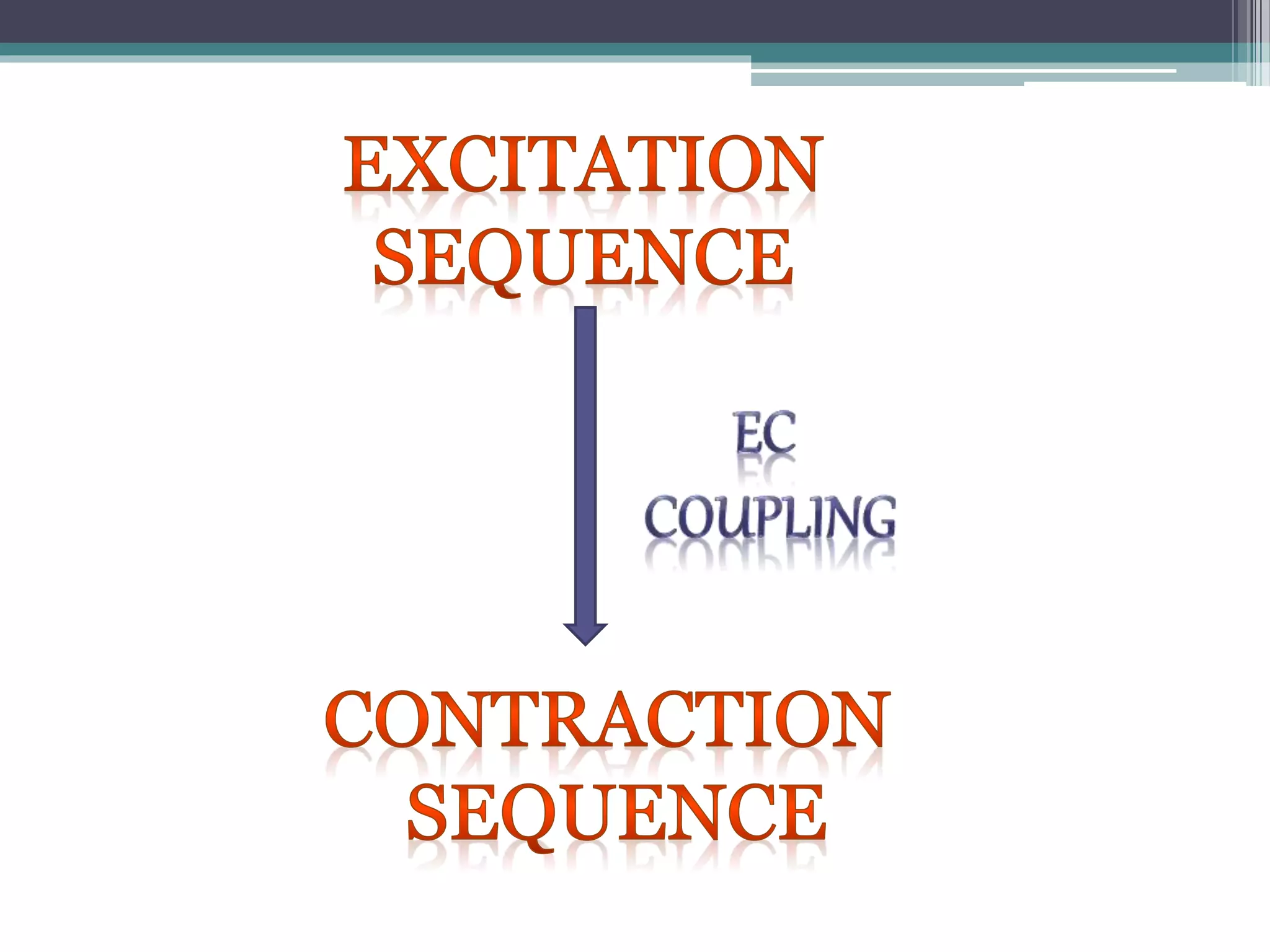
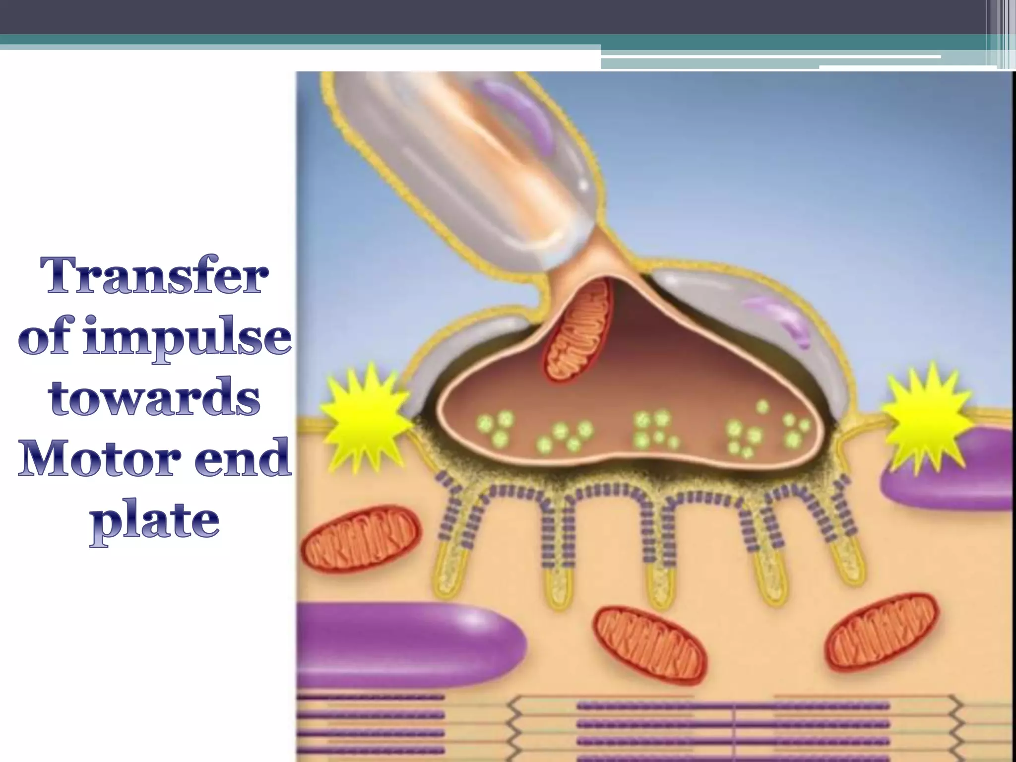
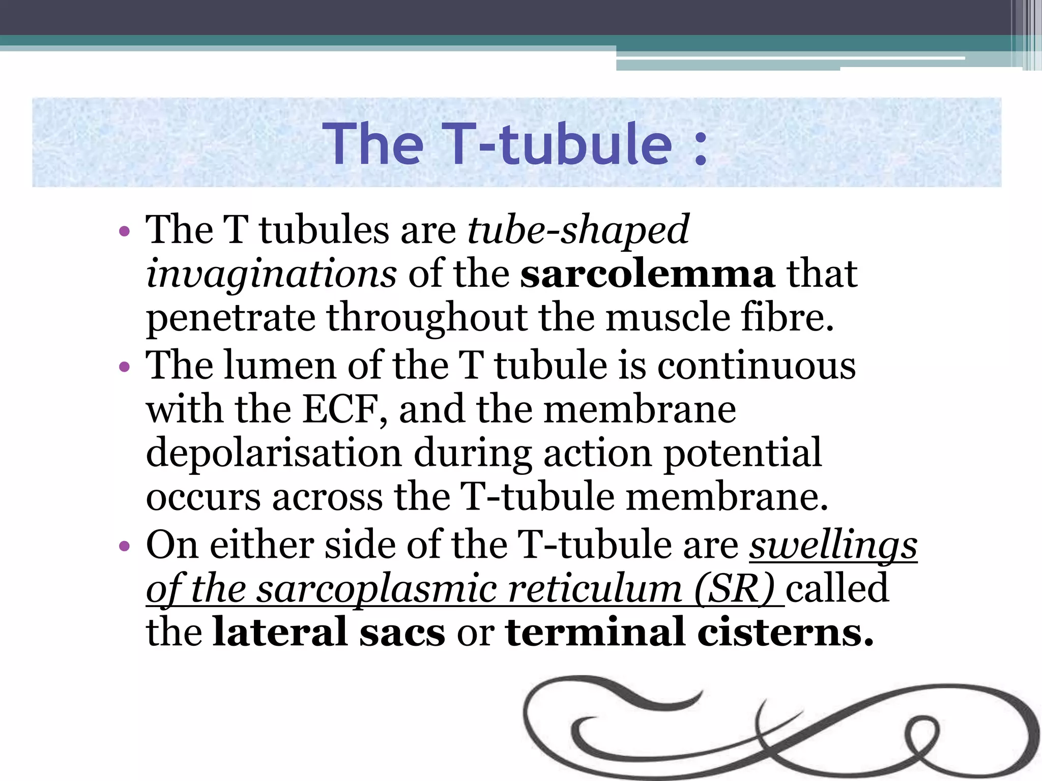
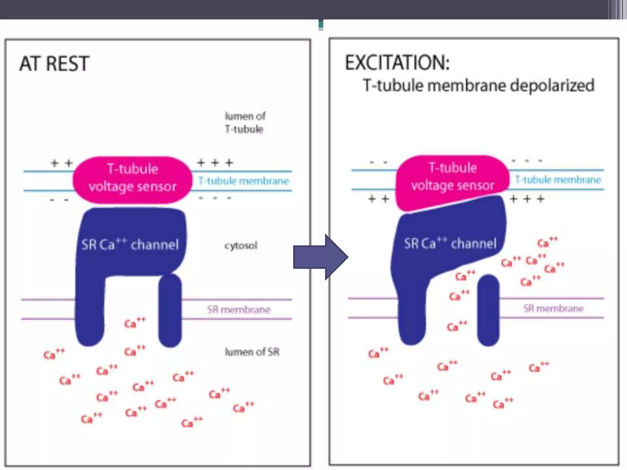
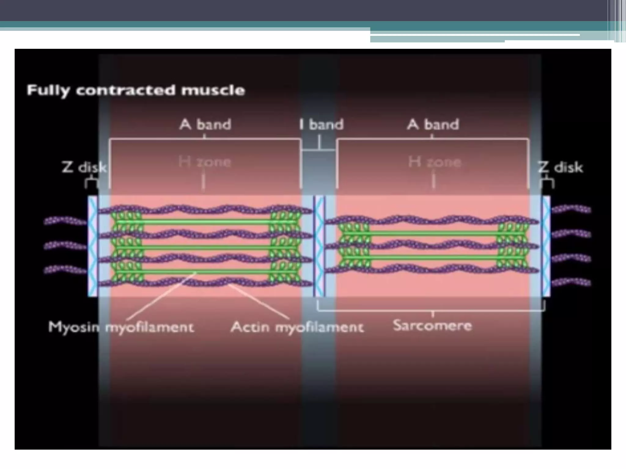
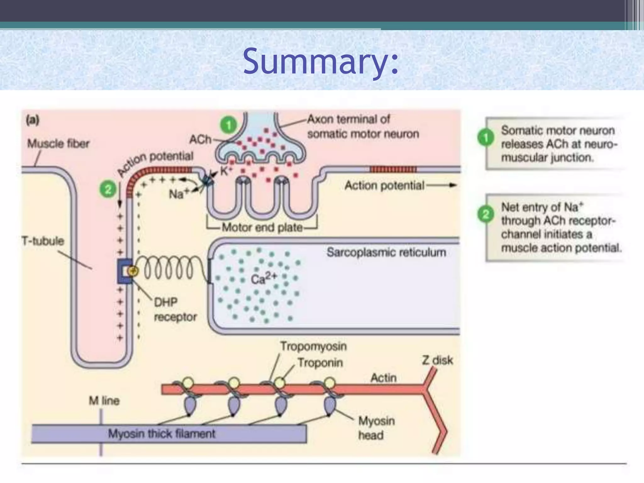
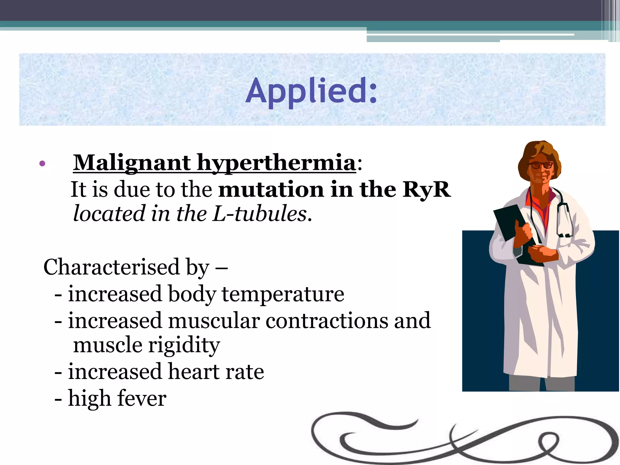
This document discusses excitation-contraction coupling (EC coupling) in skeletal muscle. It begins by defining EC coupling as the process by which an action potential triggers muscle contraction through calcium ion release. It then describes how the action potential spreads through the T-tubule system and activates the dihydropyridine and ryanodine receptors, causing calcium release from the sarcoplasmic reticulum. This triggers the contraction sequence and binding of calcium to troponin. Relaxation occurs via reuptake of calcium into the sarcoplasmic reticulum by SERCA pumps. Differences in smooth and cardiac muscle EC coupling are also summarized.
A brief overview of the physiology of the neuromuscular junction.It includes a video towards the end sourced from the internet with the copyright watermarks intact.
Receptor by Pandian M, Tutor, Dept of Physiology, DYPMCKOP, MH. This PPT for ...Pandian M
Introduction
SENSORY RECEPTORS
Structurally 3 types of receptors
Transducers
CLASSIFICATION OF RECEPTORS
A. Depending on the source of stimulus(Sherrington’s classification)
B. Depending upon type of stimulus
C. Clinical or anatomical classification of receptors
Production of receptor potential
Properties of receptors
Properties of receptor potential
Action potential By Dr. Mrs. Padmaja R Desai Physiology Dept
To study the Concept of Action Potential and describe the stages of action potential.
Ionic basis of Action Potential & its Propogation.
Properties of Action Potential.
Types action Potential
Action potential By Dr. Mrs. Padmaja R Desai Physiology Dept
To study the Concept of Action Potential and describe the stages of action potential.
Ionic basis of Action Potential & its Propogation.
Properties of Action Potential.
Types action Potential
Graduate Physiology 503 Muscle Physiology Hurley
1
MUSCLE PHYSIOLOGY
Part 2: Excitation-contraction coupling
Learning Objectives:
1. Compare the role of extracellular and intracellular calcium in excitation contraction
coupling in skeletal and cardiac muscle.
2. Describe the role of the T‐tubule and the sarcoplasmic reticulum membrane systems in
excitation‐contraction coupling.
3. Describe how calcium is removed from the cytoplasm for relaxation.
4. Describe how force can be graded in cardiac and skeletal muscles.
A. OVERVIEW
Skeletal Muscles are the effector organs of the voluntary locomotor system. The striated
appearance of both skeletal and cardiac muscle is because of the ordered arrangement of the
contractile elements within the muscle fibers. Skeletal muscle, unlike cardiac muscle, has no
intrinsic spontaneous electrical activity and therefore relied upon neural impulses to initiate
activity. Most activation of skeletal muscle takes place at specialized nerve ending called
motor end plates. There are a few exceptions: facial muscles, for example, are diffusely
innervated along the length of the muscle providing multifocal innervation of these skeletal
muscles.
B. STRUCTURE OF SKELETAL MUSCLE
The neural electrical impulse is amplified at the neuromuscular junction producing an end
plate potential that is the first step in muscle contraction.
Figure 1. Key structures of skeletal muscle fiber.
Graduate Physiology 503 Muscle Physiology Hurley
2
Like neurons, muscle fibers are excitable cells. Their plasma membranes (sarcolemma) express ion channels
and pumps necessary to support a very negative resting membrane potential as well as the voltage gated ion
channels required to generate an action potential. At any given time, the membrane potential of muscle cells
is the result of the net electrochemical gradients of ions that the membrane is permeable to. (Recall Nernst
equation; Table 1). The resting membrane potential in skeletal muscle at 37°C is similar to neurons (~-70 –
90 mV). However, there is an importance difference between the ion species that dominate the resting
membrane potential in neurons and skeletal muscle.
Question:
In contrast to skeletal muscle, what ion species dominates the resting potential of neurons? Explain.
Chloride makes a significant contribution to the resting membrane potential of skeletal
muscle. The physiological relevance of the Cl- current stems from a need to maintain
muscle activity during repeated stimulation. When muscle contracts, there is leakage of K+
from the cell. With repeated activity, there is run-down of the K+ concentration gradient
across the sarcolemma. Without the Cl- current to maintain resting membrane potential,
the muscle would not repolarize suffi ...
Properties of cm, plateau potential & pacemaker by Pandian M this PPT for I ...Pandian M
Describe the properties of cardiac muscle including its morphology, electrical, mechanical and metabolic functionsSLOs: After attending lecture & studying the assigned materials, the student will: 1.Describe the general features of cardiac muscle.2.Discuss the light and electron microscopic appearance of cardiac muscle, characteristic features of sarcotubular system.3.Enlist the electrical properties of heart muscle.4.Explain the phases of cardiac muscle action potential5.Explain the nodal action potential.6.Differentiate between cardiac muscle A.P. and nodal A.P., effect of nervous innervation and ions on AP.7.Enumerate and explain the mechanical properties of heart muscle, metabolic functions, characteristic features.
Presentation on cardiac system of human bodyitromalinda
Cross Bridge Cycle
In presence of calcium, myosin head binds to an actin filament
Changes its orientation relative to myosin filament which causes filaments to slide relative to each other - Power Stroke
During the Cross-Bridge Cycle, Contractile Proteins Convert the Energy of ATP Hydrolysis into Mechanical Energy
Each power stroke shortens sarcomere by 10nm
Cross bridge cycling is asynchronous
• 500 myosin in one thick filament, each head cycling 5 times per second
The action potential inhibits the calcium pumps, and calcium escapes from the sareophismic reticulum.
Cross Bridge Cycle
Occurs in 5 steps:-
1. Cross - Bridge formation
• cocked myosin head (perpendicular or at a 90-degree angle to the thick and thin filaments) binds to actin filament
Cocked head has the stored energy derived from the cleaved ATP
Cross Bridge Cycle
Release of Pi from the myosin
2. Dissociation of Pi from the myosin head triggers power stroke
Conformational change - myosin head bends approximately 450 about the hinge
Pulls the actin filament about 11 nm toward the tail of the myosin molecule
Generating force and motion.
Cross Bridge Cycle
ADP release –
Dissociation of ADP from myosin
Myosin head remains in the same position (45° angle with respect to the thick and thin filaments)
3. ATP binding –
ATP binding to the head of the myosin heavy chain (MHC) reduces the affinity of myosin for actin
Myosin head releases actin filament
Cross Bridge Cycle
ATP hydrolysis
Breakdown of ATP to ADP and inorganic phosphate (Pi) occurs on myosin head
Products of hydrolysis are retained on the myosin
As a result of hydrolysis, the myosin head pivots around the hinge into a "cocked" position (perpendicular or at a 900 angle to the thick and thin filaments)
Rotation causes the tip of the myosin to move about 11 nm along the actin filament so that it now lines up with a new actin monomer two monomers further along the actin filament
Cross Bridge Cycle
Cycle repeats as long as Ca, is elevated and sufficient ATP is there
Muscle cells do not regulate cross-bridge cycling by modifying [ATP]i
Instead, skeletal muscle and cardiac muscle control this cycle by preventing cross-bridge formation until the tropomyosin moves out of the way in response to an increase in [Ca']i
Steps in Relaxation
Cell may extrude Ca,- using either an Na-Ca exchanger (NCX) or a Ca' pump(PMCA)
However, would eventually deplete the cell of Ca, and is thus a minor mechanism for Ca, removal from the cytoplasm
Instead, Ca, re-uptake into the SR is the most important mechanism by which the cell returns [Ca']i to resting levels
Ca' re-uptake by the SR is mediated by a SERCA(s arcoplasmic or e ndoplasmic r eticulum C a2+8 TPase )-type Ca' pump
Steps in Relaxation
SR Ca2+-pump activity is inhibited by high [Ca2*] within the SR lumen
Inhibition of SR
Ca2+-pump activity is delayed by
Ca?+-binding proteins within the SR lumen = Buffer the Ca?* increase in the SR during Ca
1.Describe and then compare and contrast the action potentials of th.pdffathimaoptical
1.Describe and then compare and contrast the action potentials of the primary cardiac pacemaker
and primary contractile cells of the heart.
Solution
Cells within the sinoatrial (SA) node are the primary pacemaker site within the heart. These cells
are characterized as having no true resting potential, but instead generate regular, spontaneous
action potentials. Unlike non-pacemaker action potentials in the heart, and most other cells that
elicit action potentials (e.g., nerve cells, muscle cells), the depolarizing current is carried into the
cell primarily by relatively slow Ca++ currents instead of by fast Na+ currents. There are, in fact,
no fast Na+ channels and currents operating in SA nodal cells. This results in slower action
potentials in terms of how rapidly they depolarize. Therefore, these pacemaker action potentials
are sometimes referred to as \"slow response\" action potentials.
SA nodal action potentials are divided into three phases. Phase 4 is the spontaneous
depolarization (pacemaker potential) that triggers the action potential once the membrane
potential reaches threshold between -40 and -30 mV). Phase 0 is the depolarization phase of the
action potential. This is followed by phase 3 repolarization. Once the cell is completely
repolarized at about -60 mV, the cycle is spontaneously repeated.
The changes in membrane potential during the different phases are brought about by changes in
the movement of ions (principally Ca++ and K+, and to a lesser extent Na+) across the
membrane through ion channels that open and close at different times during the action potential.
When a channel is opened, there is increased electrical conductance (g) of specific ions through
that ion channel. Closure of ion channels causes ion conductance to decrease. As ions flow
through open channels, they generate electrical currents (i or I) that change the membrane
potential.
In the SA node, three ions are particularly important in generating the pacemaker action
potential. They have role of these ions in the different action potential phases.
At the end of repolarization, when the membrane potential is very negative (about -60 mV), ion
channels open that conduct slow, inward (depolarizing) Na+ currents. These currents are called
\"funny\" currents and abbreviated as \"If\". These depolarizing currents cause the membrane
potential to begin to spontaneously depolarize, thereby initiating Phase 4. As the membrane
potential reaches about -50 mV, another type of channel opens. This channel is called transient
or T-type Ca++ channel. As Ca++ enters the cell through these channels down its
electrochemical gradient, the inward directed Ca++ currents further depolarize the cell. When the
membrane depolarizes to about -40 mV, a second type of Ca++ channel opens. These are the so-
called long-lasting, or L-type Ca++ channels. Opening of these channels causes more Ca++ to
enter the cell and to further depolarize the cell until an action potential threshold is re.
A summary of skeletal muscle contraction and relaxationAyub Abdi
it consist for 4 pages and cover all the steps that occur during muscle contraction and relaxation, I does not take a time just 5 minute is enough to read. I hope it's interesting.
Muscle contraction is very important to do our daily activity every should understand how and what factors contribute for developing muscle contraction and relaxation.
in this presentation you will learns the neuromuscular junction and how excitation contraction coupling occurs.
Pulmonary Thromboembolism - etilogy, types, medical- Surgical and nursing man...VarunMahajani
Disruption of blood supply to lung alveoli due to blockage of one or more pulmonary blood vessels is called as Pulmonary thromboembolism. In this presentation we will discuss its causes, types and its management in depth.
Prix Galien International 2024 Forum ProgramLevi Shapiro
June 20, 2024, Prix Galien International and Jerusalem Ethics Forum in ROME. Detailed agenda including panels:
- ADVANCES IN CARDIOLOGY: A NEW PARADIGM IS COMING
- WOMEN’S HEALTH: FERTILITY PRESERVATION
- WHAT’S NEW IN THE TREATMENT OF INFECTIOUS,
ONCOLOGICAL AND INFLAMMATORY SKIN DISEASES?
- ARTIFICIAL INTELLIGENCE AND ETHICS
- GENE THERAPY
- BEYOND BORDERS: GLOBAL INITIATIVES FOR DEMOCRATIZING LIFE SCIENCE TECHNOLOGIES AND PROMOTING ACCESS TO HEALTHCARE
- ETHICAL CHALLENGES IN LIFE SCIENCES
- Prix Galien International Awards Ceremony
- Video recording of this lecture in English language: https://youtu.be/lK81BzxMqdo
- Video recording of this lecture in Arabic language: https://youtu.be/Ve4P0COk9OI
- Link to download the book free: https://nephrotube.blogspot.com/p/nephrotube-nephrology-books.html
- Link to NephroTube website: www.NephroTube.com
- Link to NephroTube social media accounts: https://nephrotube.blogspot.com/p/join-nephrotube-on-social-media.html
The prostate is an exocrine gland of the male mammalian reproductive system
It is a walnut-sized gland that forms part of the male reproductive system and is located in front of the rectum and just below the urinary bladder
Function is to store and secrete a clear, slightly alkaline fluid that constitutes 10-30% of the volume of the seminal fluid that along with the spermatozoa, constitutes semen
A healthy human prostate measures (4cm-vertical, by 3cm-horizontal, 2cm ant-post ).
It surrounds the urethra just below the urinary bladder. It has anterior, median, posterior and two lateral lobes
It’s work is regulated by androgens which are responsible for male sex characteristics
Generalised disease of the prostate due to hormonal derangement which leads to non malignant enlargement of the gland (increase in the number of epithelial cells and stromal tissue)to cause compression of the urethra leading to symptoms (LUTS
micro teaching on communication m.sc nursing.pdfAnurag Sharma
Microteaching is a unique model of practice teaching. It is a viable instrument for the. desired change in the teaching behavior or the behavior potential which, in specified types of real. classroom situations, tends to facilitate the achievement of specified types of objectives.
Recomendações da OMS sobre cuidados maternos e neonatais para uma experiência pós-natal positiva.
Em consonância com os ODS – Objetivos do Desenvolvimento Sustentável e a Estratégia Global para a Saúde das Mulheres, Crianças e Adolescentes, e aplicando uma abordagem baseada nos direitos humanos, os esforços de cuidados pós-natais devem expandir-se para além da cobertura e da simples sobrevivência, de modo a incluir cuidados de qualidade.
Estas diretrizes visam melhorar a qualidade dos cuidados pós-natais essenciais e de rotina prestados às mulheres e aos recém-nascidos, com o objetivo final de melhorar a saúde e o bem-estar materno e neonatal.
Uma “experiência pós-natal positiva” é um resultado importante para todas as mulheres que dão à luz e para os seus recém-nascidos, estabelecendo as bases para a melhoria da saúde e do bem-estar a curto e longo prazo. Uma experiência pós-natal positiva é definida como aquela em que as mulheres, pessoas que gestam, os recém-nascidos, os casais, os pais, os cuidadores e as famílias recebem informação consistente, garantia e apoio de profissionais de saúde motivados; e onde um sistema de saúde flexível e com recursos reconheça as necessidades das mulheres e dos bebês e respeite o seu contexto cultural.
Estas diretrizes consolidadas apresentam algumas recomendações novas e já bem fundamentadas sobre cuidados pós-natais de rotina para mulheres e neonatos que recebem cuidados no pós-parto em unidades de saúde ou na comunidade, independentemente dos recursos disponíveis.
É fornecido um conjunto abrangente de recomendações para cuidados durante o período puerperal, com ênfase nos cuidados essenciais que todas as mulheres e recém-nascidos devem receber, e com a devida atenção à qualidade dos cuidados; isto é, a entrega e a experiência do cuidado recebido. Estas diretrizes atualizam e ampliam as recomendações da OMS de 2014 sobre cuidados pós-natais da mãe e do recém-nascido e complementam as atuais diretrizes da OMS sobre a gestão de complicações pós-natais.
O estabelecimento da amamentação e o manejo das principais intercorrências é contemplada.
Recomendamos muito.
Vamos discutir essas recomendações no nosso curso de pós-graduação em Aleitamento no Instituto Ciclos.
Esta publicação só está disponível em inglês até o momento.
Prof. Marcus Renato de Carvalho
www.agostodourado.com
Lung Cancer: Artificial Intelligence, Synergetics, Complex System Analysis, S...Oleg Kshivets
RESULTS: Overall life span (LS) was 2252.1±1742.5 days and cumulative 5-year survival (5YS) reached 73.2%, 10 years – 64.8%, 20 years – 42.5%. 513 LCP lived more than 5 years (LS=3124.6±1525.6 days), 148 LCP – more than 10 years (LS=5054.4±1504.1 days).199 LCP died because of LC (LS=562.7±374.5 days). 5YS of LCP after bi/lobectomies was significantly superior in comparison with LCP after pneumonectomies (78.1% vs.63.7%, P=0.00001 by log-rank test). AT significantly improved 5YS (66.3% vs. 34.8%) (P=0.00000 by log-rank test) only for LCP with N1-2. Cox modeling displayed that 5YS of LCP significantly depended on: phase transition (PT) early-invasive LC in terms of synergetics, PT N0—N12, cell ratio factors (ratio between cancer cells- CC and blood cells subpopulations), G1-3, histology, glucose, AT, blood cell circuit, prothrombin index, heparin tolerance, recalcification time (P=0.000-0.038). Neural networks, genetic algorithm selection and bootstrap simulation revealed relationships between 5YS and PT early-invasive LC (rank=1), PT N0—N12 (rank=2), thrombocytes/CC (3), erythrocytes/CC (4), eosinophils/CC (5), healthy cells/CC (6), lymphocytes/CC (7), segmented neutrophils/CC (8), stick neutrophils/CC (9), monocytes/CC (10); leucocytes/CC (11). Correct prediction of 5YS was 100% by neural networks computing (area under ROC curve=1.0; error=0.0).
CONCLUSIONS: 5YS of LCP after radical procedures significantly depended on: 1) PT early-invasive cancer; 2) PT N0--N12; 3) cell ratio factors; 4) blood cell circuit; 5) biochemical factors; 6) hemostasis system; 7) AT; 8) LC characteristics; 9) LC cell dynamics; 10) surgery type: lobectomy/pneumonectomy; 11) anthropometric data. Optimal diagnosis and treatment strategies for LC are: 1) screening and early detection of LC; 2) availability of experienced thoracic surgeons because of complexity of radical procedures; 3) aggressive en block surgery and adequate lymph node dissection for completeness; 4) precise prediction; 5) adjuvant chemoimmunoradiotherapy for LCP with unfavorable prognosis.
Acute scrotum is a general term referring to an emergency condition affecting the contents or the wall of the scrotum.
There are a number of conditions that present acutely, predominantly with pain and/or swelling
A careful and detailed history and examination, and in some cases, investigations allow differentiation between these diagnoses. A prompt diagnosis is essential as the patient may require urgent surgical intervention
Testicular torsion refers to twisting of the spermatic cord, causing ischaemia of the testicle.
Testicular torsion results from inadequate fixation of the testis to the tunica vaginalis producing ischemia from reduced arterial inflow and venous outflow obstruction.
The prevalence of testicular torsion in adult patients hospitalized with acute scrotal pain is approximately 25 to 50 percent
Ethanol (CH3CH2OH), or beverage alcohol, is a two-carbon alcohol
that is rapidly distributed in the body and brain. Ethanol alters many
neurochemical systems and has rewarding and addictive properties. It
is the oldest recreational drug and likely contributes to more morbidity,
mortality, and public health costs than all illicit drugs combined. The
5th edition of the Diagnostic and Statistical Manual of Mental Disorders
(DSM-5) integrates alcohol abuse and alcohol dependence into a single
disorder called alcohol use disorder (AUD), with mild, moderate,
and severe subclassifications (American Psychiatric Association, 2013).
In the DSM-5, all types of substance abuse and dependence have been
combined into a single substance use disorder (SUD) on a continuum
from mild to severe. A diagnosis of AUD requires that at least two of
the 11 DSM-5 behaviors be present within a 12-month period (mild
AUD: 2–3 criteria; moderate AUD: 4–5 criteria; severe AUD: 6–11 criteria).
The four main behavioral effects of AUD are impaired control over
drinking, negative social consequences, risky use, and altered physiological
effects (tolerance, withdrawal). This chapter presents an overview
of the prevalence and harmful consequences of AUD in the U.S.,
the systemic nature of the disease, neurocircuitry and stages of AUD,
comorbidities, fetal alcohol spectrum disorders, genetic risk factors, and
pharmacotherapies for AUD.
New Directions in Targeted Therapeutic Approaches for Older Adults With Mantl...i3 Health
i3 Health is pleased to make the speaker slides from this activity available for use as a non-accredited self-study or teaching resource.
This slide deck presented by Dr. Kami Maddocks, Professor-Clinical in the Division of Hematology and
Associate Division Director for Ambulatory Operations
The Ohio State University Comprehensive Cancer Center, will provide insight into new directions in targeted therapeutic approaches for older adults with mantle cell lymphoma.
STATEMENT OF NEED
Mantle cell lymphoma (MCL) is a rare, aggressive B-cell non-Hodgkin lymphoma (NHL) accounting for 5% to 7% of all lymphomas. Its prognosis ranges from indolent disease that does not require treatment for years to very aggressive disease, which is associated with poor survival (Silkenstedt et al, 2021). Typically, MCL is diagnosed at advanced stage and in older patients who cannot tolerate intensive therapy (NCCN, 2022). Although recent advances have slightly increased remission rates, recurrence and relapse remain very common, leading to a median overall survival between 3 and 6 years (LLS, 2021). Though there are several effective options, progress is still needed towards establishing an accepted frontline approach for MCL (Castellino et al, 2022). Treatment selection and management of MCL are complicated by the heterogeneity of prognosis, advanced age and comorbidities of patients, and lack of an established standard approach for treatment, making it vital that clinicians be familiar with the latest research and advances in this area. In this activity chaired by Michael Wang, MD, Professor in the Department of Lymphoma & Myeloma at MD Anderson Cancer Center, expert faculty will discuss prognostic factors informing treatment, the promising results of recent trials in new therapeutic approaches, and the implications of treatment resistance in therapeutic selection for MCL.
Target Audience
Hematology/oncology fellows, attending faculty, and other health care professionals involved in the treatment of patients with mantle cell lymphoma (MCL).
Learning Objectives
1.) Identify clinical and biological prognostic factors that can guide treatment decision making for older adults with MCL
2.) Evaluate emerging data on targeted therapeutic approaches for treatment-naive and relapsed/refractory MCL and their applicability to older adults
3.) Assess mechanisms of resistance to targeted therapies for MCL and their implications for treatment selection
HOT NEW PRODUCT! BIG SALES FAST SHIPPING NOW FROM CHINA!! EU KU DB BK substit...GL Anaacs
Contact us if you are interested:
Email / Skype : kefaya1771@gmail.com
Threema: PXHY5PDH
New BATCH Ku !!! MUCH IN DEMAND FAST SALE EVERY BATCH HAPPY GOOD EFFECT BIG BATCH !
Contact me on Threema or skype to start big business!!
Hot-sale products:
NEW HOT EUTYLONE WHITE CRYSTAL!!
5cl-adba precursor (semi finished )
5cl-adba raw materials
ADBB precursor (semi finished )
ADBB raw materials
APVP powder
5fadb/4f-adb
Jwh018 / Jwh210
Eutylone crystal
Protonitazene (hydrochloride) CAS: 119276-01-6
Flubrotizolam CAS: 57801-95-3
Metonitazene CAS: 14680-51-4
Payment terms: Western Union,MoneyGram,Bitcoin or USDT.
Deliver Time: Usually 7-15days
Shipping method: FedEx, TNT, DHL,UPS etc.Our deliveries are 100% safe, fast, reliable and discreet.
Samples will be sent for your evaluation!If you are interested in, please contact me, let's talk details.
We specializes in exporting high quality Research chemical, medical intermediate, Pharmaceutical chemicals and so on. Products are exported to USA, Canada, France, Korea, Japan,Russia, Southeast Asia and other countries.
Tom Selleck Health: A Comprehensive Look at the Iconic Actor’s Wellness Journeygreendigital
Tom Selleck, an enduring figure in Hollywood. has captivated audiences for decades with his rugged charm, iconic moustache. and memorable roles in television and film. From his breakout role as Thomas Magnum in Magnum P.I. to his current portrayal of Frank Reagan in Blue Bloods. Selleck's career has spanned over 50 years. But beyond his professional achievements. fans have often been curious about Tom Selleck Health. especially as he has aged in the public eye.
Follow us on: Pinterest
Introduction
Many have been interested in Tom Selleck health. not only because of his enduring presence on screen but also because of the challenges. and lifestyle choices he has faced and made over the years. This article delves into the various aspects of Tom Selleck health. exploring his fitness regimen, diet, mental health. and the challenges he has encountered as he ages. We'll look at how he maintains his well-being. the health issues he has faced, and his approach to ageing .
Early Life and Career
Childhood and Athletic Beginnings
Tom Selleck was born on January 29, 1945, in Detroit, Michigan, and grew up in Sherman Oaks, California. From an early age, he was involved in sports, particularly basketball. which played a significant role in his physical development. His athletic pursuits continued into college. where he attended the University of Southern California (USC) on a basketball scholarship. This early involvement in sports laid a strong foundation for his physical health and disciplined lifestyle.
Transition to Acting
Selleck's transition from an athlete to an actor came with its physical demands. His first significant role in "Magnum P.I." required him to perform various stunts and maintain a fit appearance. This role, which he played from 1980 to 1988. necessitated a rigorous fitness routine to meet the show's demands. setting the stage for his long-term commitment to health and wellness.
Fitness Regimen
Workout Routine
Tom Selleck health and fitness regimen has evolved. adapting to his changing roles and age. During his "Magnum, P.I." days. Selleck's workouts were intense and focused on building and maintaining muscle mass. His routine included weightlifting, cardiovascular exercises. and specific training for the stunts he performed on the show.
Selleck adjusted his fitness routine as he aged to suit his body's needs. Today, his workouts focus on maintaining flexibility, strength, and cardiovascular health. He incorporates low-impact exercises such as swimming, walking, and light weightlifting. This balanced approach helps him stay fit without putting undue strain on his joints and muscles.
Importance of Flexibility and Mobility
In recent years, Selleck has emphasized the importance of flexibility and mobility in his fitness regimen. Understanding the natural decline in muscle mass and joint flexibility with age. he includes stretching and yoga in his routine. These practices help prevent injuries, improve posture, and maintain mobilit
Ozempic: Preoperative Management of Patients on GLP-1 Receptor Agonists Saeid Safari
Preoperative Management of Patients on GLP-1 Receptor Agonists like Ozempic and Semiglutide
ASA GUIDELINE
NYSORA Guideline
2 Case Reports of Gastric Ultrasound
2. Objectives:
• What is EC coupling?
• The passage of the impulse
• The T tubule
• Mechanism of Calcium Release
• The contraction sequence
• Relaxation
• Differences in smooth muscles
• Differences in cardiac muscles
• Applied aspect
3. What is EC coupling?
• An action potential triggers the contraction of a
muscle cell.
• Calcium ions can regulate whether or not
contraction can occur.
• Thus, “the link reaction which is needed to link
muscle excitation (the depolarisation of the
action potential) to Ca++ release from the
sarcoplasmic reticulum is called excitation-
contraction coupling”.
4.
5. The Passage of the Impulse
• After the conduction of the impulse through the
axons to the post-synaptic membrane via the
neuromuscular junction, there are
conformational changes leading to flux of Na+
ions inside the muscle fibre.
• Accumulation of Na+ within the cell commence
the depolarisation of the membrane, giving rise
to the end plate potential that keeps rising
towards an action potential threshold.
8. The action potential spreads throughout the fibre
and specially within the T tubules. Deep inside
the muscle fibre
9. The T-tubule :
• The T tubules are tube-shaped
invaginations of the sarcolemma that
penetrate throughout the muscle fibre.
• The lumen of the T tubule is continuous
with the ECF, and the membrane
depolarisation during action potential
occurs across the T-tubule membrane.
• On either side of the T-tubule are swellings
of the sarcoplasmic reticulum (SR) called
the lateral sacs or terminal cisterns.
11. How does depolarisation in the T-tubule
membrane open a Ca++ channel in the SR
membrane?
• Located in the T-tubule membrane, closely
associated with the foot of the SR Ca++ Channel,
is the T-tubule voltage sensor, better known
as, DHPR (dihydropyridine receptor).
• The impulse passes to the L tubules containing
another receptor – the RyR or Ryanodine
receptor.
12. • The voltage sensor, changes conformation in
response to the depolarisation of the action
potential, and causes opening of the - RyR
(Ryanodine receptor).
• This conformational change is transmitted to the
foot of the SR Ca++ Channel causing it to open,
and allowing Ca++ release.
• Thus, Ca++ ions are released and utilised during
contraction of the muscle fibres.
13.
14. The Contraction Sequence that follows:
• The Ca++ that accumulates, is the reason for
initiation and maintenance of the contraction of
sarcomere.
• The free Ca++ binds with the troponin C protein
component of the thin actin filaments
introducing the active calcium-troponin
complex.
16. There is conformational change in troponin C, which
induces alternation in the conformation of the
tropomyosin protein.
Exposure of the myosin binding sites of the actin
filament.
17. Myosin head binds to the sites on the actin filament.
The release of ADP+iP are tightly coupled to the power
stroke, thus resulting in shortening of sarcomere.
18.
19. Relaxation
• SERCA pump (Sarco- Endoplasmic Reticulum
Calcium ATPase pump) is the enzyme
responsible for relaxation.
• This enzyme pumps calcium back into the
sarcoplasmic reticulum from the cytosol.
22. Differences in Smooth Muscles:
I. Calcium channel differences:
• Different channels releasing calcium from ECF
(RyR still exists though), depends on the type
of smooth muscles.
• Channels could be voltage-gated, ligand-gated,
second messenger-gated or mechanically-
gated.
23. II. Calcium entrance options:
• From gated channels, extracellularly
• From IP3
IP3 (inositol triphosphate) is a second
messenger that opens RyR channel which
releases calcium from SR.
III. Calcium binds to calmodulin.
IV. Activation of MLCK (Myosin-light-chain-
kinase), causes phosphorylation of the myosin
head.
24. Differences in Cardiac Muscles:
• Cells are coupled as electrical syncytium due to
high expression of gap junctions between the
cells.
• Gap junctions consist of 2 connexons, each made
of 6 connexins.
• T tubules are very well developed with moe
diameter
• DIAD is present, i.e. With 1-tubule, there is only
1 cistern.
25. Applied:
• Malignant hyperthermia:
It is due to the mutation in the RyR
located in the L-tubules.
Characterised by –
- increased body temperature
- increased muscular contractions and
muscle rigidity
- increased heart rate
- high fever