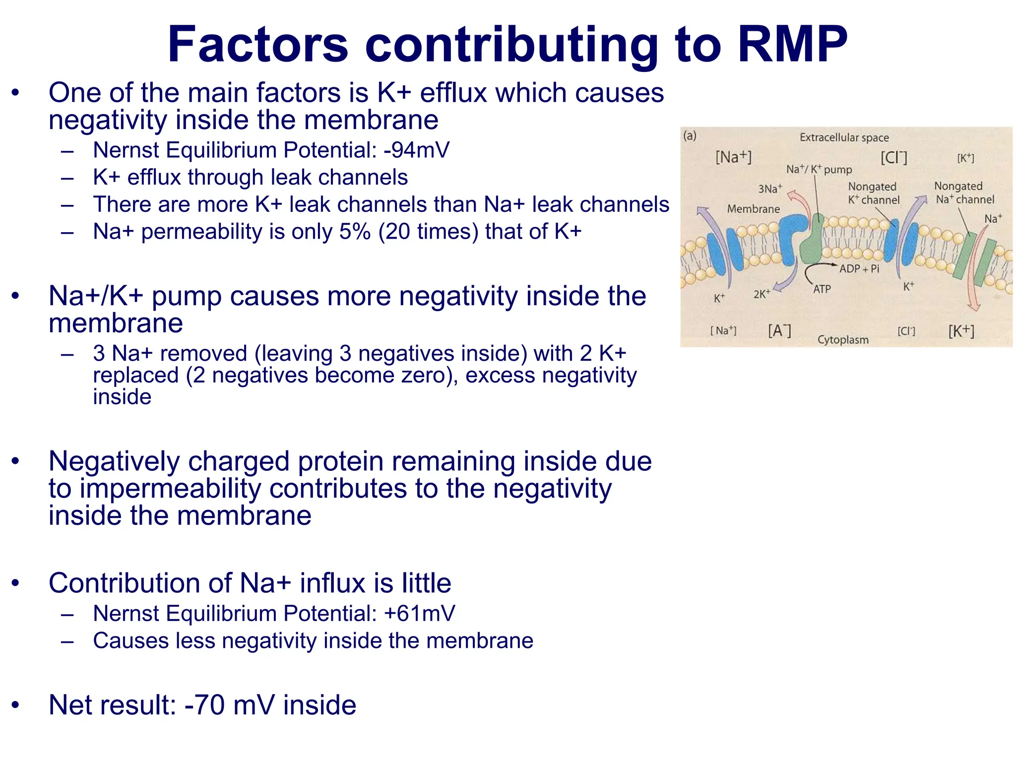
Nerve and
Membrane Potential
Prof. Vajira Weerasinghe
Senior Professor of Physiology
Department of Physiology, Faculty of Medicine,
University of Peradeniya and Consultant Neurophysiologist,
Teaching Hospital Peradeniya
www.slideshare.net/vajira54
ILO
• Describe the Classification of nerve fibers: based on structure,
diameter and functions.
• Describe the Properties of nerve fibers and basic structure of a
nerve cell
• Describe the Ionic basis for resting membrane potential
• Describe Ionic basis for action potential.
• Explain the Alteration in excitability in electrolyte imbalance
Learning resources
• Textbooks
– Ganong 26th edition
– Guyton
– Netter's Atlas of Neuroscience
• Web resources
• .edu
• .ac.uk
• .ac.lk
• www.slideshare.net
Excitable Cells
• Basic cell of the nervous system is nerve cell
• Nerve cell or neuron is an excitable cell
• Excitable cells are capable of generation and
transmission of electrochemical impulses along the
membrane
• Muscle cells are also excitable cells
Nerve cell or neuron Muscle cell
Excitable cells
Excitable Non-excitable
Red cell
GIT
neuron
muscle
•RBC
•Intestinal cells
•Fibroblasts
•Adipocytes
•Nerve
•Muscle
•Skeletal
•Cardiac
•Smooth
Basic structure of a neuron
• Cell body contains the nucleus
• Dendrites extend outward from the cell body and arborize
extensively to aid their role in receiving incoming signals,
processing the information, and then transmitting the
information to the cell body
• Axon originates from a thickened area of the cell body called
axon hillock
• First portion of the axon is called the initial segment
• Axon divides into presynaptic terminals ending in a number of
synaptic knobs that are also called terminal buttons or boutons
Neuron - functional aspect
• From a functional point of view, neurons generally
have four important zones:
• (1) a dendritic zone where multiple local potential
changes generated by synaptic connections are
integrated
• (2) a site where propagated action potentials are
generated (initial node of Ranvier)
• (3) an axonal process that transmits propagated
impulses to the nerve endings;
• (4) the nerve endings, where action potentials cause
the release of synaptic transmitters.
Neuron – myelin sheath
• Axons acquire a myelin sheath, a protein–lipid complex that is
wrapped around the axon
• Myelin forms from Schwann cells in the peripheral nervous
system or oligodendrocyte in the CNS
• Myelin sheath envelops the axon except at the nodes of
Ranvier, periodic 1-µm gaps where the axon is unmyelinated
• Myelinated axons are faster in conducting electrical impulses
– eg. Conduction velocity of unmyelinated axons 1-2 m/s whereas it is 50-
100 m/s in myelinated axons
Classification of nerve fibre types
Membrane potential
• A potential difference exists across all cell
membranes
• This is called
– Resting Membrane Potential (RMP)
Membrane potential
– Inside is negative with respect to the outside
– This is measured using microelectrodes and
oscilloscope
– This is about -70 to -90 mV
Excitable tissues
• Excitable tissues have more
negative RMP
( - 70 mV to - 90 mV)
excitable Non-excitable
Red cell
GIT
neuron
muscle
• Non-excitable tissues
have less negative RMP
-53 mV epithelial cells
-8.4 mV RBC
-20 to -30 mV fibroblasts
-58 mV adipocytes
Resting Membrane Potential
• This depends on following factors
– Ionic distribution across the membrane
– Membrane permeability
– Other factors
• Na+/K+ pump
Ionic distribution
• Major ions
– Extracellular ions
• Sodium, Chloride
– Intracellular ions
• Potassium, Proteinate
K+
Pr-
Na+ Cl-
Ionic distribution
Ion Intracellular Extracellular
Na+
10 142
K+
140 4
Cl-
4 103
Ca2+ 0 2.4
HCO3
- 10 28
Gibbs Donnan Equilibrium
• When two solutions
containing ions are
separated by membrane
that is permeable to some
of the ions and not to others
an electrochemical
equilibrium is established
• Electrical and chemical
energies on either side of
the membrane are equal
and opposite to each other
Flow of Potassium
• Potassium concentration intracellular is more
• Membrane is freely permeable to K+
• There is an efflux of K+
K+
K+
K+
K+
K+ K+
K+
K+
K+
K+
Flow of Potassium
• Entry of positive ions in to the extracellular fluid
creates positivity outside and negativity inside
K+
K+
K+
K+
K+ K+
K+
K+
K+
K+
Flow of Potassium
• Outside positivity resists efflux of K+
• (since K+ is a positive ion)
• At a certain voltage an equilibrium is reached
and K+ efflux stops
K+
K+
K+
K+
K+ K+
K+
K+
K+
K+
Nernst potential (Equilibrium potential)
• The potential level across the membrane that
will exactly prevent net diffusion of an ion
• Nernst equation determines this potential
Nernst potential (Equilibrium potential)
• The potential level across the membrane that
will exactly prevent net diffusion of an ion
Ion Intracellular Extracellular Nernst
potential
Na+
10 142 +60
K+
140 4 -90
Cl-
4 103 -89
Ca2+ 0 2.4 +129
HCO3
- 10 28 -23
(mmol/l)
Goldman Equation
• When the membrane is permeable to several ions the
equilibrium potential that develops depends on
– Polarity of each ion
– Membrane permeability
– Ionic conc
• This is calculated using Goldman Equation (or GHK
Equation)
• In the resting state
– K+ permeability is 20 times more than that of Na+
Ionic channels
• Leaky channels (leak channels)
– Allow free flow of ions
– K+ channels (large number)
– Na+ channels (fewer in number)
– Therefore membrane is more permeable to K+
Na/K pump
• Active transport system for Na+-K+
exchange using energy
• It is an electrogenic pump since 3 Na+
efflux coupled with 2 K+ influx
• Net effect of causing negative charge
inside the membrane
3 Na+
2 K+
ATP ADP
Factors contributing to RMP
• One of the main factors is K+ efflux which causes
negativity inside the membrane
– Nernst Equilibrium Potential: -94mV
– K+ efflux through leak channels
– There are more K+ leak channels than Na+ leak channels
– Na+ permeability is only 5% (20 times) that of K+
• Na+/K+ pump causes more negativity inside the
membrane
– 3 Na+ removed (leaving 3 negatives inside) with 2 K+
replaced (2 negatives become zero), excess negativity
inside
• Negatively charged protein remaining inside due
to impermeability contributes to the negativity
inside the membrane
• Contribution of Na+ influx is little
– Nernst Equilibrium Potential: +61mV
– Causes less negativity inside the membrane
• Net result: -70 mV inside
Electrochemical gradient
• At this electrochemical equilibrium, there is an exact
balance between two opposing forces:
• Chemical driving force = ratio of concentrations on 2
sides of membrane (concentration gradient)
• The concentration gradient that causes K+ to move from inside to
outside taking along positive charge and
• Electrical driving force = potential difference across
membrane
• opposing electrical gradient that increasingly tends to stop K+ from
moving across the membrane
• Equilibrium: when chemical driving force is balanced
by electrical driving force
Action potential
Action Potential (A.P.)
• When an impulse is generated
– Inside becomes positive
– Causes depolarisation
– Nerve impulses are transmitted as AP
-70
+30
RMP
Hyperpolarisation
Inside of the membrane is
• Negative
– During RMP
• Positive
– When an AP is generated
-70
+30
• Initially membrane is slowly depolarised
• Until the threshold level is reached
– (This may be caused by the stimulus)
-70
+30
Threshold level
• Then a sudden
change in polarisation
causes sharp
upstroke
(depolarisation) which
goes beyond the zero
level up to +35 mV
-70
+30
• Then a sudden
decrease in
polarisation causes
initial sharp down
stroke (repolarisation)
-70
+30
• When reaching the
Resting level rate
slows down
• Can go beyond the
resting level
– hyperpolarisation
-70
+30
• Spike potential
– Sharp upstroke and
downstroke
• Time duration of AP
– 1 msec
-70
+30
1 msec
All or none law
• Until the threshold level the potential is graded
• Once the threshold level is reached
– AP is set off and no one can stop it !
– Like a gun
All or none law
• The principle that the strength by which a nerve
or muscle fiber responds to a stimulus is not
dependent on the strength of the stimulus
• If the stimulus is any strength above threshold,
the nerve or muscle fiber will give a complete
response or otherwise no response at all
Physiological basis of AP
• When the threshold level is reached
– Voltage-gated Na+ channels open up
– Since Na conc outside is more than the inside
– Na influx will occur
– Positive ion coming inside increases the positivity of the
membrane potential and causes depolarisation
– When it reaches +30, Na+ channels closes
– Then Voltage-gated K+ channels open up
– K+ efflux occurs
– Positive ion leaving the inside causes more negativity inside
the membrane
– Repolarisation occurs
Physiological basis of AP
• Since Na+ has come in and K+ has gone out
• Membrane has become negative
• But ionic distribution has become unequal
• Na+/K+ pump restores Na+ and K+ conc slowly
– By pumping 3 Na+ ions outward and 2+ K ions
inward
VOLTAGE-GATED ION CHANNELS
• Na+ channel
– This has two gates
• Activation and inactivation gates
outside
inside
Activation gate
Inactivation gate
• At rest: the activation gate is closed
• At threshold level: activation gate opens
– Na+ influx will occur
– Na+ permeability increases to 500 fold
• when reaching +30, inactivation gate closes
– Na influx stops
• Inactivation gate will not reopen until resting membrane potential is reached
• Na+ channel opens fast
outside
inside
outside
inside
-70 Threshold level +30
Na+ Na+
outside
inside
Na+
m gate
h gate
VOLTAGE-GATED K+ Channel
• K+ channel
– This has only one gate
outside
inside
– At rest: K+ channel is closed
– At +30
• K+ channel open up slowly
• This slow activation causes K efflux
– After reaching the resting still slow K+ channels
may remain open: causing further hyperpolarisation
outside
inside
outside
inside
-70 At +30
K+ K+
n gate
Summary
Refractory Period
• Absolute refractory
period
– During this period nerve
membrane cannot be
excited again
– Because of the closure
of inactivation gate
-70
+30
outside
inside
Refractory Period
• Relative refractory
period
– During this period nerve
membrane can be
excited by supra
threshold stimuli
– At the end of
repolarisation phase
inactivation gate opens
and activation gate
closes
– This can be opened by
greater stimuli strength
-70
+30
outside
inside
Propagation of AP
• When one area is depolarised
• A potential difference exists between that site
and the adjacent membrane
• A local current flow is initiated
• Local circuit is completed by extra cellular fluid
Propagation of AP
• This local current flow will cause opening of
voltage-gated Na channel in the adjacent
membrane
• Na influx will occur
• Membrane is deloparised
Propagation of AP
• Then the previous area become repolarised
• This process continue to work
• Resulting in propagation of AP
Propagation of AP
Propagation of AP
Propagation of AP
Propagation of AP
Propagation of AP
Propagation of AP
Propagation of AP
Propagation of AP
AP propagation along myelinated
nerves
• Na channels are conc
around nodes
• Therefore depolarisation
mainly occurs at nodes
AP propagation along myelinated
nerves
• Local current will flow one node to another
• Thus propagation of A.P. is faster. Conduction
through myelinated fibres also faster.
• Known as Saltatory Conduction
• Re-establishment of Na & K conc after A.P.
– Na-K Pump is responsible for this.
– Energy is consumed
3 Na+
2 K+
ATP ADP
Muscle action potentials
• Skeletal muscle
• Smooth muscle
• Cardiac muscle
Skeletal muscle
• Similar to nerve action potential
Cardiac muscle action potential
Phases
• 0: depolarisation
• 1: short repolarisation
• 2: plateau phase
• 3: repolarisation
• 4: resting
Duration is about 250 msec
Cardiac muscle action potential
Phases
• 0: depolarisation
(Na+ influx through fast Na+
channels)
• 1: short repolarisation
(K+ efflux through K+
channels, Cl- influx as well)
• 2: plateau phase
(Ca++ influx through slow
Ca++ channels)
• 3: repolarisation
(K+ efflux through K+
channels)
• 4: resting
Smooth muscle
• Resting membrane potential may be about -55mV
• Action potential is similar to nerve AP
• But AP is not necessary for its contraction
• Smooth muscle contraction can occur by hormones
Applied physiology
• Decreasing the external Na+ concentration reduces the size of the
action potential but has little effect on the resting membrane potential
• If the extracellular level of K+ is increased (hyperkalemia),the resting
potential moves closer to the threshold for eliciting an action
potential, thus the neuron becomes more excitable
• This may inactivate Na+ channels, which increases refractory period
and may lead to cardiac arrhythmias
• If the extracellular level of K+ is decreased (hypokalemia),the
membrane potential is reduced and the neuron is hyperpolarized
• This may lead to delayed ventricular repolarisation which can
contribute to cardiac arrhythmias
• Major electrolyte abnormalities can lead to skeletal muscle spasms
and seizures
Hyperkalemia
• Causes
– Advanced renal failure
– Adrenal insufficiency
– Distal renal tubular acidosis
– Type 1 diabetes
– Dehydration
– Angiotensin II receptor blocker use
– Angiotensin-converting enzyme (ACE) inhibitor use
– Non-steroidal anti-inflammatory drugs(NSAIDs) use
– Potassium sparing diuretics
• Symptoms include
– Muscle pain
– Muscle weakness
– Numbness
– Cardiac arrhythmias
– Nausea
– Extremely high levels of plasma K+ can lead to cardiac arrest and death
Hypokalaemia
• Causes
– Diuretics
– Diabetic ketoacidosis
– Renal tubular acidosis
– Genetic conditions
• Symptoms
– Muscle weakness
– Fatigue
– Constipation
– Muscle cramping
– Palpitations
– Psychological symptoms (depression or psychosis)
– Severe hypokalemia may lead to problems with cardiac rhythmicity and can
manifest as bradycardia, tachycardia, premature beats, atrial or ventricular
fibrillation

Resting membrane and action potentials RMP AP

  • 1.
    Nerve and Membrane Potential Prof.Vajira Weerasinghe Senior Professor of Physiology Department of Physiology, Faculty of Medicine, University of Peradeniya and Consultant Neurophysiologist, Teaching Hospital Peradeniya www.slideshare.net/vajira54
  • 2.
    ILO • Describe theClassification of nerve fibers: based on structure, diameter and functions. • Describe the Properties of nerve fibers and basic structure of a nerve cell • Describe the Ionic basis for resting membrane potential • Describe Ionic basis for action potential. • Explain the Alteration in excitability in electrolyte imbalance
  • 3.
    Learning resources • Textbooks –Ganong 26th edition – Guyton – Netter's Atlas of Neuroscience • Web resources • .edu • .ac.uk • .ac.lk • www.slideshare.net
  • 4.
    Excitable Cells • Basiccell of the nervous system is nerve cell • Nerve cell or neuron is an excitable cell • Excitable cells are capable of generation and transmission of electrochemical impulses along the membrane • Muscle cells are also excitable cells Nerve cell or neuron Muscle cell
  • 5.
    Excitable cells Excitable Non-excitable Redcell GIT neuron muscle •RBC •Intestinal cells •Fibroblasts •Adipocytes •Nerve •Muscle •Skeletal •Cardiac •Smooth
  • 6.
    Basic structure ofa neuron • Cell body contains the nucleus • Dendrites extend outward from the cell body and arborize extensively to aid their role in receiving incoming signals, processing the information, and then transmitting the information to the cell body • Axon originates from a thickened area of the cell body called axon hillock • First portion of the axon is called the initial segment • Axon divides into presynaptic terminals ending in a number of synaptic knobs that are also called terminal buttons or boutons
  • 7.
    Neuron - functionalaspect • From a functional point of view, neurons generally have four important zones: • (1) a dendritic zone where multiple local potential changes generated by synaptic connections are integrated • (2) a site where propagated action potentials are generated (initial node of Ranvier) • (3) an axonal process that transmits propagated impulses to the nerve endings; • (4) the nerve endings, where action potentials cause the release of synaptic transmitters.
  • 8.
    Neuron – myelinsheath • Axons acquire a myelin sheath, a protein–lipid complex that is wrapped around the axon • Myelin forms from Schwann cells in the peripheral nervous system or oligodendrocyte in the CNS • Myelin sheath envelops the axon except at the nodes of Ranvier, periodic 1-µm gaps where the axon is unmyelinated • Myelinated axons are faster in conducting electrical impulses – eg. Conduction velocity of unmyelinated axons 1-2 m/s whereas it is 50- 100 m/s in myelinated axons
  • 9.
  • 10.
  • 11.
    Membrane potential • Apotential difference exists across all cell membranes • This is called – Resting Membrane Potential (RMP)
  • 12.
    Membrane potential – Insideis negative with respect to the outside – This is measured using microelectrodes and oscilloscope – This is about -70 to -90 mV
  • 13.
    Excitable tissues • Excitabletissues have more negative RMP ( - 70 mV to - 90 mV) excitable Non-excitable Red cell GIT neuron muscle • Non-excitable tissues have less negative RMP -53 mV epithelial cells -8.4 mV RBC -20 to -30 mV fibroblasts -58 mV adipocytes
  • 14.
    Resting Membrane Potential •This depends on following factors – Ionic distribution across the membrane – Membrane permeability – Other factors • Na+/K+ pump
  • 15.
    Ionic distribution • Majorions – Extracellular ions • Sodium, Chloride – Intracellular ions • Potassium, Proteinate K+ Pr- Na+ Cl-
  • 16.
    Ionic distribution Ion IntracellularExtracellular Na+ 10 142 K+ 140 4 Cl- 4 103 Ca2+ 0 2.4 HCO3 - 10 28
  • 17.
    Gibbs Donnan Equilibrium •When two solutions containing ions are separated by membrane that is permeable to some of the ions and not to others an electrochemical equilibrium is established • Electrical and chemical energies on either side of the membrane are equal and opposite to each other
  • 18.
    Flow of Potassium •Potassium concentration intracellular is more • Membrane is freely permeable to K+ • There is an efflux of K+ K+ K+ K+ K+ K+ K+ K+ K+ K+ K+
  • 19.
    Flow of Potassium •Entry of positive ions in to the extracellular fluid creates positivity outside and negativity inside K+ K+ K+ K+ K+ K+ K+ K+ K+ K+
  • 20.
    Flow of Potassium •Outside positivity resists efflux of K+ • (since K+ is a positive ion) • At a certain voltage an equilibrium is reached and K+ efflux stops K+ K+ K+ K+ K+ K+ K+ K+ K+ K+
  • 21.
    Nernst potential (Equilibriumpotential) • The potential level across the membrane that will exactly prevent net diffusion of an ion • Nernst equation determines this potential
  • 22.
    Nernst potential (Equilibriumpotential) • The potential level across the membrane that will exactly prevent net diffusion of an ion Ion Intracellular Extracellular Nernst potential Na+ 10 142 +60 K+ 140 4 -90 Cl- 4 103 -89 Ca2+ 0 2.4 +129 HCO3 - 10 28 -23 (mmol/l)
  • 23.
    Goldman Equation • Whenthe membrane is permeable to several ions the equilibrium potential that develops depends on – Polarity of each ion – Membrane permeability – Ionic conc • This is calculated using Goldman Equation (or GHK Equation) • In the resting state – K+ permeability is 20 times more than that of Na+
  • 24.
    Ionic channels • Leakychannels (leak channels) – Allow free flow of ions – K+ channels (large number) – Na+ channels (fewer in number) – Therefore membrane is more permeable to K+
  • 25.
    Na/K pump • Activetransport system for Na+-K+ exchange using energy • It is an electrogenic pump since 3 Na+ efflux coupled with 2 K+ influx • Net effect of causing negative charge inside the membrane 3 Na+ 2 K+ ATP ADP
  • 26.
    Factors contributing toRMP • One of the main factors is K+ efflux which causes negativity inside the membrane – Nernst Equilibrium Potential: -94mV – K+ efflux through leak channels – There are more K+ leak channels than Na+ leak channels – Na+ permeability is only 5% (20 times) that of K+ • Na+/K+ pump causes more negativity inside the membrane – 3 Na+ removed (leaving 3 negatives inside) with 2 K+ replaced (2 negatives become zero), excess negativity inside • Negatively charged protein remaining inside due to impermeability contributes to the negativity inside the membrane • Contribution of Na+ influx is little – Nernst Equilibrium Potential: +61mV – Causes less negativity inside the membrane • Net result: -70 mV inside
  • 27.
    Electrochemical gradient • Atthis electrochemical equilibrium, there is an exact balance between two opposing forces: • Chemical driving force = ratio of concentrations on 2 sides of membrane (concentration gradient) • The concentration gradient that causes K+ to move from inside to outside taking along positive charge and • Electrical driving force = potential difference across membrane • opposing electrical gradient that increasingly tends to stop K+ from moving across the membrane • Equilibrium: when chemical driving force is balanced by electrical driving force
  • 28.
  • 29.
    Action Potential (A.P.) •When an impulse is generated – Inside becomes positive – Causes depolarisation – Nerve impulses are transmitted as AP
  • 30.
  • 31.
    Inside of themembrane is • Negative – During RMP • Positive – When an AP is generated -70 +30
  • 32.
    • Initially membraneis slowly depolarised • Until the threshold level is reached – (This may be caused by the stimulus) -70 +30 Threshold level
  • 33.
    • Then asudden change in polarisation causes sharp upstroke (depolarisation) which goes beyond the zero level up to +35 mV -70 +30
  • 34.
    • Then asudden decrease in polarisation causes initial sharp down stroke (repolarisation) -70 +30
  • 35.
    • When reachingthe Resting level rate slows down • Can go beyond the resting level – hyperpolarisation -70 +30
  • 36.
    • Spike potential –Sharp upstroke and downstroke • Time duration of AP – 1 msec -70 +30 1 msec
  • 37.
    All or nonelaw • Until the threshold level the potential is graded • Once the threshold level is reached – AP is set off and no one can stop it ! – Like a gun
  • 38.
    All or nonelaw • The principle that the strength by which a nerve or muscle fiber responds to a stimulus is not dependent on the strength of the stimulus • If the stimulus is any strength above threshold, the nerve or muscle fiber will give a complete response or otherwise no response at all
  • 39.
    Physiological basis ofAP • When the threshold level is reached – Voltage-gated Na+ channels open up – Since Na conc outside is more than the inside – Na influx will occur – Positive ion coming inside increases the positivity of the membrane potential and causes depolarisation – When it reaches +30, Na+ channels closes – Then Voltage-gated K+ channels open up – K+ efflux occurs – Positive ion leaving the inside causes more negativity inside the membrane – Repolarisation occurs
  • 40.
    Physiological basis ofAP • Since Na+ has come in and K+ has gone out • Membrane has become negative • But ionic distribution has become unequal • Na+/K+ pump restores Na+ and K+ conc slowly – By pumping 3 Na+ ions outward and 2+ K ions inward
  • 41.
    VOLTAGE-GATED ION CHANNELS •Na+ channel – This has two gates • Activation and inactivation gates outside inside Activation gate Inactivation gate
  • 42.
    • At rest:the activation gate is closed • At threshold level: activation gate opens – Na+ influx will occur – Na+ permeability increases to 500 fold • when reaching +30, inactivation gate closes – Na influx stops • Inactivation gate will not reopen until resting membrane potential is reached • Na+ channel opens fast outside inside outside inside -70 Threshold level +30 Na+ Na+ outside inside Na+ m gate h gate
  • 43.
    VOLTAGE-GATED K+ Channel •K+ channel – This has only one gate outside inside
  • 44.
    – At rest:K+ channel is closed – At +30 • K+ channel open up slowly • This slow activation causes K efflux – After reaching the resting still slow K+ channels may remain open: causing further hyperpolarisation outside inside outside inside -70 At +30 K+ K+ n gate
  • 45.
  • 46.
    Refractory Period • Absoluterefractory period – During this period nerve membrane cannot be excited again – Because of the closure of inactivation gate -70 +30 outside inside
  • 47.
    Refractory Period • Relativerefractory period – During this period nerve membrane can be excited by supra threshold stimuli – At the end of repolarisation phase inactivation gate opens and activation gate closes – This can be opened by greater stimuli strength -70 +30 outside inside
  • 48.
    Propagation of AP •When one area is depolarised • A potential difference exists between that site and the adjacent membrane • A local current flow is initiated • Local circuit is completed by extra cellular fluid
  • 49.
    Propagation of AP •This local current flow will cause opening of voltage-gated Na channel in the adjacent membrane • Na influx will occur • Membrane is deloparised
  • 50.
    Propagation of AP •Then the previous area become repolarised • This process continue to work • Resulting in propagation of AP
  • 51.
  • 52.
  • 53.
  • 54.
  • 55.
  • 56.
  • 57.
  • 58.
  • 59.
    AP propagation alongmyelinated nerves • Na channels are conc around nodes • Therefore depolarisation mainly occurs at nodes
  • 60.
    AP propagation alongmyelinated nerves • Local current will flow one node to another • Thus propagation of A.P. is faster. Conduction through myelinated fibres also faster. • Known as Saltatory Conduction
  • 63.
    • Re-establishment ofNa & K conc after A.P. – Na-K Pump is responsible for this. – Energy is consumed 3 Na+ 2 K+ ATP ADP
  • 64.
    Muscle action potentials •Skeletal muscle • Smooth muscle • Cardiac muscle
  • 65.
    Skeletal muscle • Similarto nerve action potential
  • 66.
    Cardiac muscle actionpotential Phases • 0: depolarisation • 1: short repolarisation • 2: plateau phase • 3: repolarisation • 4: resting Duration is about 250 msec
  • 67.
    Cardiac muscle actionpotential Phases • 0: depolarisation (Na+ influx through fast Na+ channels) • 1: short repolarisation (K+ efflux through K+ channels, Cl- influx as well) • 2: plateau phase (Ca++ influx through slow Ca++ channels) • 3: repolarisation (K+ efflux through K+ channels) • 4: resting
  • 68.
    Smooth muscle • Restingmembrane potential may be about -55mV • Action potential is similar to nerve AP • But AP is not necessary for its contraction • Smooth muscle contraction can occur by hormones
  • 69.
    Applied physiology • Decreasingthe external Na+ concentration reduces the size of the action potential but has little effect on the resting membrane potential • If the extracellular level of K+ is increased (hyperkalemia),the resting potential moves closer to the threshold for eliciting an action potential, thus the neuron becomes more excitable • This may inactivate Na+ channels, which increases refractory period and may lead to cardiac arrhythmias • If the extracellular level of K+ is decreased (hypokalemia),the membrane potential is reduced and the neuron is hyperpolarized • This may lead to delayed ventricular repolarisation which can contribute to cardiac arrhythmias • Major electrolyte abnormalities can lead to skeletal muscle spasms and seizures
  • 70.
    Hyperkalemia • Causes – Advancedrenal failure – Adrenal insufficiency – Distal renal tubular acidosis – Type 1 diabetes – Dehydration – Angiotensin II receptor blocker use – Angiotensin-converting enzyme (ACE) inhibitor use – Non-steroidal anti-inflammatory drugs(NSAIDs) use – Potassium sparing diuretics • Symptoms include – Muscle pain – Muscle weakness – Numbness – Cardiac arrhythmias – Nausea – Extremely high levels of plasma K+ can lead to cardiac arrest and death
  • 71.
    Hypokalaemia • Causes – Diuretics –Diabetic ketoacidosis – Renal tubular acidosis – Genetic conditions • Symptoms – Muscle weakness – Fatigue – Constipation – Muscle cramping – Palpitations – Psychological symptoms (depression or psychosis) – Severe hypokalemia may lead to problems with cardiac rhythmicity and can manifest as bradycardia, tachycardia, premature beats, atrial or ventricular fibrillation